. 2018 Aug 28;115(35):8829-8834.
doi: 10.1073/pnas.1808319115. Epub 2018 Aug 13.

N6-methyladenosine modification of hepatitis B virus RNA differentially regulates the viral life cycle

Affiliations

N6-methyladenosine modification of hepatitis B virus RNA differentially regulates the viral life cycle

Hasan Imam et al. Proc Natl Acad Sci U S A. .

Abstract

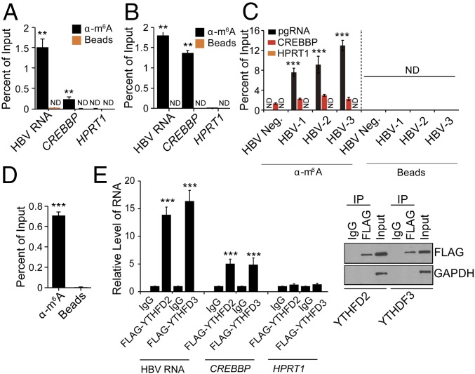
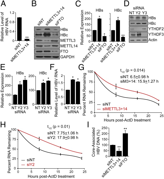
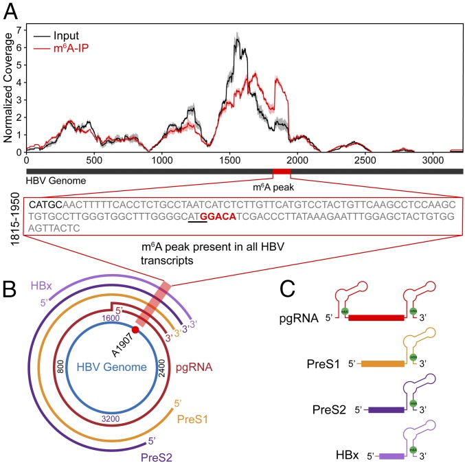
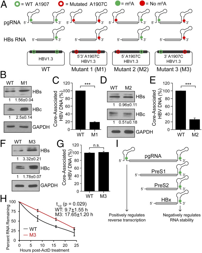
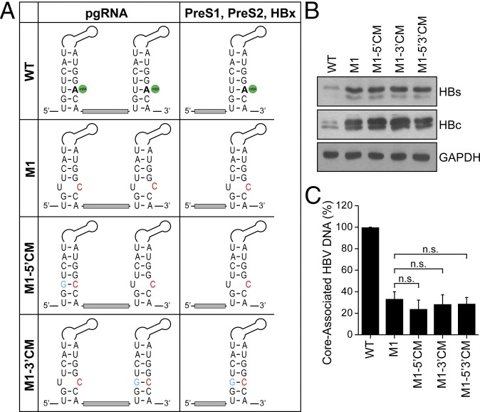
N6-methyladenosine (m6A) RNA methylation is the most abundant epitranscriptomic modification of eukaryotic messenger RNAs (mRNAs). Previous reports have found m6A on both cellular and viral transcripts and defined its role in regulating numerous biological processes, including viral infection. Here, we show that m6A and its associated machinery regulate the life cycle of hepatitis B virus (HBV). HBV is a DNA virus that completes its life cycle via an RNA intermediate, termed pregenomic RNA (pgRNA). Silencing of enzymes that catalyze the addition of m6A to RNA resulted in increased HBV protein expression, but overall reduced reverse transcription of the pgRNA. We mapped the m6A site in the HBV RNA and found that a conserved m6A consensus motif situated within the epsilon stem loop structure, is the site for m6A modification. The epsilon stem loop is located in the 3' terminus of all HBV mRNAs and at both the 5' and 3' termini of the pgRNA. Mutational analysis of the identified m6A site in the 5' epsilon stem loop of pgRNA revealed that m6A at this site is required for efficient reverse transcription of pgRNA, while m6A methylation of the 3' epsilon stem loop results in destabilization of all HBV transcripts, suggesting that m6A has dual regulatory function for HBV RNA. Overall, this study reveals molecular insights into how m6A regulates HBV gene expression and reverse transcription, leading to an increased level of understanding of the HBV life cycle.

Keywords: HBV reverse transcription; RNA methylation; epsilon loop; hepatitis B virus.


Conflict of interest statement

The authors declare no conflict of interest.

Figures

Fig. 1.
Fig. 1.
HBV transcripts contain the m6A RNA modification. MeRIP–RT-qPCR of m6A-modified HBV transcripts from total RNA extracted from (A) HepAD38 cells stably expressing HBV and (B) HepG2 cells transfected with the HBV 1.3mer genome using primers specific for the shared sequence in the 3′ UTR of all HBV RNAs. CREBBP and HPRT1 serve as positive and negative controls, respectively. (C) MeRIP–RT-qPCR analysis of total RNA from liver biopsy samples from a healthy individual (n = 1) and HBV patients (n = 3) using primers specific to pgRNA. (D) MeRIP–RT-qPCR analysis of core-associated RNA. (E) RNA immunoprecipitation (IP) from FLAG-YTHDF2 and -YTHDF3 HepAD38-HBV expression cells using an anti-FLAG antibody or IgG, with RT-qPCR analysis of HBV RNA, CREBBP, and HPRT1 were quantified as the percent of input and graphed as fold enrichment relative to IgG control. Immunoblot analysis of FLAG-YTHDF2/3 in the input and IP is shown on the Right. For AD, the fraction of m6A-modified RNA was calculated as the percent of the level present in the eluate compared with the total input RNA. The data for this figure are from three independent experiments and the bars represent the mean ± SD. **P ≤ 0.01 and ***P ≤ 0.001. ND, not detected.
Fig. 2.
Fig. 2.
Effect of depletion of methyltransferases (METTL3/14) and demethylases (FTO) on HBV protein expression, HBV RNA stability, and reverse transcription. (A) MeRIP–RT-qPCR analysis of HBV RNA harvested from HBV-induced HepAD38 cells following siRNA depletion of METTL3 and METTL14 or nontargeting control (NT). RNA was immunoprecipitated with an anti-m6A antibody and eluted RNA was quantified as a percent of input and graphed as fraction relative to the m6A level in siNT. (B) Immunoblot analysis of HBV proteins (surface antigen, HBs) and (core, HBc) from extracts of HepG2 cells expressing the HBV 1.3mer plasmid following siRNA depletion of METTL3+METTL14, and FTO, or NT at 5 d post-HBV transfection. (C) HBV proteins levels relative to the housekeeping gene GAPDH from three independent experiments, as in B, were quantified using ImageJ. (D) Immunoblot analysis of HBs and HBc from extracts of HepG2 cells expressing the HBV 1.3mer plasmid following siRNA depletion of YTHDF2 or YTHDF3, or NT at 5 d post-HBV transfection. (E) HBV protein levels relative to the housekeeping gene Actin from three independent experiments, as in D, were quantified using ImageJ. (F) RT-qPCR analysis of HBV RNA relative to GAPDH in HepG2 cells expressing the HBV 1.3mer plasmid. At 2 d post-HBV transfection, siYTHDF2 or siYTHDF3 were added and RNA was harvested at 5 d post-HBV transfection. (G) RT-qPCR analysis of HBV RNA relative to GAPDH in the HBV 1.3mer-expressing HepG2 cells. The HBV 1.3mer-transfected HepG2 cells were depleted for METTL3 and METTL14 by siRNA, following actinomycin D treatment at 24 h post-siRNA transfection. RNA was harvested at 0, 8, 16, and 24 h post actinomycin D treatment and relative levels of remaining HBV transcript were analyzed. (H) RT-qPCR analysis of HBV RNA relative to GAPDH in the HBV 1.3mer-expressing HepG2 cells. The HBV 1.3mer-transfected HepG2 cells were depleted for YTHDF2 by specific siRNA, following actinomycin D treatment at 24 h post-siRNA transfection. RNA was harvested at 0, 8, 16, and 24 h post actinomycin D treatment and relative levels of remaining HBV transcript were analyzed. (I) Following siRNA treatment for depletion of METTL3 and METTL14, FTO, or NT in HBV-expressing HepG2 cells, the core particles were isolated (Materials and Methods). The core-associated HBV DNA was then purified and quantified by qPCR assay. The values are graphed as percent relative to the siNT, which was set at 100%. All experiments were performed in triplicate. Immunoblots shown are representative of three independent experiments, and the graph bars represent the mean ± SD of these three independent experiments. *P ≤ 0.05 and **P ≤ 0.01 by unpaired Student’s t test.
Fig. 3.
Fig. 3.
Consensus m6A site within the HBV genome. (A) Map of m6A-binding sites in the HBV ayw genome by MeRIP-seq of polyA-RNA isolated from HBV expressing HepG2 cells at 5 d postinfection. Read coverage, normalized to the total number of reads mapping to the viral genome for each experiment, is in red for MeRIP-seq and in gray for input RNA-seq. Means and SDs across replicates are shown for each position in the genome. One m6A peak was identified after normalizing for coverage, indicated by the red bar within the black bar that depicts a linear representation of the HBV genome. The Inset presents nt 1815–1950 of the HBV genome, with the m6A site highlighted by red text and the ATG of core ORF underlined. (B) The location of the m6A site (A1907) in the pictorial representation of transcripts within the HBV genome is indicated by red shading. (C) Schematic showing the position of the m6A site (A1907), indicated by the green filled circle in all of the HBV transcripts. Note that it is present at both the 5′ and 3′ ends of pgRNA and only at 3′ ends of the other HBV transcripts.
Fig. 4.
Fig. 4.
Mutations of 5′ and 3′ DRACH motifs and their effect on HBV protein expression and DNA synthesis. (A) Schematics indicate the location of the A1907C mutations of 5′ and 3′ m6A sites in HBV RNAs. Open circles represent WT (green) and A1907C mutations (red) in HBV plasmid DNA. Green solid circles indicate m6A modification while red solid circles represent lack of m6A (due to A1907C mutations) in encoded RNAs. HBV-M1 having the A1907C mutation at both termini, HBV-M2 only at the 5′ end, and HBV-M3 only at the 3′ end. (BG) Immunoblot analysis of HBV proteins following transfection of indicated HBV constructs in HepG2 cells with relative quantifications below the respective blots [(B) M1, (D) M2, (F) M3] and core-associated DNA isolated from core particles and quantified by qPCR using equal amounts of purified DNA in the input. Relative levels of core-associated DNA from the various constructs [(C) M1, (E) M2, (G) M3] graphed as percent relative to HBV-WT. (H) RT-qPCR analysis of relative levels of remaining HBV transcript relative to GAPDH isolated at indicated times following 24 h of actinomycin D treatment from HBV 1.3mer (WT or M3)-expressing HepG2 cells. (I) Model for how m6A at the 5′ or 3′ epsilon stem loops of HBV transcripts differentially regulates their stability and reverse transcription. Data here are presented from three independent experiments and the bars represent mean ± SD. ***P ≤ 0.001 by unpaired Student’s t test, and n.s., not significant.
Fig. 5.
Fig. 5.
Compensatory mutations in the epsilon structure in the HBV 1.3-expressing plasmid do not restore reverse transcription. (A) The epsilon loop within HBV RNAs is depicted, with the m6A site indicated in green, to highlight the HBV base pairing (nt 1849–1861 with nt 1897–1909) in the lower stem and to show that the A1907C substitution in HBV-M1 (red) is predicted to create a bubble. To restore the base pairing with C1907 the compensatory U1851G mutation (blue) was introduced either at the 5′ end (HBV-M1-5′ CM), the 3′ end (HBV-M1-3′ CM), or both (HBV-M1-5′3′ CM). (B and C) Using these constructs, HBV protein expression (B) and core-associated DNA (C) were analyzed, as done previously. The data are the presentation of three independent experiments. The bars represent mean ± SD. n.s., not significant by unpaired Student’s t test.

Similar articles

Cited by

References

    1. Yue Y, Liu J, He C. RNA N6-methyladenosine methylation in post-transcriptional gene expression regulation. Genes Dev. 2015;29:1343–1355. - PMC - PubMed
    1. Li S, Mason CE. The pivotal regulatory landscape of RNA modifications. Annu Rev Genomics Hum Genet. 2014;15:127–150. - PubMed
    1. Saletore Y, et al. The birth of the epitranscriptome: Deciphering the function of RNA modifications. Genome Biol. 2012;13:175. - PMC - PubMed
    1. Zhou J, et al. Dynamic m(6)A mRNA methylation directs translational control of heat shock response. Nature. 2015;526:591–594. - PMC - PubMed
    1. Dimock K, Stoltzfus CM. Sequence specificity of internal methylation in B77 avian sarcoma virus RNA subunits. Biochemistry. 1977;16:471–478. - PubMed

Publication types