. 2018 Sep 28;293(39):15136-15151.
doi: 10.1074/jbc.RA118.003290. Epub 2018 Aug 13.

Bond swapping from a charge cloud allows flexible coordination of upstream signals through WASP: Multiple regulatory roles for the WASP basic region

Affiliations

Bond swapping from a charge cloud allows flexible coordination of upstream signals through WASP: Multiple regulatory roles for the WASP basic region

George J N Tetley et al. J Biol Chem. .

Abstract

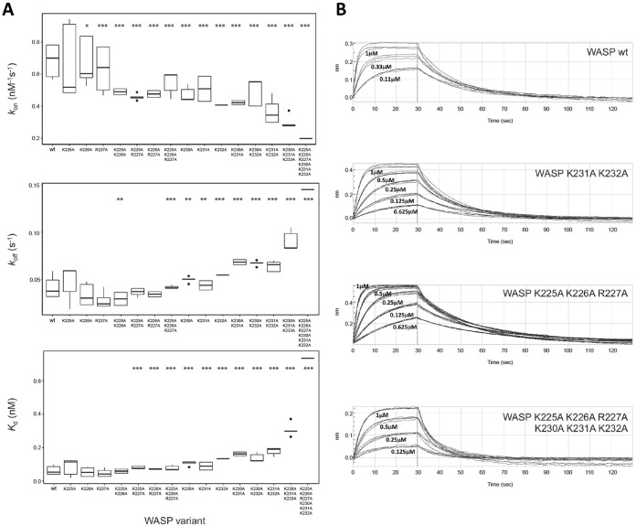
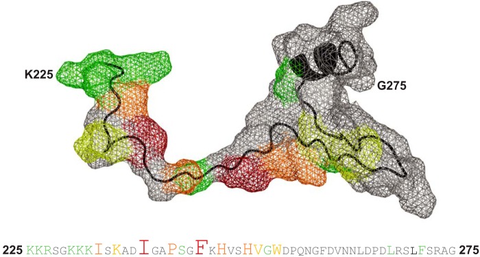
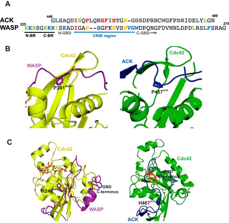
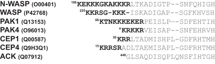
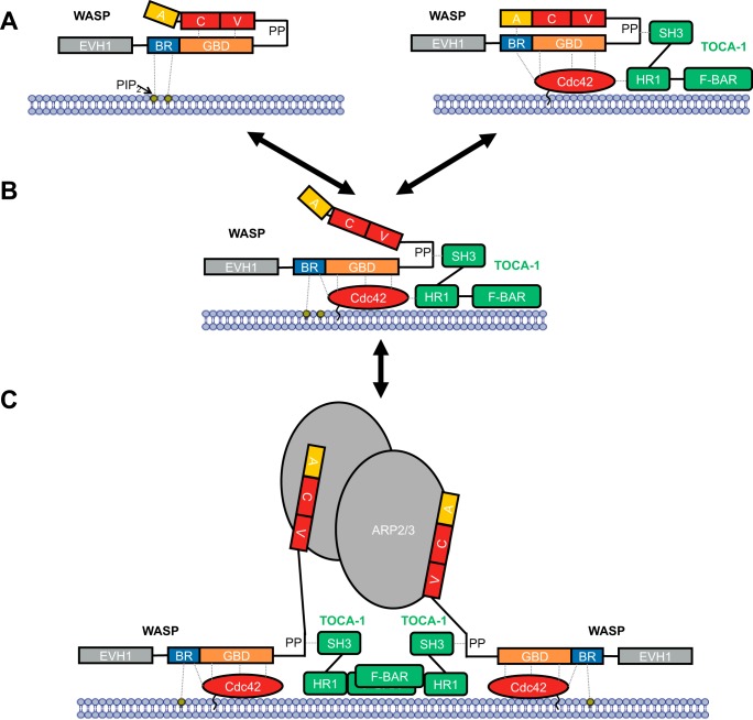
Wiskott-Aldrich syndrome protein (WASP) activates the actin-related protein 2/3 homolog (Arp2/3) complex and regulates actin polymerization in a physiological setting. Cell division cycle 42 (Cdc42) is a key activator of WASP, which binds Cdc42 through a Cdc42/Rac-interactive binding (CRIB)-containing region that defines a subset of Cdc42 effectors. Here, using site-directed mutagenesis and binding affinity determination and kinetic assays, we report the results of an investigation into the energetic contributions of individual WASP residues to both the Cdc42-WASP binding interface and the kinetics of complex formation. Our results support the previously proposed dock-and-coalesce binding mechanism, initiated by electrostatic steering driven by WASP's basic region and followed by a coalescence phase likely driven by the conserved CRIB motif. The WASP basic region, however, appears also to play a role in the final complex, as its mutation affected both on- and off-rates, suggesting a more comprehensive physiological role for this region centered on the C-terminal triad of positive residues. These results highlight the expanding roles of the basic region in WASP and other CRIB-containing effector proteins in regulating complex cellular processes and coordinating multiple input signals. The data presented improve our understanding of the Cdc42-WASP interface and also add to the body of information available for Cdc42-effector complex formation, therapeutic targeting of which has promise for Ras-driven cancers. Our findings suggest that combining high-affinity peptide-binding sequences with short electrostatic steering sequences could increase the efficacy of peptidomimetic candidates designed to interfere with Cdc42 signaling in cancer.

Keywords: CDC42; cell signaling; cytoskeleton; intrinsically disordered protein; protein conformation; protein motif; protein–protein interaction; small GTPase; structure–function; thermodynamics.


Conflict of interest statement

The authors declare that they have no conflicts of interest with the contents of this article

Figures

Figure 1.
Figure 1.
Domain architecture of WASP. Structural domains and regions with assigned function of WASP are highlighted as follows: the Ena/VASP homology domain 1 (EVH1, gray); the basic region (BR, blue); the G protein-binding domain (GBD, orange); the central proline-rich domain (PRD); the verprolin homology (V, red); central (C, red); and acidic region (A, yellow). WASP exists in an autoinhibited conformation when not bound by effectors, and the BR and A regions and the CV and GBD regions form contacts that prevent the VCA region binding and activating Arp2/3. Only WASP residues 225–275, which include the BR and residues of the GBD mutated in this study, are expanded to show their sequence below, and the residues selected for mutagenesis are highlighted. Residues colored black are CRIB consensus residues, which were all subject to mutation in this study; residues outside the CRIB region mutated in this work are colored red. The CRIB consensus sequence is also included. Secondary structure elements in WASP when bound to Cdc42 are shown above the sequence. β-Strands are denoted by arrows and the helix by a cylinder. The limits of the secondary structure elements are taken from Ref. with amendments included from Ref. . The accession number for WASP is UniProt no. P42768.
Figure 2.
Figure 2.
Direct SPA-binding data for the WASP GBD and mutant variants, with Cdc42. The indicated concentration of [3H]GTP-labeled Cdc42 was incubated with GST-tagged WASP GBD variants, as appropriate. The SPA signal was corrected by subtraction of the background signal from parallel measurements in which the effector protein was omitted. The effect of the concentration of Cdc42 on this corrected SPA signal was fitted to a binding isotherm to give an apparent Kd value and the signal at saturating Cdc42 concentrations. The data and curve fits are displayed as a percentage of this maximal signal. A, binding of representative mutants in the WASP GBD to Cdc42. B, binding of WASP BR hexa-mutant and C-terminal deletion mutant to Cdc42. 2–4 experimental replicates were performed for each WASP variant with 12 data points in each. A summary of all the binding data can be found in Table 2.
Figure 3.
Figure 3.
Structural analysis of mutational effects. A, schematic representation of part of the Cdc42/WASP–GBD structure (Ref. and PDB 1CEE) showing the contacts made by Ile-233 and Ile-238WASP. The van der Waals surfaces of relevant residues are shown either as a mesh or a semi-transparent surface (using the PyMOL default solvent radius of 1.4 Å). Cdc42 is colored green, and WASP is colored blue. B, schematic representation of part of the Cdc42/WASP–GBD structure showing the contacts made by Phe-244WASP. Coloring is as in A. C, schematic representation of part of the Cdc42/WASP–GBD structure showing the contacts made by Pro-241WASP. Coloring is as in A. D, schematic representation of part of the Cdc42/WASP–GBD structure showing the contacts made by His-246WASP and His-249WASP. Coloring is as in A. E, schematic representation of part of the Cdc42/WASP–GBD structure showing the contacts made by Val-250WASP. Coloring is as in A. F, schematic representation of part of the Cdc42/WASP–GBD structure showing the contacts made by Ser-242WASP. Coloring is as in A. G, schematic representation of part of the Cdc42/WASP–GBD structure showing the contacts made by Lys-235WASP. Coloring is as in A. H, schematic representation of part of the Cdc42/WASP–GBD structure showing the contacts made by Trp-252WASP. Coloring is as in A. Where highlighted, oxygen atoms are colored red and nitrogen in blue. Magnesium is shown as an orange sphere.
Figure 4.
Figure 4.
Direct SPA-binding data for the WASP GBD and mutant BR variants, with Cdc42. The indicated concentration of [3H]GTP-labeled Cdc42 was incubated with GST-tagged WASP GBD BR variants, as appropriate, in each SPA. The SPA signal was corrected by subtraction of the background signal from parallel measurements in which the effector protein was omitted. The effect of the concentration of Cdc42 on this corrected SPA signal was fitted to a binding isotherm to give an apparent Kd value and the signal at saturating Cdc42 concentrations. The data and curve fits are displayed as a percentage of this maximal signal. 2–4 experimental replicates were performed for each WASP variant with 12 data points in each. A summary of all the binding data can be found in Table 3.
Figure 5.
Figure 5.
Binding kinetics for WASP BR mutants measured by bio-layer interferometry. GST fusion proteins were loaded onto anti-GST sensors and dipped alternately into a concentration range of Cdc42·GMPPNP (of at least three different concentrations) and buffer to measure on- and off-rates. A, data for kon, koff, and Kd, where n = 3–9 independent experiments, plotted as boxplots showing median and quartile values with data outlying the distribution appearing as dots for all mutants. Statistical significance from WT values is indicated as follows: *, p ≤ 0.05; **, p ≤ 0.01; ***, p ≤ 0.001 as analyzed by t test with the false discovery rate controlled by the Benjamini-Hochberg method (analysis performed in R). Note: K225A/K226A value is statistically significantly lower than the WT but not than the other single mutations in the N-terminal triad; therefore, this is most likely a false discovery error. B, raw data from representative experiments. The fitted values from these data are reported in Table 4.
Figure 6.
Figure 6.
Energetic contribution of individual WASP residues to Cdc42 binding. The upper panel shows the van der Waals surface of WASP residues 225–275 as a gray mesh with the protein backbone in black. Residues involved in interaction with Cdc42 are colored with respect to the fold-change in Kd upon mutation to alanine as follows: green for 5–15-fold increase; yellow for 15–40-fold increase; orange for 40–100-fold increase; and red for a >100-fold increase. The amino acid sequence for this region is shown in the lower panel, with residues colored in the same manner and with font size also indicating relative energetic contribution to complex formation. The accession number for WASP is P42768.
Figure 7.
Figure 7.
Key differences in ACK and WASP binding to Cdc42. A, alignment of the ACK GBD and WASP BR–GBD sequences. The UniProt accession number for WASP is P42768 and for ACK is Q07912. Residues are colored according to their effect on equilibrium constant: gray, 0–5-fold increase; green, 5–10-fold increase; blue, 10–50-fold increase; orange, 50–100-fold increase; red, 100 or greater fold increase. The CRIB region, N- and C-terminal BR triads, and N- and C-terminal regions of the GBDs are labeled. B, schematic representation of part of the Cdc42–WASP GBD (yellow/magenta, PDB code 1CEE (14)) and Cdc42–ACK GBD (green/cyan, respectively, PDB code 1CF4 (12)) structures highlighting Pro-241WASP and Pro-457ACK. C, schematic representation of part of the Cdc42–WASP GBD (yellow/magenta, PDB code 1CEE (14)) and Cdc42–ACK GBD (green/cyan, respectively, PDB code 1CF4 (12)) structures highlighting the positions of the C-terminal regions of the WASP and ACK GBDs. Coloring is as in B. The nucleotide is shown as a stick representation, colored orange.
Figure 8.
Figure 8.
Alignment of the basic regions of CRIB effector proteins. Accession numbers for all proteins are shown, as are starting residue numbers. Basic region residues are highlighted in black.
Figure 9.
Figure 9.
Model for WASP activation. WASP is represented as in Fig. 1, together with binding partners PIP2 (labeled, yellow circle), Cdc42 (red oval), TOCA1 (green, composed of domains SH3, HR1, and F-BAR), and Arp2/3 (gray ovals). The cell membrane is represented by a blue bilayer with which PIP2 and Cdc42 are associated. A, WASP binding to PIP2 (left panel) or WASP binding to Cdc42 (right panel) does not stimulate VCA release alone, but binding of both can relieve autoinhibitory interactions to release the VCA region. B, this can then bind to Arp2/3, which is fully activated by binding two VCA regions. C, dimerization through scaffolds such as TOCA1 can facilitate Arp2/3 activation by two VCA regions.

References

    1. Cherfils J., and Zeghouf M. (2013) Regulation of small GTPases BY GEFs, GAPs, and GDIs. Physiol. Rev. 93, 269–309 10.1152/physrev.00003.2012 - DOI - PubMed
    1. Ridley A. J. (2015) Rho GTPase signalling in cell migration. Curr. Opin. Cell Biol. 36, 103–112 10.1016/j.ceb.2015.08.005 - DOI - PMC - PubMed
    1. Arias-Romero L. E., and Chernoff J. (2013) Targeting Cdc42 in cancer. Expert Opin. Ther. Targets 17, 1263–1273 10.1517/14728222.2013.828037 - DOI - PMC - PubMed
    1. Stengel K. R., and Zheng Y. (2012) Essential role of Cdc42 in ras-induced transformation revealed by gene targeting. PLoS ONE 7, e37317 10.1371/journal.pone.0037317 - DOI - PMC - PubMed
    1. Fritz G., Brachetti C., Bahlmann F., Schmidt M., and Kaina B. (2002) Rho GTPases in human breast tumours: expression and mutation analyses and correlation with clinical parameters. Br. J. Cancer 87, 635–644 10.1038/sj.bjc.6600510 - DOI - PMC - PubMed

Publication types

MeSH terms

LinkOut - more resources