CD147 and Cyclooxygenase Expression in Feline Oral Squamous Cell Carcinoma
- PMID: 30104530
- PMCID: PMC6163611
- DOI: 10.3390/vetsci5030072
CD147 and Cyclooxygenase Expression in Feline Oral Squamous Cell Carcinoma
Abstract
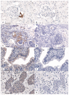
Feline oral squamous cell carcinoma (OSCC) is a highly invasive form of cancer in cats. In human OSCC, cluster of differentiation 147 (CD147) contributes to inflammation and tumor invasiveness. CD147 is a potential therapeutic target, but the expression of CD147 in feline OSCC has not been examined. Immunohistochemistry was used to determine if cyclooxygenase 2 (COX-2) and CD147 expression in feline OSCC biopsies was coordinated. Tumor cells were more likely to express COX-2 (22/43 cases or 51%) compared to stroma (8/43 or 19%) and adjacent oral epithelium (9/31 cases or 29%) (p < 0.05). CD147 was also more likely to occur in tumor cells compared to stroma and adjacent mucosa, with 21/43 (49%) of cases having >50% tumor cells with mild or moderate CD147 expression, compared to 9/28 (32%) in adjacent epithelium and only 5/43 (12%) in adjacent stroma (p < 0.05). In feline OSCC cell lines (SCCF1, SCCF2, and SCCF3), CD147 gene expression was more consistently expressed compared to COX-2, which was 60-fold higher in SCCF2 cells compared to SCCF1 cells (p < 0.05). CD147 expression did not correlate with COX-2 expression and prostaglandin E2 (PGE2) secretion, indicating that they may be independently regulated. CD147 potentially represents a novel therapeutic target for the treatment of feline OSCC and further study of CD147 is warranted.
Keywords: Basigin; CD147; COX-1; COX-2; EMMPRIN; PGE2; feline; inflammation; invasion; oral squamous cell carcinoma.
Conflict of interest statement
The authors declare no conflicts of interest.
Figures


Similar articles
-
In vitro expression of genes encoding HIF1α, VEGFA, PGE2 synthases, and PGE2 receptors in feline oral squamous cell carcinoma.J Vet Diagn Invest. 2025 Mar;37(2):223-233. doi: 10.1177/10406387251315677. Epub 2025 Feb 10. J Vet Diagn Invest. 2025. PMID: 39930728 Free PMC article.
-
Cyclooxygenase and CD147 expression in oral squamous cell carcinoma patient samples and cell lines.Oral Surg Oral Med Oral Pathol Oral Radiol. 2019 Oct;128(4):400-410.e3. doi: 10.1016/j.oooo.2019.06.005. Epub 2019 Jun 14. Oral Surg Oral Med Oral Pathol Oral Radiol. 2019. PMID: 31350224
-
Investigation of immune cell markers in feline oral squamous cell carcinoma.Vet Immunol Immunopathol. 2018 Aug;202:52-62. doi: 10.1016/j.vetimm.2018.06.011. Epub 2018 Jun 13. Vet Immunol Immunopathol. 2018. PMID: 30078599 Free PMC article.
-
Role of COX-2/PGE2 Mediated Inflammation in Oral Squamous Cell Carcinoma.Cancers (Basel). 2018 Sep 22;10(10):348. doi: 10.3390/cancers10100348. Cancers (Basel). 2018. PMID: 30248985 Free PMC article. Review.
-
The Multiple Roles of CD147 in the Development and Progression of Oral Squamous Cell Carcinoma: An Overview.Int J Mol Sci. 2022 Jul 28;23(15):8336. doi: 10.3390/ijms23158336. Int J Mol Sci. 2022. PMID: 35955471 Free PMC article. Review.
Cited by
-
Investigating Cox-2 and EGFR as Biomarkers in Canine Oral Squamous Cell Carcinoma: Implications for Diagnosis and Therapy.Curr Issues Mol Biol. 2024 Jan 4;46(1):485-497. doi: 10.3390/cimb46010031. Curr Issues Mol Biol. 2024. PMID: 38248333 Free PMC article.
-
In vitro expression of genes encoding HIF1α, VEGFA, PGE2 synthases, and PGE2 receptors in feline oral squamous cell carcinoma.J Vet Diagn Invest. 2025 Mar;37(2):223-233. doi: 10.1177/10406387251315677. Epub 2025 Feb 10. J Vet Diagn Invest. 2025. PMID: 39930728 Free PMC article.
-
Dual effects of an anti-CD147 antibody for Esophageal cancer therapy.Cancer Biol Ther. 2019;20(12):1443-1452. doi: 10.1080/15384047.2019.1647052. Epub 2019 Aug 14. Cancer Biol Ther. 2019. PMID: 31411555 Free PMC article.
-
CD147 promotes glucose metabolism, invasion and metastasis via PI3K/AKT pathway in oral squamous cell carcinomas.Transl Cancer Res. 2019 Aug;8(4):1486-1496. doi: 10.21037/tcr.2019.07.50. Transl Cancer Res. 2019. PMID: 35116891 Free PMC article.
-
Long-term survival in a cat with tonsillar squamous cell carcinoma treated with surgery and chemotherapy.JFMS Open Rep. 2021 Feb 5;7(1):2055116920984387. doi: 10.1177/2055116920984387. eCollection 2021 Jan-Jun. JFMS Open Rep. 2021. PMID: 33614106 Free PMC article.
References
-
- DiBernardi L., Dore M., Davis J.A., Owens J.G., Mohammed S.I., Guptill C.F., Knapp D.W. Study of feline oral squamous cell carcinoma: Potential target for cyclooxygenase inhibitor treatment. Prostaglandins Leukot. Essent. Fatty Acids. 2007;76:245–250. doi: 10.1016/j.plefa.2007.01.006. - DOI - PubMed
-
- Marretta J.J., Garrett L.D., Marretta S.M. Feline oral squamous cell carcinoma: An overview. Vet. Med. 2007;102:392–406.
LinkOut - more resources
Full Text Sources
Other Literature Sources
Research Materials
Miscellaneous

