. 2018 Nov;11(6):1621-1629.
doi: 10.1038/s41385-018-0068-6. Epub 2018 Aug 13.

Chronic intestinal inflammation in mice expressing viral Flip in epithelial cells

Affiliations

Chronic intestinal inflammation in mice expressing viral Flip in epithelial cells

Barbara Ruder et al. Mucosal Immunol. 2018 Nov.

Abstract

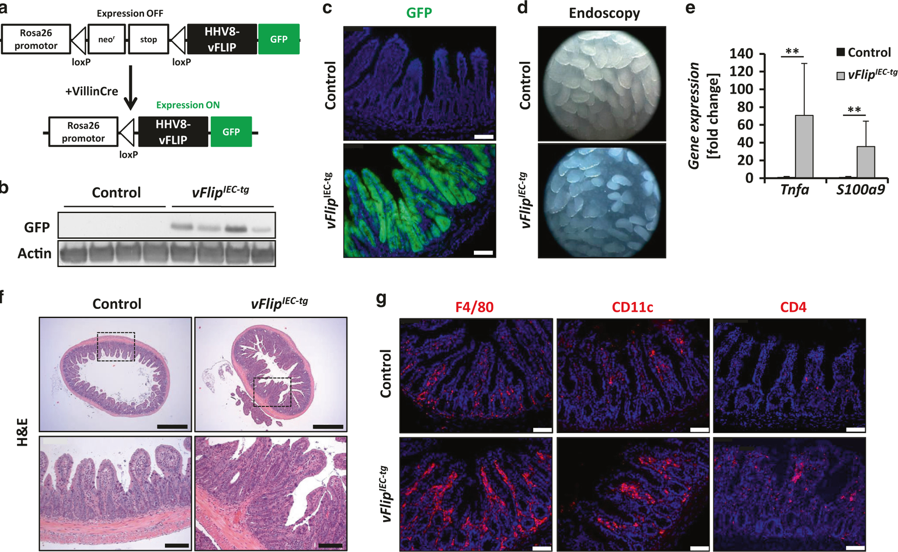
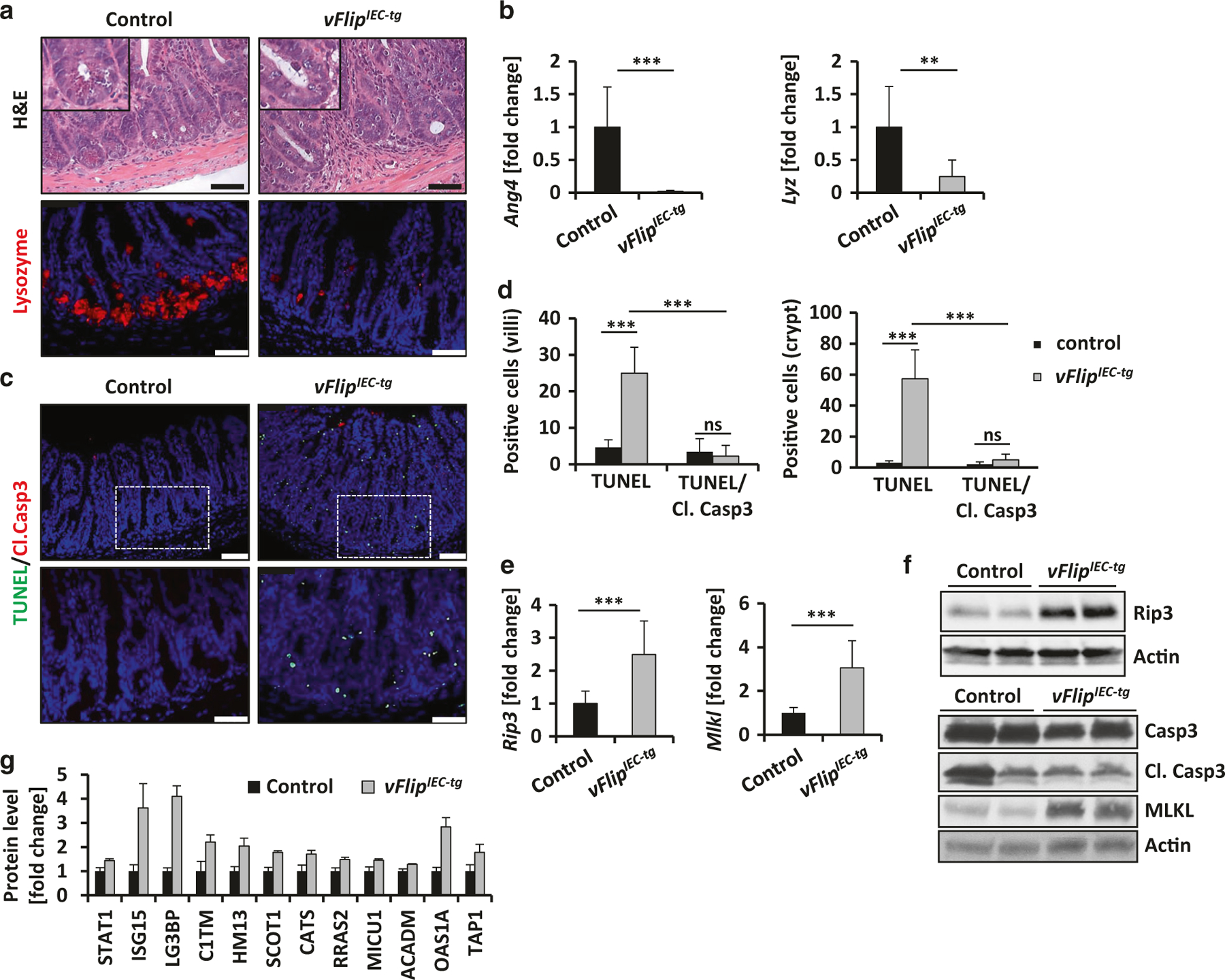
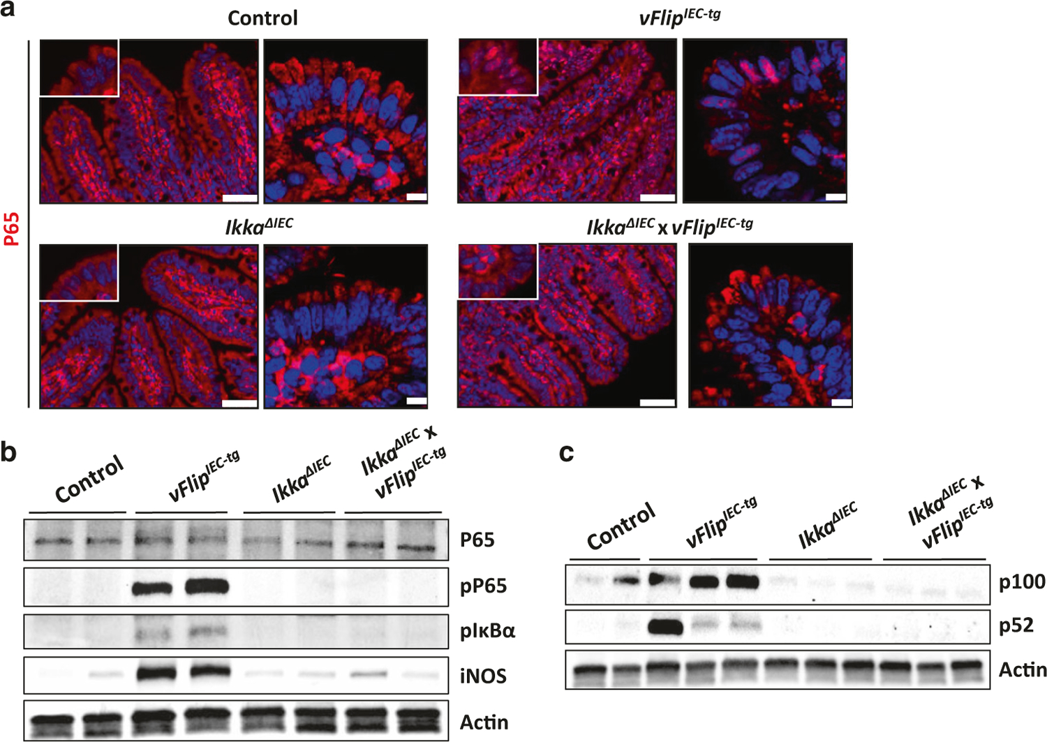
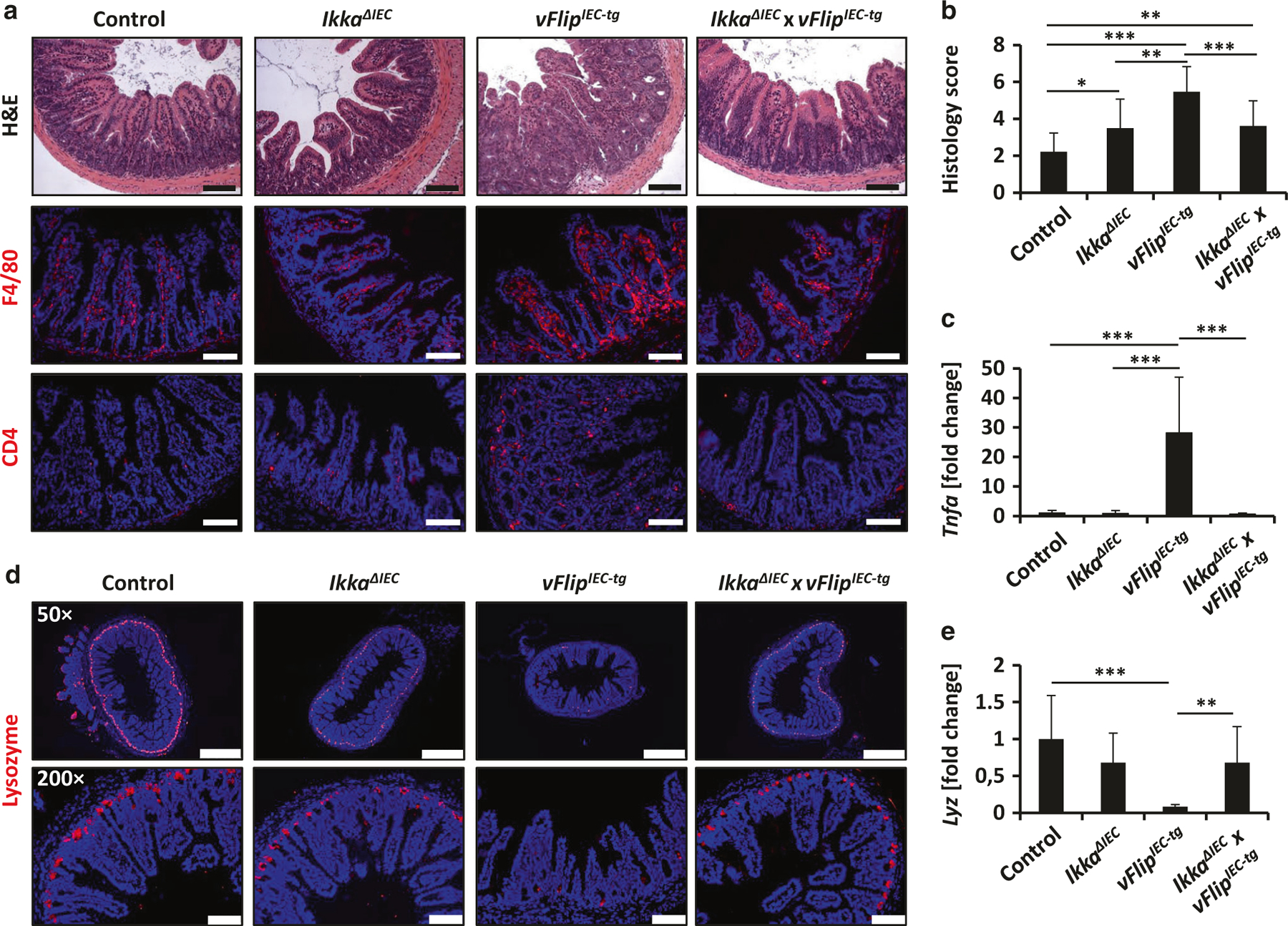
Viruses are present in the intestinal microflora and are currently discussed as a potential causative mechanism for the development of inflammatory bowel disease. A number of viruses, such as Human Herpesvirus-8, express homologs to cellular FLIPs, which are major contributors for the regulation of epithelial cell death. In this study we analyzed the consequences of constitutive expression of HHV8-viral FLIP in intestinal epithelial cells (IECs) in mice. Surprisingly, expression of vFlip disrupts tissue homeostasis and induces severe intestinal inflammation. Moreover vFlipIEC-tg mice showed reduced Paneth cell numbers, associated with excessive necrotic cell death. On a molecular level vFlip expression altered classical and alternative NFκB activation. Blocking of alternative NFκB signaling by deletion of Ikka in vivo largely protected mice from inflammation and Paneth cell loss induced by vFLIP. Collectively, our data provide functional evidence that expression of a single viral protein in IECs can be sufficient to disrupt epithelial homeostasis and to initiate chronic intestinal inflammation.


Conflict of interest statement

Competing interests: The authors declare no competing interests.

Figures

Fig. 1
Fig. 1
vFlipIEC-tg mice spontaneously develop severe intestinal inflammation. a Strategy for generating vFlipIEC-tg mice. Rosa26.vFLIP knockin mice were crossed to VillinCre mice to generate mice constitutively expressing vFlip in IECs. b Western Blot analysis of GFP using small intestinal IEC lysates from indicated mice. Actin was used as a loading control. c Representative pictures of GFP fluorescence staining of small intestinal cross sections from indicated mice. Scale bar 75 µm. d Representative endoscopic pictures from the small intestine of indicated mice. e Transcriptional analysis of Tnfa (n ≥ 10) and S100a9 (n ≥ 8) in the small intestine of control and vFlipIEC-tg mice. Values are shown + SD and were calculated relative to control mice. Hprt was used as internal standard. Data were pooled from 4 individual experiments, **p < 0.01. f Representative pictures of H&E stained small intestinal cross sections from indicated mice. Scale bar upper panel 500 µm, lower panel 100 µm. g Representative pictures of immunohistochemical stainings of small intestinal cross sections from indicated mice using antibodies against F4/80, CD11c and CD4. Scale bar 75 µm
Fig. 2
Fig. 2
vFlipIEC-tg mice are characterized by reduced Paneth cell numbers and increased cell death. a Representative pictures of H&E and immunohistochemical Lysozyme staining of small intestinal cross sections of indicated mice. Scale bar 50 µm. b Transcriptional analysis of Ang4 (n = 11) and Lyz (n = 11) in the small intestine of indicated mice. Values are shown + SD and were calculated relative to control mice. Hprt was used as internal standard. Data were pooled from 4 individual experiments, **p < 0.01, ***p < 0.001. c Representative pictures of TUNEL/Cleaved Caspase-3 doublestaining of small intestinal cross sections from indicated mice. Scale bar upper panel: 75 µm, lower panel: 50 µm. d Quantification of cell death of IECs located at the villus or the crypt area. Villi: n(control) = 5, n(vFlipIEC-tg) = 10, crypts: n(control) = 8, n(vFlipIEC-tg) = 13, ***p < 0.001. e Transcriptional analysis of Rip3 (n ≥ 9) and Mlkl (n ≥ 9) in the small intestine of indicated mice. Values are shown + SD and were calculated relative to control mice. Hprt was used as internal standard. Data were pooled from 3 individual experiments, ***p < 0,001. f Western Blot analyses of small intestinal IEC lysates from indicated mice using antibodies against RIP3, Caspase-3, its cleaved forms and MLKL. Actin was used as a loading control. g Quantitative proteome analysis of known and putative STAT1 target genes in small intestinal epithelial lysates of control and vFlipIEC-tg mice (n = 3). Values are shown + SD and were calculated relative to control mice, p < 0.05 for all target genes between both groups
Fig. 3
Fig. 3
vFlip expression activates the NFκB pathway in IECs. a Representative pictures of an immunohistochemical P65 staining of small intestinal cross sections from indicated mice. Left panel: microscopic pictures, scale bar 50 µm. Right panel: confocal images of nuclear P65 translocation. Scale bar 7.5 µm. b Western Blot analyses of P65, phospho-P65, phospho-IκBα and iNOS using proteins isolated from small intestinal IECs of indicated mice. Actin was used as a loading control. c Western Blot analyses of P100 and P52 using proteins from small intestinal IECs of indicated mice. Actin was used as a loading control
Fig. 4
Fig. 4
vFLIP-induced inflammation and Paneth cell loss depends on NFκB signaling. a Representative staining of H&E and immunohistochemical F4/80 and CD4 staining on small intestinal cross sections from indicated mice. Scale bar 100 µm. b Histology score of H&E stained small intestinal cross sections of control (n = 18), IkkaΔIEC (n = 10), vFlipIEC-tg (n = 17) and IkkaΔIEC x vFlipIEC-tg (n = 14) animals, *p < 0.05, **p < 0.01, ***p < 0.001. b Transcriptional analysis of Tnfa (n ≥ 9) in the small intestine of control and vFlipIEC-tg mice. Values are shown + SD and were calculated relative to control mice. Hprt was used as internal standard. Data were pooled from 3 individual experiments, ***p < 0.001. c Representative pictures of an immunohistochemical lysozyme staining of small intestinal cross sections from indicated mice. Scale bar upper panel: 500 µm, lower panel: 100 µm. d Transcriptional analysis of Lyz (n ≥ 9) in the small intestine of control and vFlipIEC-tg mice. Values are shown + SD and were calculated relative to control mice. Hprt was used as internal standard. Data were pooled from 3 individual experiments, **p < 0.01, ***p < 0.001

References

    1. Gunther C, Buchen B, Neurath MF & Becker C Regulation and pathophysiological role of epithelial turnover in the gut. Semin. Cell Dev. Biol 35, 40–50 (2014). - PubMed
    1. Wittkopf N et al. Cellular FLICE-like inhibitory protein secures intestinal epithelial cell survival and immune homeostasis by regulating caspase-8. Gastroenterology 145, 1369–1379 (2013). - PubMed
    1. Gunther C et al. Caspase-8 regulates TNF-alpha-induced epithelial necroptosis and terminal ileitis. Nature 477, 335–339 (2011). - PMC - PubMed
    1. Degterev A et al. Chemical inhibitor of nonapoptotic cell death with therapeutic potential for ischemic brain injury. Nat. Chem. Biol 1, 112–119 (2005). - PubMed
    1. Feoktistova M, Geserick P, Panayotova-Dimitrova D & Leverkus M Pick your poison: the Ripoptosome, a cell death platform regulating apoptosis and necroptosis. Cell Cycle 11, 460–467 (2012). - PubMed

Publication types

MeSH terms