Comparative Study
. 1986 Jun 6;232(4755):1230-7.
doi: 10.1126/science.3010466.

Deletion in cysteine-rich region of LDL receptor impedes transport to cell surface in WHHL rabbit

Comparative Study

Deletion in cysteine-rich region of LDL receptor impedes transport to cell surface in WHHL rabbit

T Yamamoto et al. Science. .

Abstract

The Watanabe heritable hyperlipidemic (WHHL) rabbit, an animal with familial hypercholesterolemia, produces a mutant receptor for plasma low-density lipoprotein (LDL) that is not transported to the cell surface at a normal rate. Cloning and sequencing of complementary DNA's from normal and WHHL rabbits, shows that this defect arises from an in-frame deletion of 12 nucleotides that eliminates four amino acids from the cysteine-rich ligand binding domain of the LDL receptor. A similar mutation, detected by S1 nuclease mapping of LDL receptor messenger RNA, occurred in a patient with familial hypercholesterolemia whose receptor also fails to be transported to the cell surface. These findings suggest that animal cells may have fail-safe mechanisms that prevent the surface expression of improperly folded proteins with unpaired or improperly bonded cysteine residues.


Figures

Fig. 1
Fig. 1
Restriction endonuclease map and sequencing strategy for normal and WHHL rabbit LDL receptor cDNA’s. Nucleotide positions in kilobases are indicated by the top scale. The coding region of the mRNA is indicated by the thick black line and the 3’ untranslated region is represented by the thin line in the restriction endonuclease map. Only selected restriction endonuclease sites are shown. The regions of the mRNA corresponding to each of the three partial cDNA clones are indicated below the map. Plasmids pLDLR-11 and pR52 are derived from mRNA of normal New Zealand White rabbits; plasmid pWRl is derived from mRNA isolated from a WHHL rabbit. Arrows emanating from vertical hatch marks indicate the direction and extent of DNA sequence established by the dideoxy chain termination method. The arrow emanating from the dot indicates a region of DNA sequenced by the chemical method. Poly(A)+ RNA was isolated from the livers of estradiol-treated (8) normal or WHHL rabbits by means of a guanidinium isothiocyanate-CsCl procedure (28) followed by oligo dT-cellulose chromatography (29). Complementary DNA libraries containing more than 106 independent transformants in E. coli HB101 were constructed (9) and size-fractionated (30). Plasmids containing LDL receptor cDNA inserts were identified by cross-hybridization under reduced stringency (11) with a 32P-labeled bovine LDL receptor cDNA probe (10). Plasmid pR52, a cDNA clone representing the 5’ end of the normal rabbit LDL receptor mRNA, was isolated by primer extension cloning (11) in a pBR322 vector. DNA sequencing was done either chemically (31) or enzymatically (32) with specific oligonucleotide primers after sub-cloning into bacteriophage Ml3 vectors (33). Routine sequencing gels contained 10 percent polyacrylamide, 100 mM tris-borate (pH 8.3), 7M urea, and 25 percent (v/v) formamide. Bacteriophage Ml3 templates giving rise to intransigent compressions under these conditions were resequenced and subjected to electrophoresis in a warm room (37°C) on 9 percent polyacrylamide gels containing 100 mM tris-borate (pH 8.3), 7M urea, and 38 percent (v/v) formamide (34).
Fig. 2
Fig. 2
Nucleotide sequence and predicted amino acid sequence of the normal rabbit LDL receptor. DNA sequences derived from the partial cDNA plasmids pLDLR-11 and pR52 were determined as described in Fig. 1. Nucleotides are numbered on the right side; position 1 is the most 5’ nucleotide cloned in pR52. Dots are placed above every tenth nucleotide in the sequence. Amino acids are numbered below the sequence; residue 1 is the alanine that is believed to constitute the NH2-terminus of the mature protein; negative numbers refer to residues in the cleaved signal sequence (boxed, NH2-terminus). Cysteine residues are circled. Three potential sites of N-linked glycosylation (Asn-X-Ser or Asn-X-Thr) are indicated by double solid underlines. Serine and threonine residues in a region that corresponds to a domain of the bovine and human LDL receptor that was previously shown to contain clustered O-linked sugars (5, 18) are indicated by dotted underlines. The 25-residue transmembrane segment located toward the COOH-terminus of the protein is boxed. A potential polyadenylation signal at the extreme 3’ end of the sequence is overlined. The sequence of the 3′-untranslated region of the rabbit LDL receptor mRNA has been described (35), and is shown here for the sake of completion.
Fig. 3
Fig. 3
Alignment of the normal rabbit and human LDL receptor protein sequences. Rabbit and human LDL receptor sequences were aligned for maximum homology with the use of a Beck-man Microgenie Align program. (A) Alignment of the amino acid sequences in the five domains of the mature rabbit and human LDL receptors. Asterisks denote identical residues. Amino acids in the respective proteins are numbered on the left and right. Dots are placed above every tenth position in a given comparison line. The single-letter amino acid code translates to the three-letter code as follows: A, Ala; C, Cys; D, Asp; E, Glu; F, Phe; G, Gly; H, His; I, Ile; K, Lys; L, Leu; M, Met; N, Asn; P, Pro; Q, Gln; R, Arg; S, Ser; T, Thr; V, Val; W, Trp; Y, Tyr. (B) Model representing five domains in the mature LDL receptor. The percentage homology between the human and rabbit proteins in a given domain is indicated above the schematic.
Fig. 4
Fig. 4
Alignment of repeat amino acid sequences in the ligand binding domain of the normal rabbit LDL receptor. The sequences of the seven cysteine-rich repeats in the ligand binding domain of the rabbit protein were aligned for maximum homology based on computer-assisted analyses and the structure of the human LDL receptor gene (13). Conserved residues are boxed. A consensus sequence representing amino acids that occur in a given position in four or more repeats is shown below the alignment. The consensus sequence derived from a similar alignment of repeats in the human LDL receptor (13) is also shown. The single-letter abbreviations for amino acid residues are given in the legend to Fig. 3.
Fig. 5
Fig. 5
Nucleotide sequences of LDL receptor cDNA’s from normal and WHHL rabbits in the region between cysteines 113 and 122. DNA sequencing was done by the dideoxy chain termination method (32) with Ml3 templates (33) containing Pst I fragments isolated from the 5′ ends of plasmids pLDLR-11 and pWRl (Fig. 1) and an LDL receptor-specific oligonucleotide primer. A 12-nt deletion is apparent in the sequence obtained from the WHHL-derived template. The consequences of this deletion for the LDL receptor protein are shown in the lower portion of the figure.
Fig. 6
Fig. 6
Ligand blotting of LDL receptors from adrenal glands of normal (lanes 1 and 2) and WHHL (lanes 3 and 4) rabbits. One adrenal gland from the indicated rabbit was disrupted by Dounce homogenization (23) in buffer containing 20 mM tris-HCl (pH 8), 2 mM CaCl2, 150 mM NaCl, 1 mM phenylmethylsulfonylfluoride, 1 mM 1,10 - phenanthroline, and 0.5 mM leupeptin in the absence (lanes 2 and 4) or presence (lanes 1 and 3) of 10 mM EGTA. Solubilized membrane fractions were prepared as described (23), and portions of each fraction (300 µg of protein) were subjected to 7 percent SDS gel electrophoresis under nonreducing conditions (23), except that the final sample buffer contained 5 percent (v/v) glycerol, 2.5 percent (w/v) sucrose, 4.9 percent (w/v) SDS, 2M urea, and 47 mM tris-HCl at pH 6.8. After electrophoretic transfer of proteins to nitrocellulose paper, the filter was incubated for 1 hour at 37°C in buffer A containing 2 mM CaCl2(22) with rabbit 125I-labeled β-VLDL (protein, 8.5 µg/ml; ~6 × 10⁵ count/min per microgram of protein), washed, dried, and exposed to x-ray film for 48 hours. Molecular size calibration was carried out as described (22).
Fig. 7
Fig. 7
Expression of LDL receptor mRNA’s in different tissues of normal, WHHL homozygote, and WHHL heterozygote rabbits. LDL receptor mRNA’s were detected with a quantitative solution hybridization-S1 nuclease assay (8, 24). The design of the experiments and the predicted results for the two mRNA’s are indicated in the lower portion of the figure. A uniformly 32P-labeled probe corresponding to nucleotides 78 to 629 in Fig. 2 plus 93 nucleotides of cloning and M13 polylinker sequences was hybridized to 25 µg of total RNA isolated from the indicated tissues of normal, WHHL heterozygous, or WHHL homozygous animals and then digested with S1 nuclease. Protected fragments were visualized by autoradiography after size-fractionation on denaturing polyacrylamide gels. Lengths of the protected fragments were estimated relative to standards generated from Hae III-digested bacteriophage φX174 DNA. The slighdy larger band above the 292-nucleotide fragment derived from the WHHL allele is a consequence of incomplete S1 digestion of poly(G) tails (generated in the cloning procedure) present at the 5’ end of the probe. Total RNA was isolated from the indicated tissue by a guanidinium isothiocyanate–CsCl procedure (28). Uniformly 32P-labeled single-stranded probes complementary to the mRNA were synthesized from bacteriophage M13 templates by the methods of Church and Gilbert (36). For hybridization, 25 µg of total RNA from various tissues was coprecipitated with the antisense probe (20,000 count/min) in 70 percent ethanol and resuspended in a buffer containing 80 percent (v/v) formamide, 400 mM NaCl, 5 mM EDTA, and 40 mM Pipes (pH 6.4). RNA-DNA hybrids were allowed to form for 16 hours at 50°C and then diluted with 470 µl of a buffer containing 250 mM NaCl, 30 mM potassium acetate (pH 4.5), 1 mM ZnCl2, and 5 percent (v/v) glycerol. Nuclease S1 (250 units, Bethesda Research Laboratories) was added and the incubation was continued for 1 hour at 37°C. Nuclease-resistant hybrids were collected by ethanol precipitation in the presence of 1 µg of calf thymus DNA, resuspended in formamide, boiled, and resolved on denaturing polyacrylamide gels. After electrophoresis, gels were fixed with trichloroacetic acid, dried, and exposed to Kodak XAR-5 film. Results were quantified by densitometric scanning of the autoradiograms. Size standards were generated by electrophoresis of a labeled Hae III digest of φX174 DNA. nt, nucleotides.
Fig. 8
Fig. 8
Kinetics of processing of LDL receptors in normal fibroblasts (left) and in fibroblasts from FH homozygote 563 (right). Fibroblasts were cultured for 6 days and induced for synthesis of LDL receptors by incubation in lipoprotein-deficient serum for 16 hours (6, 7). Cells were pulse-labeled in methionine-free Dulbecco’s modified Eagle medium (DMEM) with [35S]methionine (102 µCi/ml) for 1 hour at 37°C (6). One set of dishes from each cell strain was processed for immunoprecipitation of labeled LDL receptors (zero time-point). For the remaining dishes, the medium was switched to complete DMEM for the indicated time. LDL receptors were immunoprecipitated from detergent-solubilized cell extracts with either an irrelevant monoclonal antibody IgG-2001 (N) or with a monoclonal antibody IgG-C7 directed against the LDL receptor (immune) (6, 7). The immunoprecipitates were processed for SDS gel electrophoresis under reducing conditions and autoradiography as described (6, 7). Apparent molecular sizes of the 35S-labeled proteins were calculated from the position of migration of marker proteins (6, 7).
Fig. 9
Fig. 9
Kinetics of processing of LDL receptors in normal fibroblasts and in fibroblasts from the parents of FH homozygote 563. Cells were cultured, and pulse-labeled in methionine-free medium with [35S]methionine (125 µCi/ml) for 1 hour at 37°C; complete medium was added at the indicated time, and processed for immunoprecipitation of labeled LDL receptors with monoclonal antibody IgG-C7 and SDS gel electrophoresis as described in the legend to Fig. 8.
Fig. 10
Fig. 10
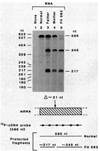
Expression of normal and mutant LDL receptor mRNA’s in human fibroblasts from FH 563 and his parents. LDL receptor mRNA’s were detected by a solution hybridization-S1 nuclease assay. A uniformly 32P-labeled probe encompassing nucleotides 422 to 1007 of the human receptor mRNA (11) was prepared as described in the legend to Fig. 7 and annealed to the indicated RNA at 39°C for 16 hours. After digestion with 700 units of S1 nuclease for 1 hour at 37°C and electrophoresis on denaturing polyacrylamide gels, nuclease-resistant hybrids were visualized by autoradiography. Each hybridization reaction contained RNA isolated from the following cell strains induced for LDL receptor expression (11): (lane 1) no RNA; (lane 2) 10 µg of total RNA from SV40-transformed normal human fibroblasts; (lane 3) 15 µg of total RNA from fibroblasts of the father of FH 563; (lane 4) 15 µg of total RNA from fibroblasts from the mother of FH 563; (lane 5) 10 µg of total RNA from fibroblasts of FH 563. The dried gel was exposed to Kodak XAR 5 film for 24 hours at −70°C with a Dupont Cronex Lightning Plus screen. Size standards were generated by electrophoresis of a labeled Msp I digest of pBR322 DNA.

Similar articles

Cited by

References

    1. Sabatini DD, Kriebich G, Morimoto T, Adesnik M. J. Cell. Biol. 1982;92:1. - PMC - PubMed
    2. Farquhar MG. Annu. Rev. Cell. Biol. 1985;1:447. - PubMed
    1. Gething M-J, Doyle C, Roth M, Sambrook J. Curr. Top. Membr. Tramp. 1985;23:17.
    2. Guan J-L, Rose JK. Cell. 1984;37:779. - PubMed
    3. McQueen NL, Nayak DP, Jones LV, Compans RW. Proc. Natl. Acad. Sci. U.S.A. 1984;81:395. - PMC - PubMed
    1. Goldstein JL, Brown MS. In: in The Metabolic Basis of Inherited Disease. 5. Stanbury JB, Wyngaarden JB, Fredrickson DS, Goldstein JL, Brown MS, editors. New York: McGraw-Hill; 1983. pp. 672–712.
    1. Watanabe Y. Atherosclerosis. 1980;36:261. - PubMed
    2. Goldstein JL, Kita T, Brown MS. N. Engl. J. Med. 1983;309:288. - PubMed
    1. Goldstein JL, Brown MS, Anderson RGW, Russell DW, Schneider WJ. Annu. Rev. Cell Biol. 1985;1:1. - PubMed

Publication types

Associated data

LinkOut - more resources