. 2018 Aug 13;8(1):12056.
doi: 10.1038/s41598-018-30561-z.

Distribution of oxidized DJ-1 in Parkinson's disease-related sites in the brain and in the peripheral tissues: effects of aging and a neurotoxin

Affiliations

Distribution of oxidized DJ-1 in Parkinson's disease-related sites in the brain and in the peripheral tissues: effects of aging and a neurotoxin

Yuichiro Mita et al. Sci Rep. .

Abstract

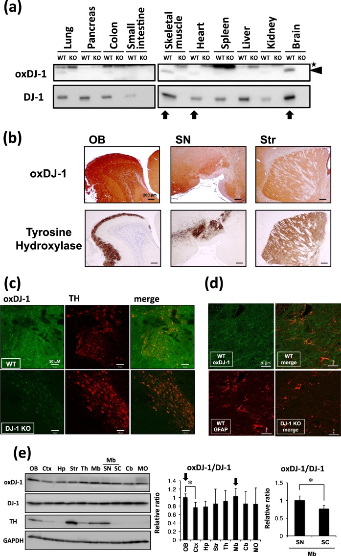
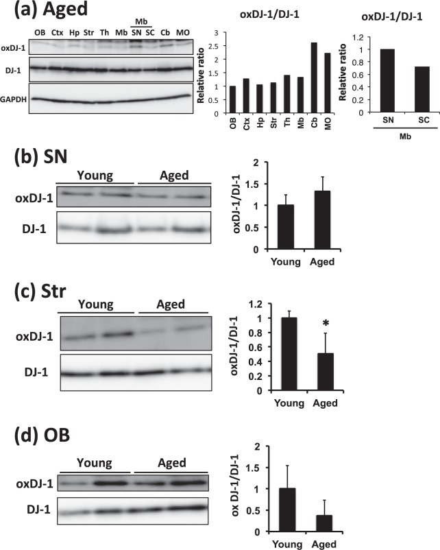
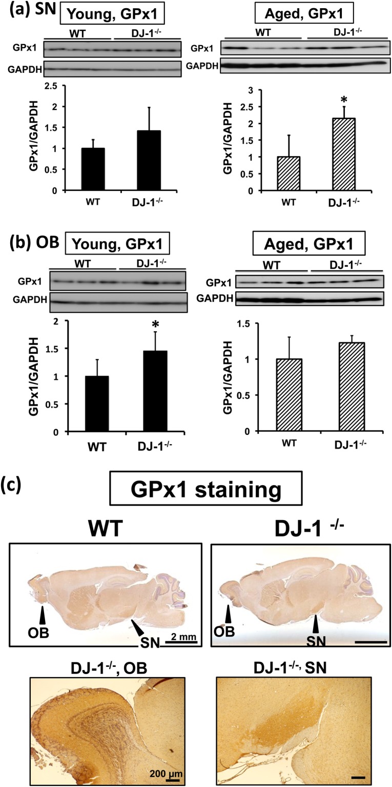
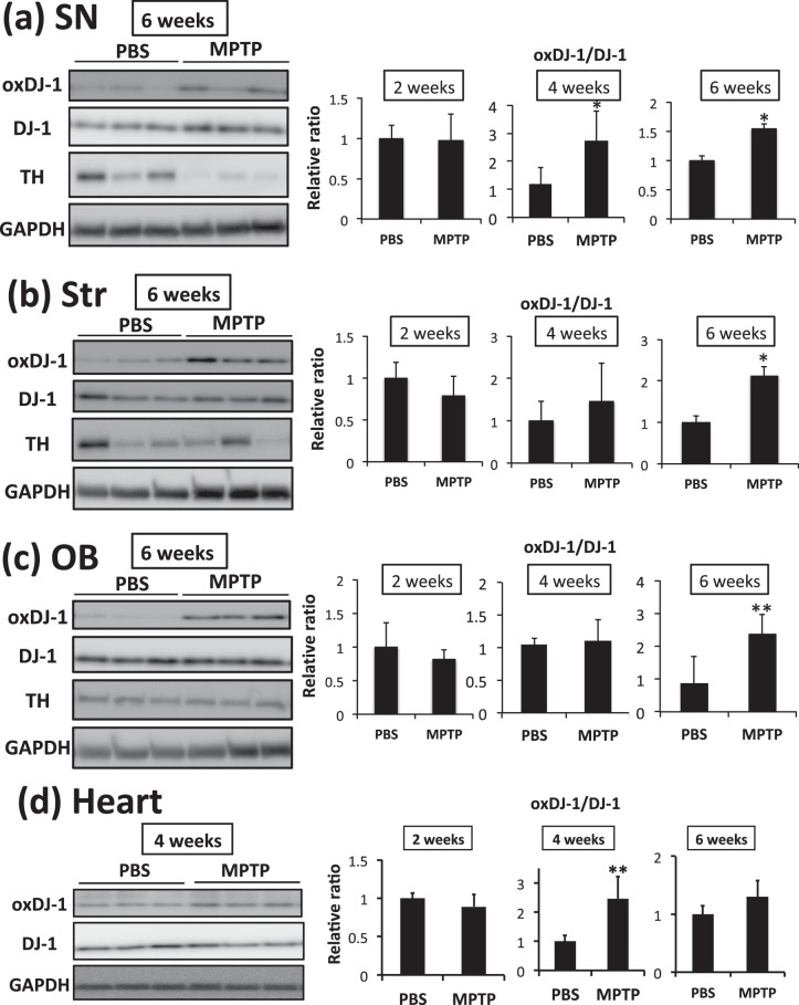
DJ-1 plays an important role in antioxidant defenses, and a reactive cysteine at position 106 (Cys106) of DJ-1, a critical residue of its biological function, is oxidized under oxidative stress. DJ-1 oxidation has been reported in patients with Parkinson's disease (PD), but the relationship between DJ-1 oxidation and PD is still unclear. In the present study using specific antibody for Cys106-oxidized DJ-1 (oxDJ-1), we analyzed oxDJ-1 levels in the brain and peripheral tissues in young and aged mice and in a mouse model of PD induced using 1-methyl-4-phenyl-1,2,3,6-tetrahydropyridine (MPTP). OxDJ-1 levels in the brain, heart, and skeletal muscle were high compared with other tissues. In the brain, oxDJ-1 was detected in PD-related brain sites such as the substantia nigra (SN) of the midbrain, olfactory bulb (OB), and striatum. In aged wild-type mice, oxDJ-1 levels in the OB, striatum, and heart tended to decrease, while those in the skeletal muscle increased significantly. Expression of dopamine-metabolizing enzymes significantly increased in the SN and OB of aged DJ-1-/- mice, accompanied by a complementary increase in glutathione peroxidase 1. MPTP treatment concordantly changed oxDJ-1 levels in PD-related brain sites and heart. These results indicate that the effects of physiological metabolism, aging, and neurotoxin change oxDJ-1 levels in PD-related brain sites, heart, and skeletal muscle where mitochondrial load is high, suggesting a substantial role of DJ-1 in antioxidant defenses and/or dopamine metabolism in these tissues.


Conflict of interest statement

The authors declare no competing interests.

Figures

Figure 1
Figure 1
Distribution of oxidized DJ-1 in mouse brain and peripheral tissues. (a) Lysates from each tissue of young wild-type (WT) and DJ-1−/− mice were subjected to western blotting using anti-oxDJ-1 and anti-DJ-1 Abs. The specific oxDJ-1 band is indicated by a black arrowhead and a nonspecific band is indicated by an asterisk. (b) Comparison of oxDJ-1 immunoreactivity with TH immunostaining in the olfactory bulb (OB), substantia nigra (SN), and striatum (Str). Sagittal sections from wild-type C57BL/6J mice were stained with anti-oxDJ-1 Ab and anti-TH Ab. Oxidized DJ-1 IR was observed around TH-positive neurons. (c,d) Confocal images of oxDJ-1 in SN dopaminergic neurons and astrocytes. A section containing the SN was immunostained with anti-oxDJ-1 Ab and anti-TH Ab (c) or anti-glial fibrillary acidic protein (GFAP) Ab (d) and then visualized using fluorescence confocal microscopy. A scale bar is shown in each figure. (e) Western blot analyses of oxDJ-1 in separated brain tissues. Protein lysates of each brain site were subjected to western blot analyses using Abs against DJ-1, oxDJ-1, TH, and glyceraldehyde-3-phosphate dehydrogenase (GAPDH). The relative band densities of oxDJ-1 normalized to DJ-1 were calculated and are presented as mean ± SD (n = 6). *P < 0.05, Tukey-Kramer test, ANOVA (middle panel) and Student’s t-test (right panel). Ctx, cortex; Hp, hippocampus; Th, thalamus; Mb, midbrain; SC, superior colliculus; Cb, cerebellum; MO, medulla oblongata.
Figure 2
Figure 2
The effects of aging on oxDJ-1 levels in the brain. (a) Western blot analyses of oxDJ-1 in separated brain tissues of aged mice. Protein lysates of each brain site were subjected to western blot analyses. The relative band densities of oxDJ-1 normalized to DJ-1 were calculated and are presented as mean (n = 2). Abbreviations are same with Fig. 1. (bd) Protein lysates of the substantia nigra (SN, b), striatum (Str, c), and olfactory bulb (OB, d) of young (9 weeks) and aged (130 weeks) WT mice were separated by SDS-PAGE and then subjected to western blotting using each Ab. The densities of each band were determined and the relative densities of oxDJ-1 relative to DJ-1 were calculated and are presented as mean ± SD (n = 3). *P < 0.05, Student’s t-test.
Figure 3
Figure 3
Change of oxDJ-1 levels in the heart and skeletal muscle of aged mice. (a) Lysates from each tissue of young (9 weeks) and aged (130 weeks) wild-type (WT) mice were subjected to western blotting using anti-oxDJ-1 and anti-DJ-1 Abs. The change in oxDJ-1 levels in the skeletal muscle and heart is shown. (b–e) Proteins in lysates from hearts (b and c) and skeletal muscle (d and e) of young and aged WT mice were separated by SDS-PAGE (b and d) or 2D-PAGE (c and e) and then subjected to western blotting using the indicated Ab. The densities of each band were determined and the relative densities of oxDJ-1 compared with DJ-1 were calculated and are presented as mean ± SD (b, n = 3). In 2D-PAGE, black, white, and striped arrowheads indicate native DJ-1, oxDJ-1, and unknown modified DJ-1 (UmDJ-1). The densities of each spot were determined and the relative densities of oxDJ-1 and UmDJ-1 relative to DJ-1 were calculated and are presented as mean ± SD (C, n = 3). *P < 0.05, Student’s t-test.
Figure 4
Figure 4
Elevation of glutathione peroxidase 1 (GPx1) levels in Parkinson’s disease-related brain sites of DJ-1−/− (DJ-1 KO) mice. (a,b) Young (9 weeks of age, left panel) and aged (more than 100 weeks of age, right panel) mice were analyzed. Protein lysates of the substantia nigra (SN, a) and olfactory bulb (OB, b) of wild-type (WT) and DJ-1 KO mice were subjected to western blot analyses using anti-GPx1 Ab. The relative band densities of GPx1 per GAPDH were calculated and presented as mean ± SD (n = 3–5). *P < 0.05, Student’s t-test. (c) Immunohistochemical distribution of the GPx1 protein in the mouse brain. Sagittal sections from young WT and DJ-1 KO mice brains were stained with anti-GPx1 Ab. GPx1 immunoreactivity was present throughout the brain from both mice, and was dense in the OB and SN of mouse brain. A scale bar is shown in each figure part.
Figure 5
Figure 5
The alteration of dopamine metabolism in DJ-1−/− (DJ-1 KO) and aged mice. (a–c) Striatal levels of dopamine (a) and the substantia nigra levels of tyrosine hydroxylase (TH, b) and monoamine oxidase-B (MAO-B, c) in wild-type (WT) and DJ-1 KO mice were determined in young (9 weeks of age, left panel) and aged (more than 100 weeks of age, right panel) mice (n = 4–5). The relative band densities of TH or MAO-B relative to GAPDH were calculated. *P < 0.05, Student’s t-test.
Figure 6
Figure 6
The change in DJ-1 oxidation in the substantia nigra (SN) of MPTP-treated mice. After the administration of PBS and MPTP (15 mg/kg, 3 times i.p.), each brain was separated and subjected to analysis. (a) Striatal levels of dopamine in mice were determined 3 days after MPTP administration (n = 5). **P < 0.01, Student’s t-test. (b) Protein lysates of the SN of MPTP-treated mice were subjected to western blot analyses using each specific Ab. The relative band densities of oxDJ-1 relative to DJ-1 and tyrosine hydroxylase (TH) relative to GAPDH were calculated and are presented as mean ± SD (n = 4). **P < 0.01, *P < 0.05, Tukey-Kramer test, ANOVA, when compared with day 0. (c) Modification of DJ-1 in the SN of MPTP-treated mice was evaluated using 2D-PAGE and western blotting for DJ-1. The filled triangles and open triangles indicate native and oxidized DJ-1, respectively. The striped triangles A and B indicate DJ-1 with an unknown modification. The relative densities of each spot were estimated. Graphs display the relative densities of each spot compared with PBS control as mean ± SD (n = 5). **P < 0.01, Student’s t-test.
Figure 7
Figure 7
Elevation of oxidized DJ-1 in the brain and heart of MPTP-treated mice. After the administration of PBS and MPTP (15 mg/kg, 3 times i.p.), each brain and heart were separated and subjected to analysis. (a–d) Protein lysates of the substantia nigra (SN, a), striatum (Str, b), olfactory bulb (OB, c), and heart (d) of MPTP-treated mice were subjected to western blot analyses using each specific Ab. The relative band densities of oxDJ-1 relative to DJ-1 were calculated and are presented as mean ± SD (n = 6). **P < 0.01, *P < 0.05, Student’s t-test.

Similar articles

Cited by

References

    1. Bonifati V, et al. Mutations in the DJ-1 gene associated with autosomal recessive early-onset parkinsonism. Science. 2003;299:256–259. doi: 10.1126/science.1077209. - DOI - PubMed
    1. Taira T, et al. DJ-1 has a role in antioxidative stress to prevent cell death. EMBO Rep. 2004;5:213–218. doi: 10.1038/sj.embor.7400074. - DOI - PMC - PubMed
    1. Le HT, et al. YajL, prokaryotic homolog of parkinsonism-associated protein DJ-1, functions as a covalent chaperone for thiol proteome. J. Biol. Chem. 2012;287:5861–5870. doi: 10.1074/jbc.M111.299198. - DOI - PMC - PubMed
    1. Ariga, H. & Iguchi-Ariga, M. M. S. DJ-1/PARK7 Protein. Springer Nature Shingapore: Adv. Exp. Med. Biol. 1037 (2017)
    1. Raninga PV, Di Trapani G, Tonissen KF. The Multifaceted Roles of DJ-1 as an Antioxidant. Adv. Exp. Med. Biol. 2017;1037:67–87. doi: 10.1007/978-981-10-6583-5_6. - DOI - PubMed

Publication types

MeSH terms

LinkOut - more resources