. 2019 Jan;38(3):445-453.
doi: 10.1038/s41388-018-0439-1. Epub 2018 Aug 13.

Overexpression of miR-489 derails mammary hierarchy structure and inhibits HER2/neu-induced tumorigenesis

Affiliations

Overexpression of miR-489 derails mammary hierarchy structure and inhibits HER2/neu-induced tumorigenesis

Y Patel et al. Oncogene. 2019 Jan.

Erratum in

Abstract

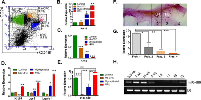
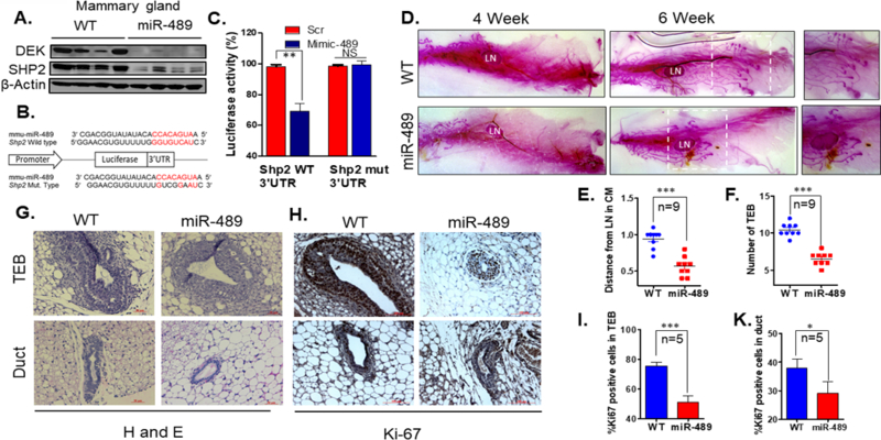
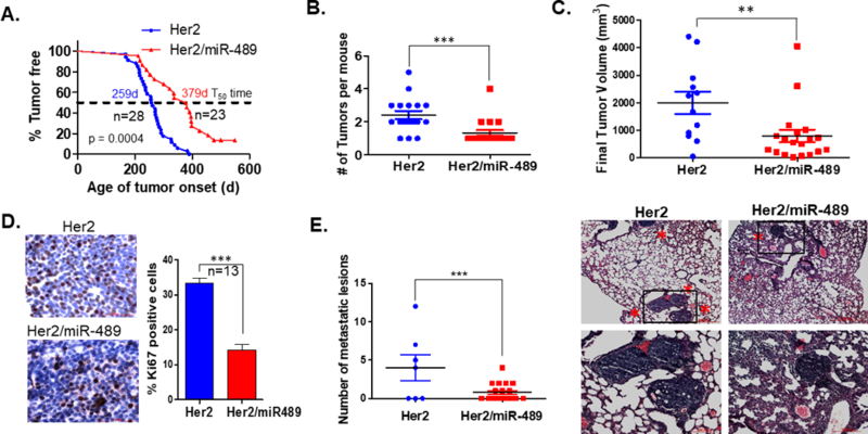
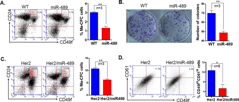
Although it has been demonstrated that transformed progenitor cell population can contribute to tumor initiation, factors contributing to this malignant transformation are poorly known. Using in vitro and xenograft-based models, previous studies demonstrated that miR-489 acts as a tumor suppressor miRNA by targeting various oncogenic pathways. It has been demonstrated that miR-489 directly targets HER2 and inhibits the HER2 signaling pathway; however, its role in mammary gland development and HER2-induced tumor initiation hasn't been studied. To dissect the role of miR-489, we sorted different populations of mammary epithelial cells and determined that miR-489 was highly expressed in mammary stem cells. MMTV-miR-489 mice that overexpressed miR-489 in mammary epithelial cells were developed and these mice exhibited an inhibition of mammary gland development in early ages with a specific impact on highly proliferative cells. Double transgenic MMTV-Her2-miR489 mice were then generated to observe how miR-489 overexpression affects HER2-induced tumorigenesis. miR-489 overexpression delayed HER2-induced tumor initiation significantly. Moreover, miR-489 overexpression inhibited tumor growth and lung metastasis. miR-489 overexpression reduced mammary progenitor cell population significantly in preneoplastic mammary glands of MMTV-Her2 mice which showed a putative transformed population in HER2-induced tumorigenesis. The miR-489 overexpression reduced CD49fhiCD61hi populations in tumors that have stem-like properties, and miR-489 overexpression altered the HER2 signaling pathway in mammary tumors. Altogether, these data indicate that the inhibition of HER2-induced tumorigenesis by miR-489 overexpression was due to altering progenitor cell populations while decreasing tumor growth and metastasis via influencing tumor promoting genes DEK and SHP2.


Conflict of interest statement

CONFLICT OF INTEREST

The authors declare no conflict of interest.

Figures

Figure 1
Figure 1. miR-489 expression in different sub population of mammary epithelial cells and in the mammary gland during purberty, pregnancy, lactation, and early involution.
(A) Representative FACS profile for mouse mammary epithelial cells after eliminating hematopoit stained with CD24-PE and CD49f-FITC (B) Keratin14 was assayed by qRT-PCR in luminal, Ma-CFC, myoepthelial cells and MRU. (Lum vs Ma-CFC p=0.0264, Lum vs Myo p=0.0243, Lum vs MRU p=0.0014) (C) Keratin18 was assayed by qRT-PCR in luminal, Ma-CFC, MRU and MRU. (Lum vs Ma-CFC p=0.0003, Lum vs Myo p=0.0039, Lum vs MRU p<0.0001) (D) Krt15, Lgr5 and Lgals1 were measured in different subpopulation of mammary epithelial cells. (For Keratin15 Lum vs MRU p=0.0080 and Myo vs MRU p=0.0056, Lgr5 Lum vs MRU p=0.0008 and Myo vs MRU p=0.0055, Lgals1 Lum vs MRU p=0.0053 and Myo vs MRU p=0.0205) (E) Luminal, Ma-CFC, MRU and myoepithelial subpopulations were collected and assayed for expression of miR-489. (Sorting of cells have been performed 3 independent times and each time 4 sister mice were used, Lum vs MRU p=0.0012, Lum vs Ma-CFC p<0.0001, Myo vs MRU p=0.0011, MRU vs Ma-CFC p=0.0017, Myo vs Ma-CFC p=0.0396) (F) Different fractions of the mammary gland has been collected from 6-week-old WT female as demonstrated. Whole mount image was taken at 4x magnification. (G) RNA was isolated from each fraction and examined for expression of miR-489 (three mice were used for this experiment, Frac1 vs Frac4 p=0.0004, Frac2 vs Frac4 p=0.0034, Frac3 vs Frac4 p=0.0481). (H) RT-PCR data demonstrate miR-489 expression in different stages of mammary gland development. RNA was isolated from three independent mice for each stage. Method: All mice experiments and breeding were performed with approval of the University of South Carolina IACUC. All mice used in this study are on FVB background. For sorting experiment, 6-week-old WT mice were used for this experiment. For each experiment 4 sister mice were used. Thoracic and inguinal mammary glands were used. Glands were minced and digested for 4–6 h at 37°C in DMEM/F12 medium supplemented with 750U/ml Collagenase and 250U/ml hyaluronidase. After this step cells organoids were collected by centrifugation then subjected to ammonium chloride to lyse erythrocytes. After that cells were collected and subjected to separate trypsin (0.5%), dispase (5 mg/ml) and DNase treatment. Cells were passed through a 40µM strainer and subjected for hematopoietic cell depletion. Mouse epithelial cell enrichment kits were obtained from Stem Cell Technologies (catalog#19758) and their protocol was followed. Cells were stained by anti- CD49f (2µl antibody for 1million cells) and anti-CD24 (1.5µl antibody for 1million cells) antibody for 30min on ice. CD49f was obtained from Bio-Legend (cat#313606) and anti-CD24 was obtained from BD bioscience (cat#553262). Different fractions were sorted by using BD FACS aria II sorter. RNA was isolated using Trizole reagent from mammary epithelial cells derived from 3.5, 7 and 14 weeks old nulliparous WT mice. RT-PCR was performed as described in supplementary method to quantitate miR-489 expression with U6 as an internal control. For rest of the stage female mice were kept with males. For Pregnancy day 16, female mice were kept with male and copulation was confirmed by plug visualization and males removed post coitum. For involution and lactation, pups were removed on day 9 of lactation and mammary glands were collected on day 1, 3 and 5 after the removal of pups. Following primers were used for qRT-PCR; mmu-miR-489–3p 5’-AATGACACCACATATATGGCAGC-3’ and U6 F-5’ GACGTGAGGCGACAAGATGG3’ and R-5’AAAGCCCTTCAGGATACCGC3’. All statistical tests were performed by student t-test using prism (GraphPad software, Inc., San Diego, CA, USA). For qRT-PCR statistical analyses were performed on 2-CT values.
Figure 2
Figure 2. Targeted overexpression of miR-489 in mouse mammary epithelial cells impaired mammary gland development in early pubertal age
(A) Western blot analysis was performed on protein lysate isolated from mammary epithelial cells from WT and MMTV-miR-489 mice and analyzed for DEK, SHP2 and beta-actin. (n=4 pair of female mice were used) (B) A schematic representation of the Shp2 mRNA with putative miR-489 binding site in the 3’UTR, where seed region is highlighted. (C) H605 cells were co-transfected with either of these vectors with Scramble or miR-489 mimic and renilla expressing vector for 48 h. Firefly luciferase was measured for each condition and normalized with renilla luciferase. (p=0.0096) (D) Whole-mount carmine staining of mammary glands from 4 week and 6 week old virgin mice. Representative images from a sister pair are shown. Whole mount images were taken at 4X magnification. (E, F) Number of Terminal End Bud and distance of TEB from lymph node were measured for analysis purpose (n=9 pair of female mice were used, for number of TEBs p<0.0001 and for distance p<0.0001). (G) Hematoxylin and eosin (H&E) stained representative image of WT and MMTV-miR-489 mouse mammary gland. (H, I, K) Ki-67 stained TEB, duct and histogram of WT and MMTV-miR-489 mammary glands have been demonstrated. (n=5 pair of female mice were used; TEB p<0.0001 and Duct p=0.0441). Method: The full-length mouse pre miR-489 cDNA was amplified by PCR and inserted into a pMMTV vector (a kind gift from Dr. Jeffrey Rosen at Baylor College of Medicine) to generate the plasmid MMTV-miR-489. Pronuclear microinjection to generate transgenic mice was performed by the core facility at MUSC. Transgenic progeny was identified by southern blot analysis and PCR also was performed with genomic DNA isolated from tail biopsy. The following primer pairs were used to perform genotyping: F-5’ACCAGGTCCTGTTGTAGGTTC3’ and R-5’TACTCTGAGTCCAAACCGGG3’. RNA and proteins were isolated from mammary epithelial cells from WT and MMTV-miR-489 mice. For qRT-PCR analysis 9 pair of sister mice were used and performed as described above. Western blot was performed with a total of 4 pairs of sister mice. DEK and SHP2 antibodies were obtained from Proteintech. Dissected 4th inguinal mammary glands were spread on glass slides and exposed to kahle’s fixative overnight. Glands were stained overnight with carmine alum. Adipose was removed by exposure to xylene overnight. All whole mount images were taken by using Stemi 305 dissecting microscope by using 4X magnification. For whole mount analysis, a total of 9 pairs of sister mice were used. H&E and Ki-67 staining was performed as described in supplementary method. For Ki-67 positive cell counting, 3 independent ducts and TEBs were counted from each mammary gland tissue. Total of 5 pairs of sister mice were used.
Figure 3
Figure 3. Overexpression of miR-489 impedes tumor initiation, growth and lung metastasis in HER2 induced mammary tumors
(A) Kaplan-Meier Curves of mammary tumor free survival of virgin MMTV-Her2 (n=15) and MMTV-Her2-miR489 mice (n=20). The p value was calculated using the long rank-rank test (p<0.0001) (B) Mammary tumor multiplicity between MMTV-Her2 (n=17) and MMTV-Her2-miR489 (n=18) mice (P=0.0008) (C) Tumor growth was monitored every 7 days. Final tumor sizes of both group were compared (P=0.0086) (D) Proliferation rate was assessed by Ki-67 immunostaining comparing tumors from MMTV-Her2 (n=10) and MMTV-Her2-miR489 (n=15). At least 500 cells have been counted per mammary tumor. (p<0.0001) (E) Quantitation of the number of metastatic foci in the lungs of MTTV-Her2 and MMTV-Her2-miR489 mice after 8-week post tumor onset. H and E staining of lungs of MMTV-Her2 and MMTV-Her2-miR489 mice. Images were taken at 4x and 10x magnification(n=17) (p=0.0035). Method: MMTV-Neu mice were purchased from Jackson laboratory. MMTV-Neu mice were crossed with MMTV-miR489 mice to generate MMTV-Her2-miR489 double transgenic mice. For analysis of mammary tumor incidence, we monitored adult female mice starting at 4 months of age and once per week. We monitored the mice weekly and noted the time of tumor appearance, tumor size, the number of tumors and lung metastasis in each mouse. After detecting palpable tumor, tumor size was measured every week. Tumor free survival curves were evaluated using Kaplan-meier method and differences between the survival curves were evaluated by a log-rank test. Tumor volume of each primary tumor was calculated by modified ellipsoidal formula (1/2 (LxW2)). Tumor volumes were plotted using graph-pad prism software. For Ki-67 counting 1000 tumor cells were counted from 13 mammary tumors and the percentage of positive staining cells are presented in the figures.
Figure 4
Figure 4. Overexpression of miR-489 in mammary epithelial cells has reduced Ma-CFC population
(A) FACS analysis was performed to examine the luminal progenitor cell population (CD49fmedCD24hi) in mammary epithelial cells from 6-week-old WT and MMTV-miR489 transgenic mice (n=3). P=0.0016 (unpaired student t test). (B) Colony formation assay was performed from mammary epithelial cells from 6-week-old WT and MMTV-miR489 transgenic mice (n=3). P=0.0038 (unpaired student t test). (C) Mammary epithelial cells were isolated from 24-week-old Her2 and Her2-miR489 mice and stained with CD49f-FITC and CD24-PE and analyzed for Ma-CFC population (n=3). P=0.0015 (unpaired student t test). (D) FACS analysis of the putative CSC cells (CD49fhiCD61hi population) in the first mammary tumor of each animal (n=8). P=0.0337 (unpaired student t test) Method: Thoracic and inguinal mammary glands were isolated from 6-week-old WT and MMTV-miR-489 mice, single cell mammary epithelial cells suspension were prepared and stained with CD49f and CD24 antibody as described above. Cells were analyzed with BD Accuri C6 flow cytometer for different mammary epithelial cell population. A total of 35,000 events were collected for each sample. Mammary epithelial cells were seeded in plates for Ma-CFC colony formation assay as described in supplementary method. Mammary tumors were harvested, minced and incubated in digestion media to generate single cell suspensions (described above) at 37°C for 2h. Cells were stained with CD49f and CD61 on ice for 30 min. CD61 was obtained from BioLegend (cat#104308).

Similar articles

Cited by

References

    1. Boras-Granic K, Dann P, Wysolmerski JJ. Embryonic cells contribute directly to the quiescent stem cell population in the adult mouse mammary gland. Breast cancer research : BCR 2014; 16: 487. - PMC - PubMed
    1. Cai S, Kalisky T, Sahoo D, Dalerba P, Feng W, Lin Y et al. A Quiescent Bcl11b High Stem Cell Population Is Required for Maintenance of the Mammary Gland. Cell stem cell 2017; 20: 247–260 e245. - PMC - PubMed
    1. Celia-Terrassa T, Liu DD, Choudhury A, Hang X, Wei Y, Zamalloa J et al. Normal and cancerous mammary stem cells evade interferon-induced constraint through the miR-199a-LCOR axis. Nature cell biology 2017; 19: 711–723. - PMC - PubMed
    1. Chang TH, Kunasegaran K, Tarulli GA, De Silva D, Voorhoeve PM, Pietersen AM. New insights into lineage restriction of mammary gland epithelium using parity-identified mammary epithelial cells. Breast cancer research : BCR 2014; 16: R1. - PMC - PubMed
    1. Chen H, Soni M, Patel Y, Markoutsa E, Jie C, Liu S et al. Autophagy, Cell Viability and Chemo-resistance are Regulated by miR-489 in Breast Cancer. Mol Cancer Res 2018. - PMC - PubMed

Publication types

MeSH terms