. 2018 Sep;21(9):1260-1271.
doi: 10.1038/s41593-018-0203-4. Epub 2018 Aug 13.

Mapping projections of molecularly defined dopamine neuron subtypes using intersectional genetic approaches

Affiliations

Mapping projections of molecularly defined dopamine neuron subtypes using intersectional genetic approaches

Jean-Francois Poulin et al. Nat Neurosci. 2018 Sep.

Abstract

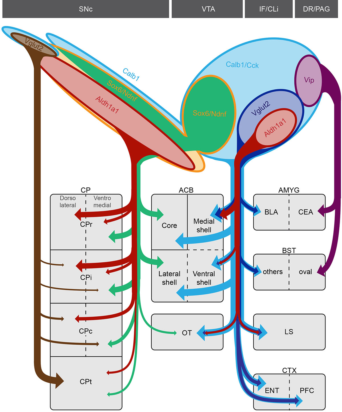
Midbrain dopamine (DA) neurons have diverse functions that can in part be explained by their heterogeneity. Although molecularly distinct subtypes of DA neurons have been identified by single-cell gene expression profiling, fundamental features such as their projection patterns have not been elucidated. Progress in this regard has been hindered by the lack of genetic tools for studying DA neuron subtypes. Here we develop intersectional genetic labeling strategies, based on combinatorial gene expression, to map the projections of molecularly defined DA neuron subtypes. We reveal distinct genetically defined dopaminergic pathways arising from the substantia nigra pars compacta and from the ventral tegmental area that innervate specific regions of the caudate putamen, nucleus accumbens and amygdala. Together, the genetic toolbox and DA neuron subtype projections presented here constitute a resource that will accelerate the investigation of this clinically significant neurotransmitter system.


Figures

Figure 1.
Figure 1.. Generation and validation of Th–2A–Flpo
(A) We used dual Recombinase Mediated Cassette Exchange (dRMCE) to reengineer a previously generated Th–LacZ allele in which lacZ expression is driven by the tyrosine hydroxylase (Th) transcription by virtue of a splice acceptor (SA) located in the intron between the 6th and 7th exon. We converted this allele and replaced lacZ by exons 7 to 13 of the Th coding sequence, followed by the P2A peptide, and the recombinase Flpo.(B) Simplified schematic of Th–2A–Flpo crossed to reporter mouse RC::FA, designed to activate nlacZ expression in midbrain DA neuron clusters resulting in nuclear βgal expression (black triangles = FRT sites). (C) In Th–2A–Flpo; RC::FA midbrain, the large majority of TH+ neurons (green) of the substantia nigra pars compacta (SNc; 99.9% (±0.1)), the ventral tegmental area (VTA; 100.0%), interfascicular nucleus (IF; 100.0%), retrorubral area (RR; 100.0%), caudal linear nucleus (CLi; 100.0%), and periaqueductal gray/dorsal raphe regions (PAG/DR; 99.6% (±0.2); not shown) are βgal+ (red). We observed some βgal+ cells are TH– (examples are indicated with arrowhead), particularly in the rostral linear nucleus (RLi), interpeduncular nucIeus (IPN), and CLi. The fact that TH protein synthesis is required for Flpo production, because of effective P2A “autocatalytic” activity, suggests that these recombined cells produced TH protein, at least at some point during their developmental history. However, the majority of βgal+ cells also expressed TH in all DA neuron clusters: SNc (98.9% ±0.3), VTA (97.5% ±1.2), IF (85.8% ±5.8), CLi (88.1% ±2.4), RR (97.6% ±1.5) and the PAG/DR (68.4% ±5.1; not shown). n = 3.. PN = paranigral region, PBP = parabrachial pigmented area. Scale bar = 100 μm
Figure 2.
Figure 2.. Three intersectional strategies to genetically target dopamine neuron subtypes
(A) A first strategy is to use subtype-specific Cre drivers in combination with Th–2A–Flpo and the intersectional reporter mouse Ai65 (or equivalent), which results in tdTomato expression only in cells expressing both Cre and Flpo recombinases. (B) A second strategy is to use subtype-specific Cre drivers, in combination with both Th–2AvFlpo and the INTRSECT viral system. This virus leads to EYFP expression in neurons expressing both Cre and Flpo recombinases. A simplified schematic of the INTRSECT virus is shown. (C) A third strategy is to use Cre drivers in conjunction with Dat–tTA and Ai82 intersectional reporter mice. In this reporter, EGFP expression is driven by Dat–tTA, following Cre recombination. (D) Validating Strategy I, we only detected tdTomato expression in midbrain DA neurons in mice with both Dat–ires–Cre and Th–2A–Flpo alleles, whereas no tdTomato was observed in the absence of Dat–ires–Cre or Th–2A–Flpo. (E) Validating strategy II, we observed strong EYFP fluorescence after viral injection of hSyn–CreON,FlpON–EYFP in mice with both Datvires–Cre and Th–2A–Flpo alleles, whereas no EYFP was observed in the absence of Dat–ires–Cre or Th–2A–Flpo. (F) For Strategy III, we observed EGFP labeling in Ai82+ mice having both Dat–ires–Cre and Dat–tTA alleles, whereas no fluorescence was observed in controls. (G) Using Dat–ires–Cre;Th–2A–Flpo;Ai65 mice to provide a comprehensive picture midbrain DA neuron projections, we observed tdTomato labeled fibers in the caudate putamen (CP), nucleus accumbens (ACB), olfactory tubercule (OT), prefrontal cortex (PFC), lateral septum (LS), bed nucleus of the stria terminalis (BST), basolateral amygdala (BLA), and external globus pallidus (GPe). We also observed very sparse tdTomato+ fibers in the lateral habenula, motor cortical areas, and rostral pontine regions (not shown). Grey triangles = loxP sites, grey triangle outline = lox2722, grey striped triangle = loxN, black triangles = FRT sites, black triangle outline = F3 sites, black striped triangle = F5. See Table S1 for number of animal replicates for each experiment. Scale bars = 400 μm
Figure 3.
Figure 3.. Dopamine neuron subtype projections to the caudate putamen
(A) Distribution of DA neuron subtypes used in the intersectional genetic experiments to trace projections in the caudate putamen (CP; each dot = 3 neurons; yellow = Ndnf experiment shown in B, red = Aldh1a1 experiment shown in C, green = Calb1 experiment shown in D, blue = Vglut2 experiment shown in E). (B) In Ndnf–dgCre;Dat–tTA;Ai82 mice, EGFP+ cell bodies are located throughout the SNc, the dorsolateral VTA and RR. The vast majority of these cells express TH and SOX6 (arrowheads). (C) Example of a Aldh1a1–CreERT2 brain injected in the SNc with AAV–CreON–EYFP virus. EYFP cells were positive for TH and ALDH1A1. (D) Injection of Calb1–Cre;Th–2A–Flpo mouse with a AAV–CreON,FlpON–EYFP virus in the SNc. Most EYFP+ cells did not express ALDH1A1. (E) Injection of a AAV–CreON,FlpON–EYFP virus in the SNc of Vglut2–Cre;Th–2A–Flpo mouse yields labeled neurons in the dorsolateral part of the SNc (corresponding in part to the pars lateralis). A small fraction of these neurons is ALDH1A1+ (arrowhead). (F) Distinctive areas of the dorsal striatum are innervated in the experiments described above, shown at different rostrocaudal levels (CPr = rostral, CPi = intermediate, CPc = caudal, CPt = tail). (G-J) Representative images from these experiments (See Table S1 for number of animal replicates for each experiment). Scale bars: B-E low mag. = 200 μm, high mag. = 40 μm; G-J =200 μm.
Figure 4.
Figure 4.. Dopamine neuron subtype projections to the nucleus accumbens and olfactory tubercle
(A) Distribution of genetically labeled DA neurons with Vglut2–Cre (dark blue), Sox6–FSF–Cre (yellow), Cck–Cre (cyan) and Aldh1a1–CreERT2 (red) (each dot = 3 neurons). (B) Example of a Cck–Cre;Th–2A–Flpo mouse injected with AAV–CreON,FlpON–EYFP. EYFP+ cells either colocalized with SOX6 or OTX2 (arrowheads). (C) Example of a Sox6–FSF–Cre;Th–2AvFlpo mouse injected with AAV–CreON,FlpON–EYFP. In this mouse, EYFP+ cells colocalize only with SOX6 (white arrowhead) and not with OTX2. (D) Example of a Vglut2–Cre;Th–2A–Flpo mouse injected in the VTA with AAV–CreON,FlpON–EYFP. EYFP+ cells mostly express OTX2 (white arrowhead) and very few cells express SOX6. (E) An example of Aldh1a1–CreERT2 injected in the VTA with AAV–CreON–EYFP. EYFP+ cells are OTX2+ and SOX6–. (F) Projections of molecularly defined DA neurons to the nucleus accumbens at different rostral (ACBr), intermediate (ACBi), and caudal (ACBc) levels. Representative images are shown for Sox6–FSF–Cre (G), Aldh1a1–CreERT2 (H), Vglut2–Cre (I), and Cck–Cre (J) experiments. See Table S1 for number of animal replicates for each experiment. Scale bars: B-E low mag. = 200 μm, high mag. = 30 μm; G-J = 200 μm.
Figure 5.
Figure 5.. Dopamine neuron subtype projections to the amygdala
(A) Projections to the amygdala, as observed in Dat–ires–Cre;Th–2A–Flpo;Ai65 brains. (B) Distribution of DA neuron subtypes found to project to the amygdala labeled by Vip–Cre (red), Vglut2–Cre (blue), and Cck–Cre (green). (C) Labeled neurons in the Vip–Cre;Th–2A–Flpo;Ai65. (D) Labeled neurons by intersectional viral injections in the SNc of the Vglut2–Cre;Th–2A–Flpo. (E) EYFP+ labeled neurons by an injection in the VTA of a Cck–Cre;Th–2A–Flpo mouse. (F) Projections of molecularly defined DA neurons labeled with Vip–Cre (red), Vglut2–Cre (blue), and Cck–Cre (green). (G-I) Representative images from the experiments described above (See Table S1 for number of animal replicates for each experiment). Scale bars: A = 100 μm; C-E = 200 μm; G-I = 100 μm.
Figure 6.
Figure 6.. Dopamine neuron subtype projections to other brain regions
Intersectional genetic Strategy I with Dat–ires–Cre revealed DAergic projections to (A) the prefrontal cortex (PFC), (B) the lateral septum (LS), (C) the bed nucleus of the stria terminalis (BST) and (D) the entorhinal cortex (ENT). Using Strategy II in the VTA of Cck–Cre;Th–2A–Flpo mice we observed also an innervation of the PFC (E), LS (F), BST (G) and ENT (H). (I) EYFP+ fibers are observed in the PFC of Vglut2–Cre;Th–2A–Flpo mice after injection of AAV–CreON,FlpON–EYFP in the VTA. (J) EYFP+ fibers are observed in the LS of Vglut2–Cre;Th–2A–Flpo after injection of AAV–CreON,FlpON–EYFP in the VTA. (K) tdTomato+ fibers are observed in the BST of Vip–Cre;Th–2A–Flpo;Ai65 brains. (L) EYFP+ fibers are observed in the ENT of Vglut2–Cre;Th–2A–Flpo after injection of AAV–CreON,FlpON–EYFP in the VTA. See Table S1 for number of animal replicates for each experiment. Scale bar: A-L = 100 μm.
Figure 7.
Figure 7.. Remarkable specificity of genetically-defined dopamine neuron subtype projections within target regions
Top caption shows the approximate distribution of DA subtypes analyzed in this study. Vglut2+/Calb1+ DA neurons located in the SNc (brown) project most densely to the tail of the caudate putamen (CPt) extending ventrally to the capsular region of the amygdala (not depicted). Sox6+/Aldh1a1+ DA neurons of the SNc (red) project mainly to the rostral and intermediate caudate putamen (CPr and CPi respectively). Within the CP, Aldh1a1+ fibers are mainly observed in the dorsolateral region. The Calb1+/Sox6+ population of the SNc and dorsolateral VTA (green) send projections to the medial CPr and ventromedial CPi and CPc. This population also contributes projections to the lateral shell and core region of the nucleus accumbens. In the VTA, Aldh1a1+ DA neurons (red), which are also Cck+/Vglut2+, project to the medial shell of the ACB, olfactory tubercle (OT) and lateral septum (LS). Cck+/Vglut2+ (dark blue), but Aldh1a1-, contribute to the prefrontal (PFC) and entorhinal (ENT) cortices as well as the BLA. Finally, a Vip+ DA neuron population (purple) located in the dorsal raphe (DR) and periaqueductal gray (PAG) sends projections to the lateral part of the central amygdala (CEA) and the oval nucleus of the bed nucleus of the stria terminalis (BST). The Slc32a1+ subtype from Poulin et al. and La Manno et al. was not analyzed due to lack of unambiguous access.

Similar articles

Cited by

References

    1. Berke JD What does dopamine mean? Nat Neurosci 1 (2018). doi:10.1038/s41593-018-0152-y - DOI - PMC - PubMed
    1. Björklund AA & Dunnett SB Dopamine neuron systems in the brain: an update. Trends Neurosci 30, 194–202 (2007). - PubMed
    1. Lammel S, Lim BK & Malenka RC Reward and aversion in a heterogeneous midbrain dopamine system. Neuropharmacology 76 Pt B, 351–359 (2014). - PMC - PubMed
    1. Roeper J Dissecting the diversity of midbrain dopamine neurons. Trends Neurosci 36, 336–342 (2013). - PubMed
    1. Morales M & Margolis EB Ventral tegmental area: cellular heterogeneity, connectivity and behaviour. Nat Rev Neurosci 18, 73–85 (2017). - PubMed

Methods reference list

    1. Skarnes WC et al. A conditional knockout resource for the genome-wide study of mouse gene function. Nature 474, 337–342 (2011). - PMC - PubMed
    1. Osterwalder M et al. Dual RMCE for efficient re-engineering of mouse mutant alleles. Nat Methods 7, 893–895 (2010). - PMC - PubMed
    1. Donnelly ML et al. Analysis of the aphthovirus 2A/2B polyprotein ‘cleavage’ mechanism indicates not a proteolytic reaction, but a novel translational effect: a putative ribosomal ‘skip’. J. Gen. Virol 82, 1013–1025 (2001). - PubMed
    1. Anastassiadis K et al. Dre recombinase, like Cre, is a highly efficient site-specific recombinase in E. coli, mammalian cells and mice. Dis Model Mech 2, 508–515 (2009). - PubMed
    1. Wallén A et al. Fate of mesencephalic AHD2-expressing dopamine progenitor cells in NURR1 mutant mice. Exp. Cell Res. 253, 737–746 (1999). - PubMed

Publication types