. 2018 Sep;21(9):1229-1238.
doi: 10.1038/s41593-018-0204-3. Epub 2018 Aug 13.

Voluntary urination control by brainstem neurons that relax the urethral sphincter

Affiliations

Voluntary urination control by brainstem neurons that relax the urethral sphincter

Jason A Keller et al. Nat Neurosci. 2018 Sep.

Abstract

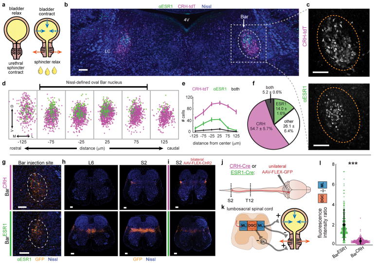
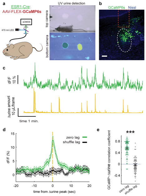
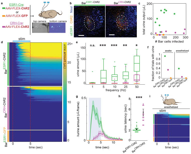
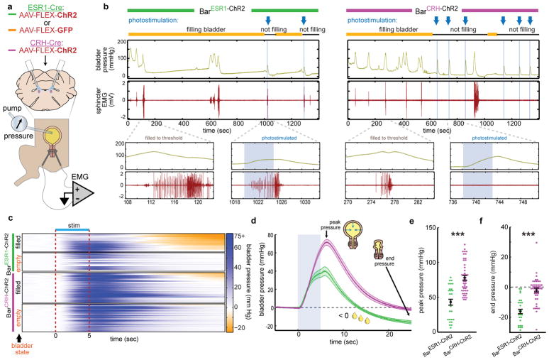
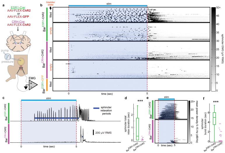
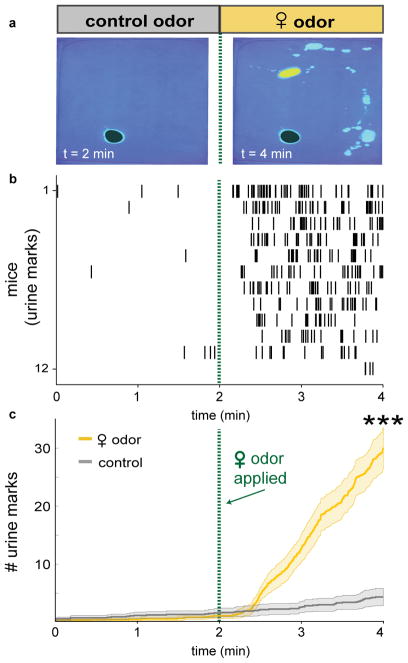
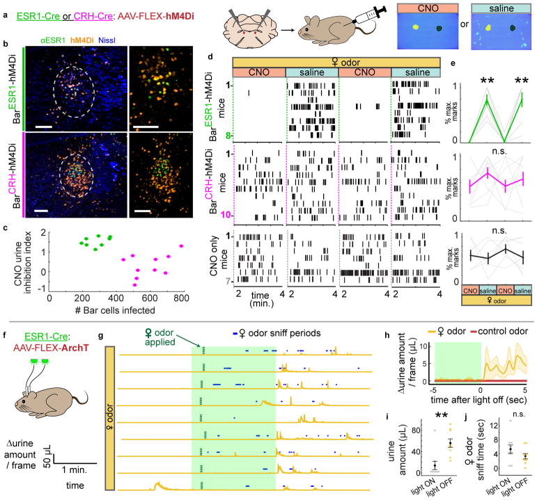
Voluntary urination ensures that waste is eliminated when safe and socially appropriate, even without a pressing urge. Uncontrolled urination, or incontinence, is a common problem with few treatment options. Normal urine release requires a small region in the brainstem known as Barrington's nucleus (Bar), but specific neurons that relax the urethral sphincter and enable urine flow are unknown. Here we identify a small subset of Bar neurons that control the urethral sphincter in mice. These excitatory neurons express estrogen receptor 1 (BarESR1), project to sphincter-relaxing interneurons in the spinal cord and are active during natural urination. Optogenetic stimulation of BarESR1 neurons rapidly initiates sphincter bursting and efficient voiding in anesthetized and behaving animals. Conversely, optogenetic and chemogenetic inhibition reveals their necessity in motivated urination behavior. The identification of these cells provides an expanded model for the control of urination and its dysfunction.


Conflict of interest statement

Competing financial interests statement

The authors declare no competing interests.

Figures

Fig. 1
Fig. 1. A novel cell type in Barrington’s nucleus with projections biased to sphincter-inhibiting interneurons
a, Urination requires sphincter relaxation. b, ESR1-immunostaining in Bar (dotted oval) in CRH-tdT mouse. LC = locus coeruleus, 4V = 4th ventricle. c, Larger view of CRH-tdT (top) and αESR1 (bottom) channels from (b). d, Rostrocaudal overlay of αESR1 cells (green) in Bar registered to centroid of CRH-tdT cells (magenta). e, Cell counts, and f, cell percentages in Bar (mean ± s.e.m., n=6 mice). g, GFP expression at Bar injection site in CRH-Cre (top) or ESR1-Cre (bottom) individuals. h, Axonal projections in lumbosacral spinal cord (right L6, left S2) for injections in (g). i, Axonal projections in lumbosacral S2 spinal cord for injection sites in Fig. 3b. j, Schematic for identifying Bar cell type axonal projections to spinal cord. k, Simplified urinary circuitry in the lumbosacral spinal cord. ML = mediolateral column, DGC = dorsal grey commissure, DL = dorsolateral nucleus. l, Quantification of BarESR1 and BarCRH axonal projections in lumbosacral spinal cord. Points are individual sections, thick black line is mean ± s.e.m for BarCRH (magenta, n=10 mice), BarESR1 (green, n=10 mice). Scale bars = 100 μm. ***p=0.00018 (Mann-Whitney U test).
Fig. 2
Fig. 2. BarESR1 activity increases during urination events
a, Schematic of fiber photometry experiment and example urine quantification with control odor (black shading) and female odor (yellow shading) on bottom camera view. b, Example GCaMP6s expression in Bar (grey dotted oval) of ESR1-Cre mouse. Dotted orange rectangle shows approximate fiber location. c, Example BarESR1-GCaMP6s fluorescence (top green) and derivative of urine detection (Δurine, bottom yellow). c, Example GCaMP6s expression in Bar (grey dotted oval) of ESR1-Cre mouse. Dotted orange rectangle shows approximate fiber location. d, GCaMP6s fluorescence synchronized to Δurine peaks (green) or at shuffled times (black) for all mice (thick line and shading are mean ± s.e.m., n=76 urination events from 7 mice). e, Correlation coefficient between GCaMP6s and Δurine traces at zero lag (green) and random lag (grey) for all mice (mean ± s.e.m., same events as panel n). Scale bar = 100 μm. ***p=1.1E-40 (Mann-Whitney U test).
Fig. 3
Fig. 3. Photostimulation of BarESR1 neurons induces efficient urination in awake and anesthetized animals
a, Schematic of optogenetic stimulation and example urine detection. b, Example ChR2 expression in ESR1-Cre (left) or CRH-Cre (right) individuals. c, Total urine output across all trials for each individual versus ChR2 expression. d, Heatmap of urine output following awake photostimulation for all trials >10Hz (n=10 BarESR1-ChR2, 10 BarCRH-ChR2, 3 BarESR1-GFP mice), sorted by decreasing total urine amount. e, Urine amounts at different photostimulation frequencies: boxplots show median, 25th/75th quartiles, 1.5x interquartile ranges, and outlier dots outside these ranges (n=20 trials from 10 mice, p=0.42, 0.00049, 0.00096, 0.0021, 0.011 for 1, 5, 10, 25, 50 Hz, Mann-Whitney U test for BarESR1-ChR2 compared to BarCRH-ChR2 at each frequency). Quartile range boxes are condensed at zero when most trials do not have any urine. f, Fraction of trials with photostimulated urine detected in panels (d), awake, and (i), anesthetized. g, Δurine amount around 50 Hz photostimulation (blue shading; same mice as panel d; thick line and shading are mean ± s.e.m, n=20 trials from 10 mice). h, Urination latency after 50 Hz photostimulation (same trials as panel g), mean ± s.e.m, p=1.7E-6, Mann-Whitney U test. i, Heatmap of urine output around anesthetized photostimulation for all trials (n=7 BarESR1-ChR2, 8 BarCRH-ChR2, 3 BarESR1-GFP mice). Scale bars = 100 μm. Colors for all panels: green = BarESR1- ChR2, magenta = BarCRH-ChR2, orange = BarESR1- GFP.
Fig. 4
Fig. 4. BarESR1 neurons facilitate urine release during cystometry
a, Schematic for optogenetic Bar stimulation during cystometry. b, Representative raw bladder pressure and EUS EMG traces for BarESR1-ChR2 (left) and BarCRH-ChR2 (right) individuals. Blue arrows and shading indicate photostimulation times, and yellow/black lines denote cystometry pump on/off. Top traces are 20 minutes; bottom traces show 15 second detail when the bladder is filled to threshold (no photostimulation) versus when Bar is photostimulated (blue shading). c, Heatmap of bladder pressure, sorted by increasing end pressure (decreasing urine release), around photostimulation for both filled and empty bladder trials (n=3 BarESR1-ChR2, top, and 5 BarCRH-ChR2, bottom, mice). d, Bladder pressure for ‘filled’ bladder data in panel (c), showing peak and end pressure where negative pressure indicates urine release (thick line and shading are mean ± s.e.m., green = BarESR1- ChR2, magenta = BarCRH-ChR2). e, Peak and f, end bladder pressure from (d), mean ± s.e.m,p=6.8E-6 and p=4.7E-8, respectively (Mann-Whitney U test).
Fig. 5
Fig. 5. BarESR1 neurons relax the urethral sphincter by promoting bursting activity
a, Schematic for optogenetic Bar stimulation during cystometry and urethral EMG recording. b, Heatmap of EUS EMG, sorted by increasing mean voltage, around photostimulation for both filled and empty bladder trials (n=6 BarESR1-ChR2, top, 5 BarCRH-ChR2, middle, and 3 BarESR1-GFP, bottom, mice). c, Example RMS EMG traces from single ‘filled’ bladder trials in (b), showing calculated sphincter relaxation periods (dark blue) between bursts. d, Total sphincter relaxation time boxplot (median, 25th/75th quartiles, 1.5x interquartile ranges, and outlier dots outside these ranges) for ‘filled’ bladder trials in (b), n=33 BarESR1-ChR2 trials and n=48 BarCRH-ChR2 trials, ***p=2.4E-8 (Mann-Whitney U test). e, Heatmap of mean EMG power density at bursting frequencies (5–15 Hz) for ‘filled’ bladder trials in (b) (BarESR1-ChR2, top, and BarCRH-ChR2, bottom). f, Sphincter burst duration boxplot for trials in (e), n=33 BarESR1-ChR2 trials and n=48 BarCRH-ChR2 trials, ***p=1.1E-11 (Mann-Whitney U test). Quartile range boxes in (d) and (f) are condensed at zero when most trials do not have any bursting.
Fig. 6
Fig. 6. Naïve male mice rapidly and robustly scent mark to female odor cues
a, Scent marking behavior in wild-type mice. Left: after 2 min. exposure to control odor (black shading), right: after additional 2 min. with female odor (yellow shading). b, Raster plot of urine marks detected. c, Urine marks during habituation with control odor only (grey) or with female odor (yellow) (thick line and shading are mean ± s.e.m., n=12 mice). ***p=0.00049 (Wilcoxon signed rank) for number of urine marks at 2 min. and 4 min.
Fig. 7
Fig. 7. Chemogenetic and optogenetic inhibition of BarESR1 neurons impairs voluntary scent marking urination
a, Schematic of chemogenetic inhibition of Bar during scent marking urination. b, Example hM4Di expression in Bar of ESR1-Cre, top, and CRH-Cre, bottom, mice; larger views minus Nissl on the right. c, Number of Bar cells infected with hM4Di virus versus CNO urine inhibition index (see Methods) for all mice (green for ESR1-Cre, magenta for CRH-Cre). d, Raster plots of urine marks on consecutive days with either CNO or saline (BarESR1-hM4Di, top, BarCRH-hM4Di, middle, CNO-only control, bottom). e, Percentage of maximum urine marks across all CNO or saline days for, top, BarESR1-hM4Di (n=8, p=0.00013 Friedman’s test, **day 2 saline p=0.0058, day 4 saline p=0.011 Dunn-Sidak posthoc differences from CNO days 1 & 3, middle, BarCRH-hM4Di (n=10, p=0.29 Friedman’s test) and, bottom, CNO control (n=7, p=0.86 Friedman’s test) mice (thin lines individual mice, thick lines mean ± s.e.m.). f, Schematic of optogenetic inhibition of BarESR1 during scent marking urination. g, Δurine amount around 2 min. photoinhibition period. Female odor presented within 15 seconds of light on, and subsequent sniff periods shown in blue. n=9 trials from 3 mice. h, Δurine amount ± 5 sec. from end of photoinhibition for control odor and female odor (thick line and shading are mean ± s.e.m., n=9 total trials from 3 mice). i, Urine amount (**p=0.0039), and, j, female odor sniff time (p=0.20) during 2 min. photoinhibition period and 2 min. immediately following (mean ± s.e.m., same trials as h, Wilcoxon signed rank tests). Green shading denotes photoinhibition periods. Scale bars = 100 μm.

Comment in

Similar articles

Cited by

References

    1. de Groat WC, Griffiths D, Yoshimura N. Neural Control of the Lower Urinary Tract. In: Terjung R, editor. Comprehensive Physiology. John Wiley & Sons, Inc; 2014. pp. 327–396. - PMC - PubMed
    1. Valentino RJ, Wood SK, Wein AJ, Zderic SA. The bladder–brain connection: putative role of corticotropin-releasing factor. Nat Rev Urol. 2011;8:19–28. - PMC - PubMed
    1. Holstege G. The emotional motor system and micturition control. Neurourol Urodyn. 2010;29:42–48. - PubMed
    1. Minassian VA, Drutz HP, Al-Badr A. Urinary incontinence as a worldwide problem. Int J Gynecol Obstet. 2003;82:327–338. - PubMed
    1. Bayani D-M, Taborsky M, Frommen JG. To pee or not to pee: urine signals mediate aggressive interactions in the cooperatively breeding cichlid Neolamprologus pulcher. Behav Ecol Sociobiol. 2017;71

Publication types

Substances