. 2018 Jul 30:9:814.
doi: 10.3389/fphar.2018.00814. eCollection 2018.

Gambogic Acid and Its Analogs Inhibit Gap Junctional Intercellular Communication

Affiliations

Gambogic Acid and Its Analogs Inhibit Gap Junctional Intercellular Communication

Eun J Choi et al. Front Pharmacol. .

Abstract

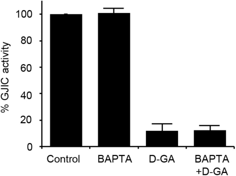
Gap junctions (GJs) are intercellular channels composed of connexins. Cellular molecules smaller than 1 kDa can diffuse through GJs by a process termed gap junctional intercellular communication (GJIC), which plays essential roles in various pathological and physiological conditions. Gambogic acid (GA), a major component of a natural yellow dye, has been used as traditional medicine and has been reported to have various therapeutic effects, including an anti-cancer effect. In this study, two different GJ assay methods showed that GA and its analogs inhibited GJIC. The inhibition was rapidly reversible and was not mediated by changes in surface expression or S368 phosphorylation of Cx43, cellular calcium concentration, or redox state. We also developed an assay system to measure the intercellular communication induced by Cx40, Cx30, and Cx43. Dihydrogambogic acid (D-GA) potently inhibited GJIC by Cx40 (IC50 = 5.1 μM), whereas the IC50 value of carbenoxolone, which is known as a broad spectrum GJIC inhibitor, was 105.2 μM. Thus, D-GA can act as a pharmacological tool for the inhibition of Cx40.

Keywords: Cx40; connexin; dihydrogambogic acid; gambogic acid; gap junction; tetrahydrogambogic acid.


Figures

FIGURE 1
FIGURE 1
Chemical structures of GA, D-GA, and T-GA. The red circles indicate the structural differences in the three compounds.
FIGURE 2
FIGURE 2
Inhibition of GJIC by GA, D-GA, and T-GA. I-YFP GJIC assay (see Materials and Methods) was conducted using LN215-I and LN215-YFP cells. The vehicle, GA (A), D-GA (B), or T-GA (C) was diluted in C-solution and applied to cells at the concentrations indicated above for 10 min before the GJIC assay. The mean % of GJIC activity ± SD of three independent experiments is presented as bar graphs.
FIGURE 3
FIGURE 3
Inhibition of GJIC by D-GA as demonstrated by the Gap-FRAP assay. FRT-Cx43 cells pre-loaded with calcein-AM were treated with vehicle, 1, 2, 5, or 10 μM D-GA before the FRAP assay. Representative images of the gap-FRAP assay are presented in (A). The percentage of fluorescence recovery was plotted as the mean ± SD (n = 10) against incubation time after photobleaching (B).
FIGURE 4
FIGURE 4
Reversible inhibition of GJIC by D-GA. The 2:1 mixture of LN215-I and LN215-YFP cells were plated on four separate 96-well plates. After incubation for 24 h, the cells were treated as indicated above. D-GA was treated at 20 μM for 10 min. For the wash and incubation groups, the cells were washed with growth media twice and further incubated for 20 or 60 min before the I-YFP GJIC assay was performed. The control group (without D-GA treatment) was used to calculate the percentage of GJIC activity, which was presented as the mean ± SD of three independent experiments in a bar graph.
FIGURE 5
FIGURE 5
Effect of D-GA treatment on surface expression and S368 phosphorylation of Cx43. LN215-Cx43 cells were grown to 80% confluency in 100 mm plates before treatment with vehicle, 100 ng/mL PMA combined with 50 ng/mL EGF, 1 μg/mL BFA combined with 10 μg/mL CHX or 20 μM D-GA. The treatments were conducted for 10 min except for BFA + CHX, which was treated for 6 h. The whole cell lysates were prepared after surface biotinylation. Biotinylated proteins from 500 μg protein of the whole cell lysates and 20 μg protein of the whole cell lysates were used for immunoblotting of anti-phospho-S368 and total Cx43, anti-Na+–K+ ATPase, and anti-actin antibodies. Three independent experiments were conducted, and representative blots are presented in (A). The images before cropping are also presented in Supplementary Figures 1A–E. The relative phospho-S368 Cx43 band intensity, divided by the total Cx43 band intensity, is reflective of Cx43 S368-phosphorylation and was calculated and presented as bar graphs (B). The data represent the mean ± SD (n = 3). The band intensity of total Cx43 bands was normalized to Na+–K+ ATPase and presented as a bar graph (C). The data represent the mean ± SD (n = 3). P < 0.05 (Student’s t-test); Veh, vehicle.
FIGURE 6
FIGURE 6
Effect of intracellular Ca2+ chelation on GJIC inhibition by D-GA. The LN215-I and LN215-YFP cells were mixed at a 2:1 ratio and plated in 96-well plates. After incubation for 24 h, the cells were pre-treated with vehicle or 5 μM BAPTA-AM in 100 μL of C-solution for 30 min. For the treatment of D-GA at 20 μM, D-GA or its vehicle was added at 20 min of the BAPTA-AM pre-treatment period. Subsequently, the I-YFP GJIC assay was conducted. The data represent the means ± SD (n = 3).
FIGURE 7
FIGURE 7
No ROS generation results from D-GA treatment in LN215 cells. LN215 cells were plated in 96-well plates and grown to full confluency. After washing with C-solution, the cells were loaded with 10 μM DCFH-DA diluted in C-solution for 1 h before fluorometry. H2O2 or D-GA was added after 30 min or 10 min before the measurement, respectively. The data are the mean ± SD (n = 3). p < 0.05 (Student’s t-test).
FIGURE 8
FIGURE 8
Validation of GJA1-null LN215 cells. The genomic DNA sequence alignment of the original and GJA1-null LN215 cells in the GJA1 region targeted by the Cas9 system (A). Ten TA-clones were read, and three types of alleles were identified. Dashes and lower-case letters indicate deleted and inserted residues, respectively. xN, +N, and –N represent the numbers of the TA-clones among the 10 clones inserted, and the deleted bases, respectively. A1, A2, or A3 are three types of alleles. The inverted triangle indicates the cleavage site by Cas9 in GJA1. WT, wild-type. The immunoblots showing Cx43 and actin expression of the original and GJA1-null LN215 cells (B). The images before cropping are also presented in Supplementary Figures 2A,B. I-YFP GJIC assay using the original (C, left) and GJA1-null (C, right) LN215 cells. The acceptor cells only or the 2:1 mixture of donor and acceptor cells were plated on 96-well plates before the I-YFP assay. The percentage of YFP fluorescence was plotted against assay time. The open and filled circles indicated the acceptor only and donor + acceptor groups, respectively. The data are the mean ± SD (n = 3).
FIGURE 9
FIGURE 9
The I-YFP assay measuring GJs composed of Cx43, Cx40, Cx31, or Cx30. The I-YFP GJIC assay was conducted with the acceptor only (formula image) or donor + acceptor cells (formula image) expressing Cx43 (A), Cx40 (B), Cx31 (C), or Cx30 (D) (see Materials and Methods for detailed information about the cells) for 10 s. The percentage of YFP fluorescence was plotted against assay time. The data represent the mean ± SD (n = 3).
FIGURE 10
FIGURE 10
IC50 values of D-GA and CBX inhibition of GJs composed of Cx43, Cx40, or Cx30. The 2:1 mixture of donor and acceptor cells originating from LN215-Cx43, -Cx40, or -Cx30 cells were plated, grown for 24 h, and then treated with various concentrations of D-GA (A) or CBX (B) for 10 min before the I-YFP assay. The percentage of GJIC inhibition was calculated and plotted against the log [compound] (μM). The data represent the mean ± SD (n = 3). The IC50 values were calculated using GraphPad Prism 5.

References

    1. Alexander D. B., Goldberg G. S. (2003). Transfer of biologically important molecules between cells through gap junction channels. Curr. Med. Chem. 10 2045–2058. 10.2174/0929867033456927 - DOI - PubMed
    1. Bastide B., Neyses L., Ganten D., Paul M., Willecke K., Traub O. (1993). Gap junction protein connexin40 is preferentially expressed in vascular endothelium and conductive bundles of rat myocardium and is increased under hypertensive conditions. Circ. Res. 73 1138–1149. 10.1161/01.RES.73.6.1138 - DOI - PubMed
    1. Brissette J. L., Kumar N. M., Gilula N. B., Dotto G. P. (1991). The tumor promoter 12-O-tetradecanoylphorbol-13-acetate and the ras oncogene modulate expression and phosphorylation of gap junction proteins. Mol. Cell. Biol. 11 5364–5371. 10.1128/MCB.11.10.5364 - DOI - PMC - PubMed
    1. Chen J., Gu H. Y., Lu N., Yang Y., Liu W., Qi Q., et al. (2008). Microtubule depolymerization and phosphorylation of C-Jun N-terminal kinase-1 and p38 were involved in gambogic acid induced cell cycle arrest and apoptosis in human breast carcinoma MCF-7 cells. Life Sci. 83 103–109. 10.1016/j.lfs.2008.05.003 - DOI - PubMed
    1. Garcia-Dorado D., Rodriguez-Sinovas A., Ruiz-Meana M. (2004). Gap junction-mediated spread of cell injury and death during myocardial ischemia-reperfusion. Cardiovasc. Res. 61 386–401. 10.1016/j.cardiores.2003.11.039 - DOI - PubMed

LinkOut - more resources