. 2018 Jul 9:2018:9463295.
doi: 10.1155/2018/9463295. eCollection 2018.

Microbial Diversity of Chronic Wound and Successful Management of Traditional Chinese Medicine

Affiliations

Microbial Diversity of Chronic Wound and Successful Management of Traditional Chinese Medicine

Minfeng Wu et al. Evid Based Complement Alternat Med. .

Abstract

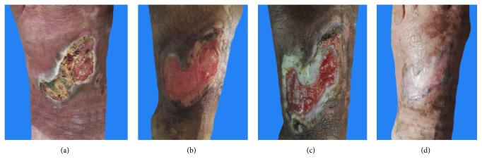
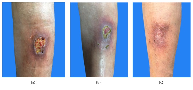
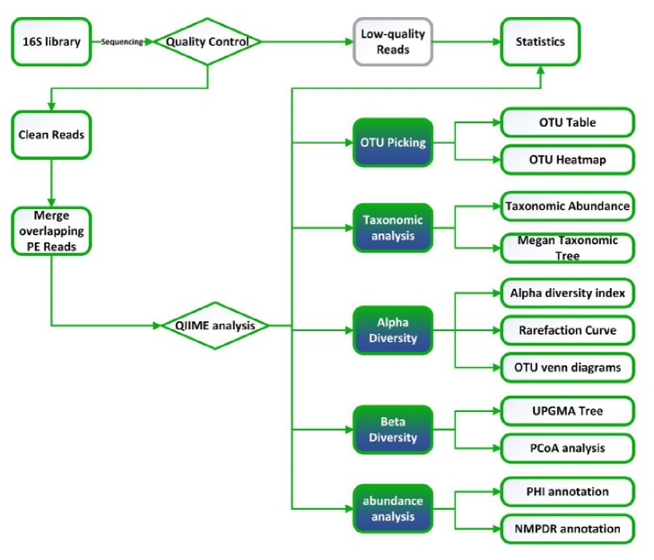
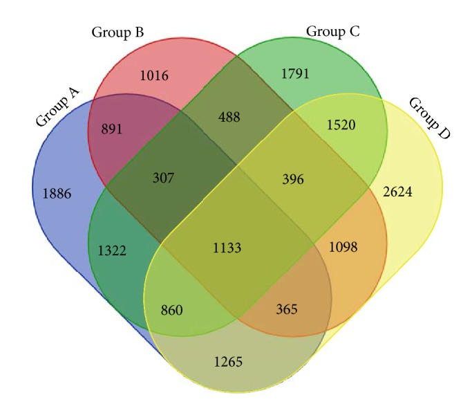
Chronic ulcer, including diabetic ulcer, varicose ulcer, and pressure ulcer, negatively affects patients' quality of life. As microbiology plays an important role in the mechanism of pathology for chronic wound healing, this study concentrates on microecology environment of the wound and how Traditional Chinese Medicine (TCM) regulates wound bacteria. Method. The study took wound samples from 35 patients and analyzed bacteria variation before and after TCM treatment by 16s rRNA sequencing. All samples were evaluated from aspects of α-diversity, β-diversity, and Simpson's Diversity index. Result. After total DNA extraction, PCR, and 16S rRNA sequencing of wound bacteria from 35 individuals, it was discovered that younger patients with shorter course of disease have a higher microbial diversity and were easier to recover from ulcers. Additionally, gender also played a vital role in wound healing, and a significant microbial diversity existed between male and female patients. Conclusion. Patients with chronic ulcers achieved a positive effect after TCM treatment (skin-producing ointment). Mechanistically, TCM helped promote wound healing by regulating the wound microbiota.


Figures

Figure 1
Figure 1
Case presentation of a 69-year-old female patient with chronic ulcer and lesion changes during TCM treatment.
Figure 2
Figure 2
Case presentation of a 56-year-old female patient with chronic ulcer and lesion changes during TCM treatment.
Figure 3
Figure 3
Flow diagram of biological information analysis.
Figure 4
Figure 4
Venn diagram: number of common and specific wound microbes before and after TCM treatment.
Figure 5
Figure 5
Diversity index “observed species” of species rarefaction curve.
Figure 6
Figure 6
Microbial diversity and bacteria proportion in wound and skin around wound before and after TCM treatment.
Figure 7
Figure 7
Microbial alteration of wound as well as skin around wound before and after TCM treatment, and its relationship with gender. (Note: A, B: lesions around wound before treatment (male and female); C, D: lesions around wound after treatment (male and female); E, F: lesions within wound before treatment (male and female); G, H: lesions within wound after treatment (male and female). All the bacteria were observed at the biological level of order.)
Figure 8
Figure 8
Microbial alteration of wound as well as skin around wound before and after TCM treatment and its relationship with ages. (Notes: A, B: lesions around wound before treatment (over 65 years old and under 65 years old); C, D: lesions around wound after treatment (over 65 and under 65); E, F: lesions within wound before treatment (over 65 and under 65); G, H: lesions within wound after treatment (over 65 and under 65). All the bacteria were observed at the biological level of order.)
Figure 9
Figure 9
Microbial alteration of wound as well as skin around wound before and after TCM treatment and its relationship with disease courses. (Notes: A, B: lesions around wound before treatment (disease course over and below half year); C, D: lesions around wound after treatment (disease course over and below half year); E, F: lesions within wound before treatment (disease course over and below half year); G, H: lesions within wound after treatment (disease course over and below half year). All the bacteria were observed at the biological level of order.)
Figure 10
Figure 10
PCoA results based on different algorithms: (a) Weighted UniFrac (Consider sequencing amounts when analyzing); (b) Unweighted UniFrac.

References

    1. Nassiri S., Zakeri I., Weingarten M. S., Spiller K. L. Relative expression of proinflammatory and antiinflammatory genes reveals differences between healing and nonhealing human chronic diabetic foot ulcers. Journal of Investigative Dermatology. 2015;135(6):1700–1703. doi: 10.1038/jid.2015.30. - DOI - PubMed
    1. Podbielska A., Galkowska H., Stelmach E., Mlynarczyk G., Olszewski W. L. Slime production by staphylococcus aureus and staphylococcus epidermidis strains isolated from patients with diabetic foot ulcers. Archivum Immunologiae et Therapia Experimentalis. 2010;58(4):321–324. doi: 10.1007/s00005-010-0079-9. - DOI - PubMed
    1. Grover G., Tanase A., Elstone A., Ashley S. Chronic venous leg ulcers: Effects of foam sclerotherapy on healing and recurrence. Phlebology. 2016;31(1):34–41. doi: 10.1177/0268355514557854. - DOI - PubMed
    1. Jiang Y., Huang S., Fu X., et al. Epidemiology of chronic cutaneous wounds in China. Wound Repair and Regeneration. 2011;19(2):181–188. doi: 10.1111/j.1524-475X.2010.00666.x. - DOI - PubMed
    1. Mantovani A., Perrone F., Stoico V., et al. Primary cutaneous B-cell lymphoma and chronic leg ulcers in a patient with type 2 diabetes. Endocrinology, diabetes & metabolism case reports. doi: 10.1530/EDM-17-0032. - DOI - PMC - PubMed

LinkOut - more resources