Case Reports
. 2018 Jul 11:2018:7813175.
doi: 10.1155/2018/7813175. eCollection 2018.

Fulminant Cerebral Fat Embolism: Case Description and Review of the Literature

Affiliations
Case Reports

Fulminant Cerebral Fat Embolism: Case Description and Review of the Literature

Giorgio Berlot et al. Case Rep Crit Care. .

Abstract

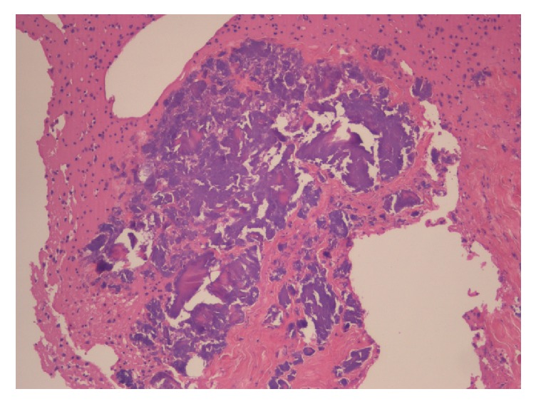
The release of fat and bone marrow fragments is a common occurrence following traumatic and nontraumatic events. In most cases, they go symptomless or cause only minor disturbances, but occasionally they can determine a multiorgan dysfunction whose severity ranges from mild to fatal. The authors describe the case of a patient who became deeply comatose and ultimately died after a traffic accident in which he suffered the exposed right femoral and tibial fracture in the absence of other injuries. He underwent the external fixation of the fractured bones 2 hours after the admission under general anesthesia. Three hours later, he failed to awake at the suspension of the anesthetic agents and became anisocoric; a CT scan demonstrated a diffuse cerebral edema with the herniation of the cerebellar tonsils; these abnormalities were unresponsive to the treatment and the brain death was one day later. The causes, the mechanisms, the symptoms, the prevention, and the treatment of the syndrome are reviewed and discussed.


Figures

Figure 1
Figure 1
a (left): brain CT scan showing a diffuse edema with the disappearance of the gray/white matter limitation; b (right): herniation of the cerebellar tonsils.
Figure 2
Figure 2
Large vascular thrombus in the cerebellum (H & E, 2.5).
Figure 3
Figure 3
A cerebral vein completely obliterated by normally noncirculating bone marrow components (promyelocytes and myelocytes) (H & E, 40).
Figure 4
Figure 4
Venular occlusion by erythrocytes, promyelocytes, and myelocytes (arrows) (H & E, 20).
Figure 5
Figure 5
Coarse calcification in the cerebral parenchyma (H & E, 10).

References

    1. Newbigin K., Souza C. A., Torres C., et al. Fat embolism syndrome: State-of-the-art review focused on pulmonary imaging findings. Respiratory Medicine. 2015 doi: 10.1016/j.rmed.2016.01.018. - DOI - PubMed
    1. Stein P. D., Yaekoub A. Y., Matta F., Kleerekoper M. Fat embolism syndrome. The American Journal of the Medical Sciences. 2008;336(6):472–477. doi: 10.1097/MAJ.0b013e318172f5d2. - DOI - PubMed
    1. Gangaraju R., Reddy V. V. B., Marques M. B. Fat embolism syndrome secondary to bone marrow necrosis in patients with hemoglobinopathies. Southern Medical Journal. 2016;109(9):549–553. doi: 10.14423/SMJ.0000000000000520. - DOI - PubMed
    1. Akhtar S. Fat Embolism. Anesthesiology Clinics. 2009;27(3):533–550. doi: 10.1016/j.anclin.2009.07.018. - DOI - PubMed
    1. Husebye E. E., Lyberg T., Røise O. Bone marrow fat in the circulation: clinical entities and pathophysiological mechanisms. Injury. 2006;37(4):p. -S18. doi: 10.1016/j.injury.2006.08.036. - DOI - PubMed

Publication types

LinkOut - more resources