

Rapid malignant progression of an intraparenchymal choroid plexus papillomas
- PMID: 30105129
- PMCID: PMC6044141
- DOI: 10.4103/sni.sni_434_17
Rapid malignant progression of an intraparenchymal choroid plexus papillomas
Abstract
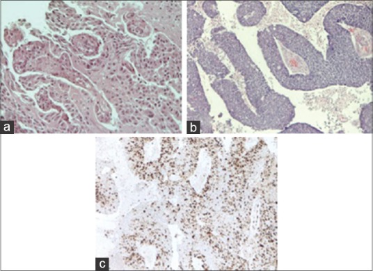
Background: Choroid plexus tumors (CPTs) are rare neoplasms accounting for only 0.3-0.6% of all brain tumors in adults and 2-5% in children. The World Health Organization (WHO) classification describes three histological grades: grade I is choroid plexus papilloma (CPP), grade II is atypical papilloma, and grade III is the malignant form of carcinoma. In adults, CPTs rarely have a supratentorial localization.
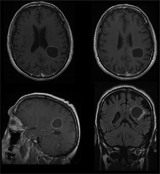
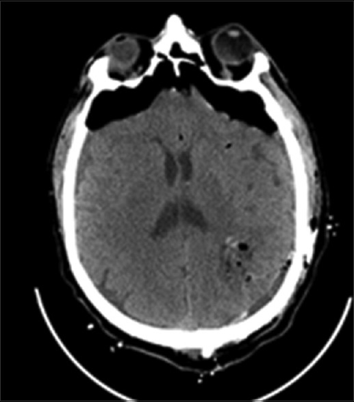
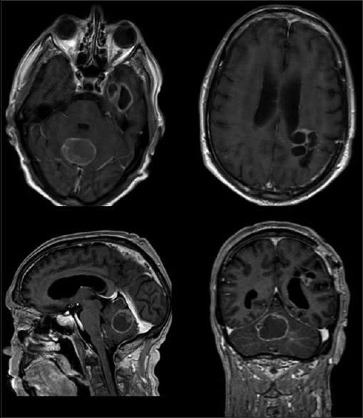
Case description: Here we report a very rare case of an intraparenchymal parietal CPP with a rapid histological transition from grade I to grade III WHO in a 67-year-old man, in <7 months.
Conclusion: Because of the rarity of these oncotypes, descriptions of each new case are useful, mostly to consider this diagnostic entity in extraventricular brain tumors of adults, despite an unusual location.
Keywords: Choroid plexus atypical papilloma; World Health Organization classification; choroid plexus carcinoma; choroid plexus papillomas; malignant progression.
Conflict of interest statement
There are no conflicts of interest.
Figures
References
-
- Bahar M, Hashem H, Tekautz T, Worley S, Tang A, de Blank P, et al. Choroid plexus tumors in adult and pediatric populations: The Cleveland Clinic and University Hospitals experience. J Neurooncol. 2017;132:427–32. - PubMed
-
- Cannon DM, Mohindra P, Gondi V, Kruser TJ, Kozak KR. Choroid plexus tumor epidemiology and outcomes: Implications for surgical and radiotherapeutic management. J Neurooncol. 2015;121:151–7. - PubMed
-
- Carter AB, Price DL, Jr, Tucci KA, Lewis GK, Mewborne J, Singh HK. Choroid plexus carcinoma presenting as an intraparenchymal mass. J Neurosurg. 2001;95:1040–4. - PubMed
-
- Dhillon RS, Wang YY, McKelvie PA, O’Brien B. Progression of choroid plexus papilloma. J Clin Neurosci. 2013;20:1775–8. - PubMed
Publication types
LinkOut - more resources
Full Text Sources
Other Literature Sources