

Addition of ionophore A23187 increases the efficiency of Cocos nucifera somatic embryogenesis
- PMID: 30105191
- PMCID: PMC6086808
- DOI: 10.1007/s13205-018-1392-y
Addition of ionophore A23187 increases the efficiency of Cocos nucifera somatic embryogenesis
Abstract
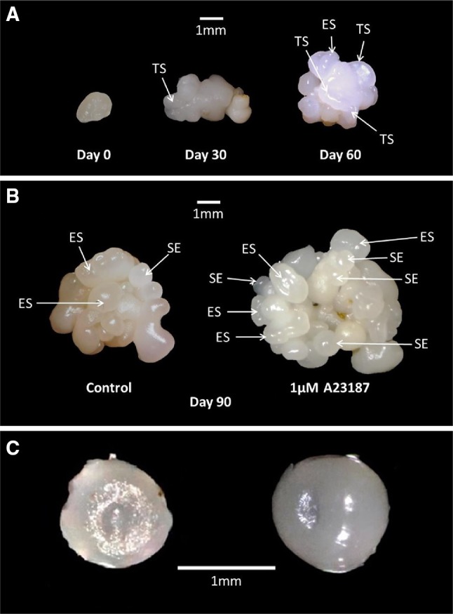
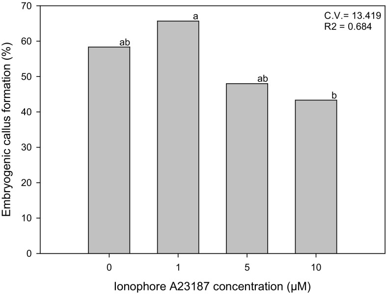
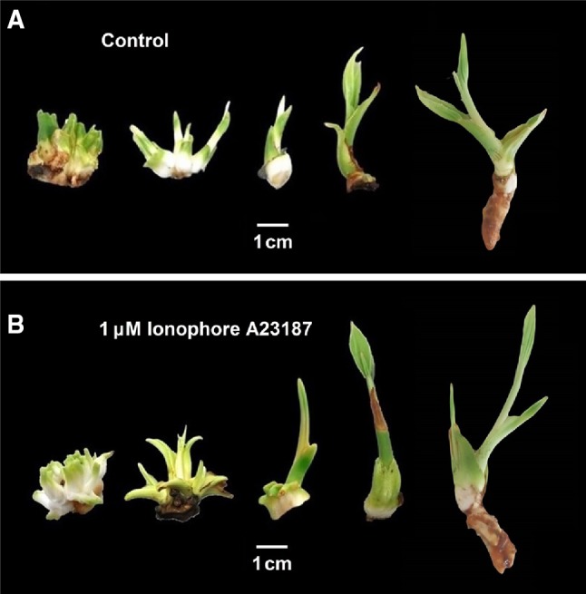
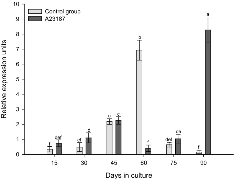
The present study reports the effect of treatment of coconut embryogenic structure explants (derived from embryogenic callus) with the calcium ionophore A23187 (0, 1, 5, 10 µM) to promote somatic embryogenesis under in vitro conditions. The results showed no significant increase in the percentage of explants forming embryogenic callus, but with 1 µM ionophore there were significant increases in the formation of embryogenic structures per callus (2.8-fold), of somatic embryos per callus (1.5-fold) and also a greater absolute number (1.5-fold) of developing plantlets per callus. The ionophore treatment also promoted a change of pattern of the expression of the CnSERK gene during embryogenic callus formation. It is proposed that if the use of ionophore A23187 treatment is coupled with an embryogenic callus multiplication process there could be a potentially greater increase in the efficiency of the formation of somatic embryos and plantlets of coconut.
Keywords: Calcium ionophore A23187; Cn SERK; Coconut; Somatic embryogenesis.
Conflict of interest statement
Compliance with ethical standardsThe authors declare that they have no conflict of interest in publication of this paper. This article does not contain any studies with human subjects or animal performed by any of the authors.
Figures
Similar articles
-
Cloning Coconut via Somatic Embryogenesis: A Review of the Current Status and Future Prospects.Plants (Basel). 2021 Sep 29;10(10):2050. doi: 10.3390/plants10102050. Plants (Basel). 2021. PMID: 34685859 Free PMC article. Review.
-
Detection of a SERK-like gene in coconut and analysis of its expression during the formation of embryogenic callus and somatic embryos.Plant Cell Rep. 2009 Jan;28(1):11-9. doi: 10.1007/s00299-008-0616-8. Epub 2008 Sep 26. Plant Cell Rep. 2009. PMID: 18818928
-
De novo assembly and characterization of global transcriptome of coconut palm (Cocos nucifera L.) embryogenic calli using Illumina paired-end sequencing.Protoplasma. 2016 May;253(3):913-928. doi: 10.1007/s00709-015-0856-8. Epub 2015 Jul 26. Protoplasma. 2016. PMID: 26210639
-
Endogenous cytokinins in Cocos nucifera L. in vitro cultures obtained from plumular explants.Plant Cell Rep. 2010 Nov;29(11):1227-34. doi: 10.1007/s00299-010-0906-9. Epub 2010 Aug 6. Plant Cell Rep. 2010. PMID: 20690021
-
Plant regeneration via somatic embryogenesis and shoot organogenesis from immature cotyledons of Camellia nitidissima Chi.J Plant Physiol. 2013 Sep 1;170(13):1202-11. doi: 10.1016/j.jplph.2013.03.019. Epub 2013 Jun 20. J Plant Physiol. 2013. PMID: 23790533
Cited by
-
Calcium dynamics and modulation in carrot somatic embryogenesis.Front Plant Sci. 2023 Mar 31;14:1150198. doi: 10.3389/fpls.2023.1150198. eCollection 2023. Front Plant Sci. 2023. PMID: 37063186 Free PMC article.
-
Calcium levels modulate embryo yield in Brassica napus microspore embryogenesis.Front Plant Sci. 2025 Jan 16;15:1512500. doi: 10.3389/fpls.2024.1512500. eCollection 2024. Front Plant Sci. 2025. PMID: 39886677 Free PMC article.
-
Calcium Dynamics, WUSCHEL Expression and Callose Deposition during Somatic Embryogenesis in Arabidopsis thaliana Immature Zygotic Embryos.Plants (Basel). 2023 Feb 23;12(5):1021. doi: 10.3390/plants12051021. Plants (Basel). 2023. PMID: 36903882 Free PMC article.
-
Cloning Coconut via Somatic Embryogenesis: A Review of the Current Status and Future Prospects.Plants (Basel). 2021 Sep 29;10(10):2050. doi: 10.3390/plants10102050. Plants (Basel). 2021. PMID: 34685859 Free PMC article. Review.
References
-
- Altamura MM, Rovere FD, Fattorini L, D’, Angeli S, Falasca G. Recent advances on genetic and physiological bases on in vitro somatic embryo formation. In: Germanà MA, Lambardi M, editors. In vitro embryogenesis in higher plants. New York: Humana Press; 2016. p. 558. - PubMed
-
- Azpeitia A, Chan JL, Sáenz L, Oropeza C. Effect of 22(S)-homobrassinolide in somatic embryogenesis in plumule explants of Cocos nucifera (L.) cultured in vitro. J Hortic Sci Biotech. 2003;78:591–596. doi: 10.1080/14620316.2003.11511669. - DOI
-
- Batugal P, Rao Ramanatha V, Oliver J (2005) Coconut genetic resources. International Plant Genetic Resources Institute - Regional office for Asia, the Pacific and Oceania. IPGRI-APO, Serdang
-
- Buffard-Morel J, Verdeil JL, Pannetier C. Embryogenèse somatique du cocotier (Cocos nucifera L.) à partir d´explants foliaires: étude histologique. Can J Bot. 1992;70:735–741. doi: 10.1139/b92-094. - DOI
LinkOut - more resources
Full Text Sources
Other Literature Sources
