

Macular ganglion cells alteration in a patient with left homonymous hemianopia subsequent to surgical excision of an arteriovenous malformation
- PMID: 30105312
- PMCID: PMC6085272
- DOI: 10.1016/j.ajoc.2018.07.009
Macular ganglion cells alteration in a patient with left homonymous hemianopia subsequent to surgical excision of an arteriovenous malformation
Abstract
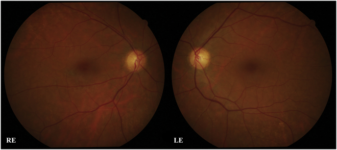
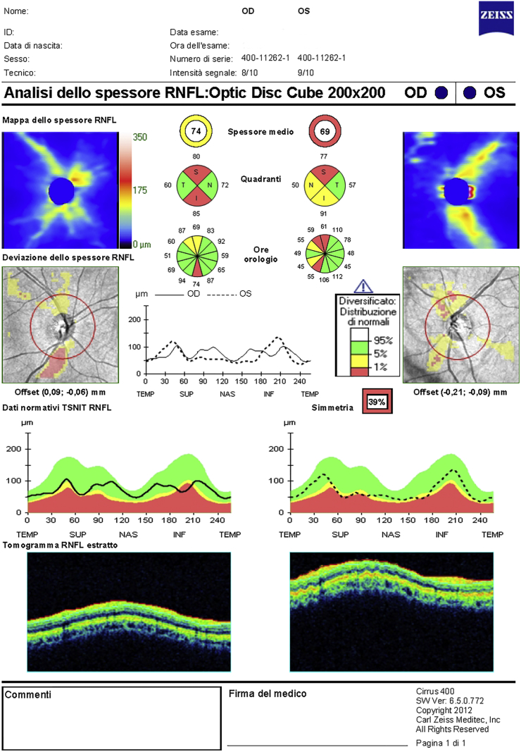
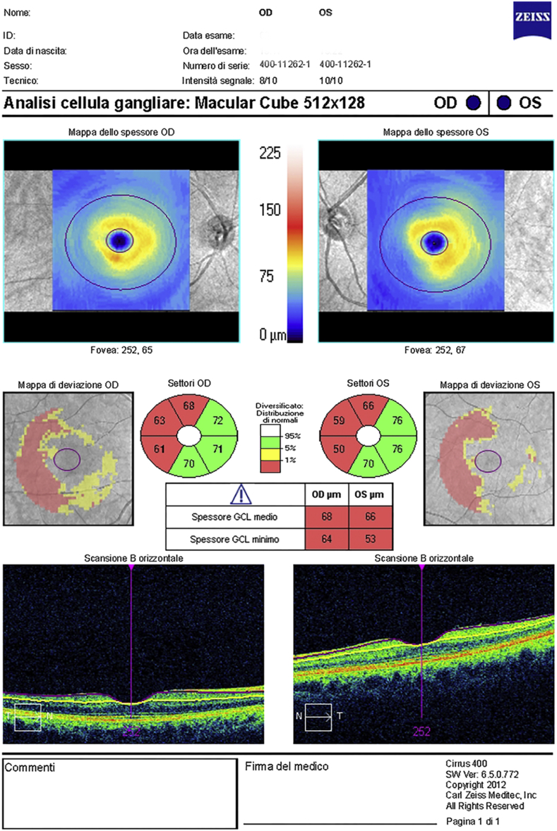
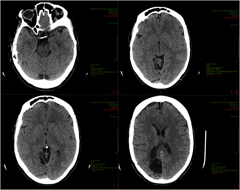
Purpose: To report the case of a 57 years old woman who showed a macular ganglion cell complex (GCC), that is a combination of ganglion cell layer and inner plexiform layer, and peripapillary Retinal Nerve Fiber Layer (pRNFL) thickness reduction in association with left homonymous hemianopia subsequent to surgical excision of an arteriovenous malformation in the cerebral right occipital lobe 37 years before.
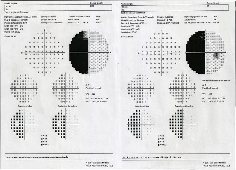
Observations: One patient with left homonymous hemianopia due to surgical excision of an arteriovenous malformation in the right cerebral occipital lobe came to our attention for transient blurred vision.Measurement of the GCC and pRNFL thickness was performed using spectral domain optical coherence tomography (SDOCT; Cirrus HD-OCT model 400). Visual field (VF) defects were assessed using Humphrey field analyzer using the central 30-2 Swedish Interactive Threshold Algorithm (SITA) program with appropriate trial lenses (Humphrey Field Analyzer II, Carl Zeiss Meditech, Inc, Dublin, California).The average pRNFL thickness was bilaterally reduced, showing a symmetry value of 39%. The patients showed a significant GCC thinning in the projecting sector of the retina mapping to the brain lesion. Corresponding VF defects were found.
Conclusions and importance: These findings show SDOCT potentials in the field of neuro-ophthalmology, supporting the usefulness of GCC thickness as a possible imaging marker before and after brain surgery, and, possibly, in the diagnosis of neurodegenerative conditions.
Keywords: Arteriovenous malformation; GCC; Hemianopia; Neuro-ophthalmology; Neurodegenerative diseases; OCT; Retrograde transneural degeneration; pRNFL.
Figures
Similar articles
-
Sectoral analysis of the retinal nerve fiber layer thinning and its association with visual field loss in homonymous hemianopia caused by post-geniculate lesions using spectral-domain optical coherence tomography.Graefes Arch Clin Exp Ophthalmol. 2016 Apr;254(4):745-56. doi: 10.1007/s00417-015-3181-1. Epub 2015 Oct 7. Graefes Arch Clin Exp Ophthalmol. 2016. PMID: 26446718 Free PMC article.
-
Reduced Peripapillary and Macular Vessel Density in Unilateral Postgeniculate Lesions With Retrograde Transsynaptic Degeneration.J Neuroophthalmol. 2019 Dec;39(4):462-469. doi: 10.1097/WNO.0000000000000794. J Neuroophthalmol. 2019. PMID: 31658224
-
Retina ganglion cell/inner plexiform layer and peripapillary nerve fiber layer thickness in patients with acromegaly.Int Ophthalmol. 2017 Jun;37(3):591-598. doi: 10.1007/s10792-016-0310-8. Epub 2016 Aug 4. Int Ophthalmol. 2017. PMID: 27492731
-
Visual fields and optical coherence tomography (OCT) in neuro-ophthalmology: Structure-function correlation.J Neurol Sci. 2021 Oct 15;429:118064. doi: 10.1016/j.jns.2021.118064. Epub 2021 Sep 1. J Neurol Sci. 2021. PMID: 34488042 Review.
-
[New insights into the study of optic nerve diseases].Nippon Ganka Gakkai Zasshi. 2013 Mar;117(3):187-210; discussion 211. Nippon Ganka Gakkai Zasshi. 2013. PMID: 23631254 Review. Japanese.
Cited by
-
MRI and Clinical Biomarkers Overlap between Glaucoma and Alzheimer's Disease.Int J Mol Sci. 2023 Oct 5;24(19):14932. doi: 10.3390/ijms241914932. Int J Mol Sci. 2023. PMID: 37834380 Free PMC article. Review.
-
Interobserver reproducibility and interocular symmetry of the macular ganglion cell complex: assessment in healthy children using optical coherence tomography.BMC Ophthalmol. 2020 May 24;20(1):197. doi: 10.1186/s12886-020-01379-z. BMC Ophthalmol. 2020. PMID: 32448232 Free PMC article.
-
Translational perspective: is cinnamon a suitable agent for cognitive impairment and Alzheimer's disease associated with brain trauma?Neural Regen Res. 2019 Aug;14(8):1372-1373. doi: 10.4103/1673-5374.253518. Neural Regen Res. 2019. PMID: 30964057 Free PMC article. No abstract available.
-
Correlation between serum biomarkers, brain volume, and retinal neuronal loss in early-onset Alzheimer's disease.Neurol Sci. 2024 Jun;45(6):2615-2623. doi: 10.1007/s10072-023-07256-z. Epub 2024 Jan 13. Neurol Sci. 2024. PMID: 38216851
References
-
- Cowey A., Alexander I., Stoerig P. Transneuronal retrograde degeneration of retinal ganglion cells and optic tract in hemianopic monkeys and humans. Brain. 2011 Jul;134(Pt 7):2149–2157. - PubMed
-
- Dinkin M. Trans-synaptic retrograde degeneration in the human visual system: slow, silent, and real. Curr Neurol Neurosci Rep. 2017 Feb;17(2):16. - PubMed
-
- Shin H.Y., Park H.Y., Choi J.A., Park C.K. Macular ganglion cell-inner plexiform layer thinning in patients with visual field defect that respects the vertical meridian. Graefes Arch Clin Exp Ophthalmol. 2014 Sep;252(9):1501–1507. - PubMed
Publication types
LinkOut - more resources
Full Text Sources
Other Literature Sources
