

Telmisartan Protects a Microglia Cell Line from LPS Injury Beyond AT1 Receptor Blockade or PPARγ Activation
- PMID: 30105672
- PMCID: PMC6677563
- DOI: 10.1007/s12035-018-1300-9
Telmisartan Protects a Microglia Cell Line from LPS Injury Beyond AT1 Receptor Blockade or PPARγ Activation
Abstract
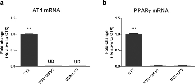
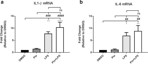
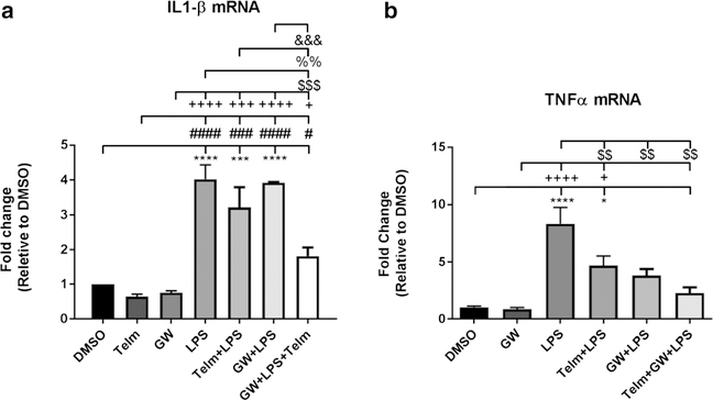
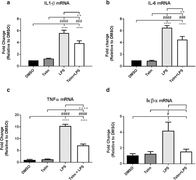
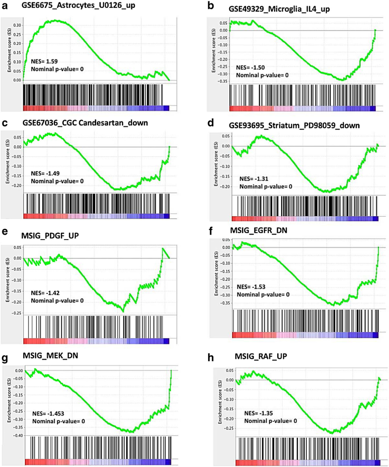
The Angiotensin II Receptor Blocker (ARB) Telmisartan reduces inflammation through Angiotensin II AT1 receptor blockade and peroxisome proliferator-activated receptor gamma (PPARγ) activation. However, in a mouse microglia-like BV2 cell line, imitating primary microglia responses with high fidelity and devoid of AT1 receptor gene expression or PPARγ activation, Telmisartan reduced gene expression of pro-injury factors, enhanced that of anti-inflammatory genes, and prevented LPS-induced increase in inflammatory markers. Using global gene expression profiling and pathways analysis, we revealed that Telmisartan normalized the expression of hundreds of genes upregulated by LPS and linked with inflammation, apoptosis and neurodegenerative disorders, while downregulating the expression of genes associated with oncological, neurodegenerative and viral diseases. The PPARγ full agonist Pioglitazone had no neuroprotective effects. Surprisingly, the PPARγ antagonists GW9662 and T0070907 were neuroprotective and enhanced Telmisartan effects. GW9226 alone significantly reduced LPS toxic effects and enhanced Telmisartan neuroprotection, including downregulation of pro-inflammatory TLR2 gene expression. Telmisartan and GW9662 effects on LPS injury negatively correlated with pro-inflammatory factors and upstream regulators, including TLR2, and positively with known neuroprotective factors and upstream regulators. Gene Set Enrichment Analysis (GSEA) of the Telmisartan and GW9662 data revealed negative correlations with sets of genes associated with neurodegenerative and metabolic disorders and toxic treatments in cultured systems, while demonstrating positive correlations with gene sets associated with neuroprotection and kinase inhibition. Our results strongly suggest that novel neuroprotective effects of Telmisartan and GW9662, beyond AT1 receptor blockade or PPARγ activation, include downregulation of the TLR2 signaling pathway, findings that may have translational relevance.
Keywords: Angiotensin receptor blockers; Inflammation; Neuroprotection, microglia; PPARγ; TLR2.
Conflict of interest statement
Figures
Similar articles
-
Telmisartan ameliorates Aβ oligomer-induced inflammation via PPARγ/PTEN pathway in BV2 microglial cells.Biochem Pharmacol. 2020 Jan;171:113674. doi: 10.1016/j.bcp.2019.113674. Epub 2019 Oct 18. Biochem Pharmacol. 2020. PMID: 31634455
-
Involvement of PPAR-γ in the neuroprotective and anti-inflammatory effects of angiotensin type 1 receptor inhibition: effects of the receptor antagonist telmisartan and receptor deletion in a mouse MPTP model of Parkinson's disease.J Neuroinflammation. 2012 Feb 22;9:38. doi: 10.1186/1742-2094-9-38. J Neuroinflammation. 2012. PMID: 22356806 Free PMC article.
-
Telmisartan directly ameliorates the neuronal inflammatory response to IL-1β partly through the JNK/c-Jun and NADPH oxidase pathways.J Neuroinflammation. 2012 May 29;9:102. doi: 10.1186/1742-2094-9-102. J Neuroinflammation. 2012. PMID: 22642771 Free PMC article.
-
Angiotensin II AT(1) receptor blockers as treatments for inflammatory brain disorders.Clin Sci (Lond). 2012 Nov;123(10):567-90. doi: 10.1042/CS20120078. Clin Sci (Lond). 2012. PMID: 22827472 Free PMC article. Review.
-
Blockade of brain angiotensin II AT1 receptors ameliorates stress, anxiety, brain inflammation and ischemia: Therapeutic implications.Psychoneuroendocrinology. 2011 Jan;36(1):1-18. doi: 10.1016/j.psyneuen.2010.10.001. Epub 2010 Oct 29. Psychoneuroendocrinology. 2011. PMID: 21035950 Free PMC article. Review.
Cited by
-
Amorfrutin B Compromises Hypoxia/Ischemia-induced Activation of Human Microglia in a PPARγ-dependent Manner: Effects on Inflammation, Proliferation Potential, and Mitochondrial Status.J Neuroimmune Pharmacol. 2024 Jul 1;19(1):34. doi: 10.1007/s11481-024-10135-9. J Neuroimmune Pharmacol. 2024. PMID: 38949694 Free PMC article.
-
Telmisartan Inhibits TNFα-Induced Leukocyte Adhesion by Blocking ICAM-1 Expression in Astroglial Cells but Not in Endothelial Cells.Biomol Ther (Seoul). 2020 Sep 1;28(5):423-430. doi: 10.4062/biomolther.2020.119. Biomol Ther (Seoul). 2020. PMID: 32782234 Free PMC article.
-
The application of telmisartan in central nervous system disorders.Pharmacol Rep. 2025 Jun 19. doi: 10.1007/s43440-025-00737-2. Online ahead of print. Pharmacol Rep. 2025. PMID: 40536710 Review.
-
Candesartan Neuroprotection in Rat Primary Neurons Negatively Correlates with Aging and Senescence: a Transcriptomic Analysis.Mol Neurobiol. 2020 Mar;57(3):1656-1673. doi: 10.1007/s12035-019-01800-9. Epub 2019 Dec 7. Mol Neurobiol. 2020. PMID: 31811565 Free PMC article.
-
Addressing Peroxisome Proliferator-Activated Receptor-gamma in 3-Nitropropionic Acid-Induced Striatal Neurotoxicity in Rats.Mol Neurobiol. 2022 Jul;59(7):4368-4383. doi: 10.1007/s12035-022-02856-w. Epub 2022 May 12. Mol Neurobiol. 2022. PMID: 35553009 Free PMC article.
References
MeSH terms
Substances
Grants and funding
LinkOut - more resources
Full Text Sources
Other Literature Sources
Molecular Biology Databases
Research Materials
