. 2018 Aug;13(8):1396-1407.
doi: 10.4103/1673-5374.235293.

Scutellarin protects oxygen/glucose-deprived astrocytes and reduces focal cerebral ischemic injury

Affiliations

Scutellarin protects oxygen/glucose-deprived astrocytes and reduces focal cerebral ischemic injury

Jing-Bo Sun et al. Neural Regen Res. 2018 Aug.

Abstract

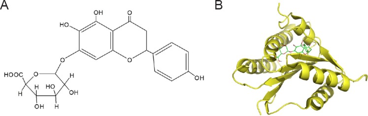
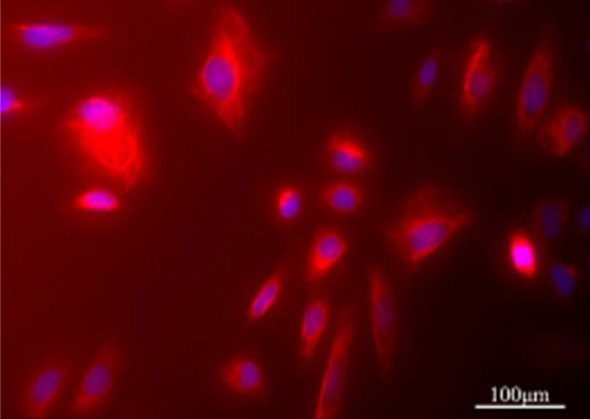
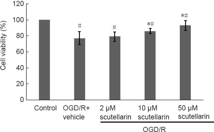
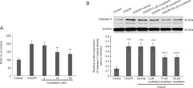
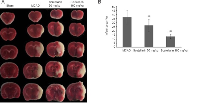
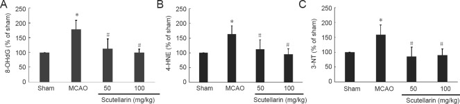
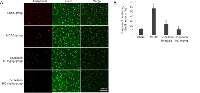
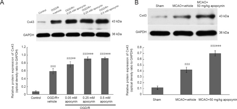
Scutellarin, a bioactive flavone isolated from Scutellaria baicalensis, has anti-inflammatory, anti-neurotoxic, anti-apoptotic and anti-oxidative effects and has been used to treat cardiovascular and cerebrovascular diseases in China. However, the mechanisms by which scutellarin mediates neuroprotection in cerebral ischemia remain unclear. The interaction between scutellarin and nicotinamide adenine dinucleotide phosphate oxidase 2 (NOX2) was assessed by molecular docking study, which showed that scutellarin selectively binds to NOX2 with high affinity. Cultures of primary astrocytes isolated from the cerebral cortex of neonatal Sprague-Dawley rats were pretreated with 2, 10 or 50 μM scutellarin for 30 minutes. The astrocytes were then subjected to oxygen/glucose deprivation by incubation for 2 hours in glucose-free Dulbecco's modified Eagle's medium in a 95% N2/5% CO2 incubator, followed by simulated reperfusion for 22 hours. Cell viability was assessed by cell counting kit-8 assay. Expression levels of NOX2, connexin 43 and caspase-3 were assessed by western blot assay. Reactive oxygen species were measured spectrophotometrically. Pretreatment with 10 or 50 μM scutellarin substantially increased viability, reduced the expression of NOX2 and caspase-3, increased the expression of connexin 43, and diminished the levels of reactive oxygen species in astrocytes subjected to ischemia-reperfusion. We also assessed the effects of scutellarin in vivo in the rat transient middle cerebral artery occlusion model of cerebral ischemia-reperfusion injury. Rats were given intraperitoneal injection of 100 mg/kg scutellarin 2 hours before surgery. The Bederson scale was used to assess neurological deficit, and 2,3,5-triphenyltetrazolium chloride staining was used to measure infarct size. Western blot assay was used to assess expression of NOX2 and connexin 43 in brain tissue. Enzyme-linked immunosorbent assay was used to detect 8-hydroxydeoxyguanosine (8-OHdG), 4-hydroxy-2-nonenal (4-HNE) and 3-nitrotyrosin (3-NT) in brain tissue. Immunofluorescence double staining was used to determine the co-expression of caspase-3 and NeuN. Pretreatment with scutellarin improved the neurological function of rats with focal cerebral ischemia, reduced infarct size, diminished the expression of NOX2, reduced levels of 8-OHdG, 4-HNE and 3-NT, and reduced the number of cells co-expressing caspase-3 and NeuN in the injured brain tissue. Furthermore, we examined the effect of the NOX2 inhibitor apocynin. Apocynin substantially increased connexin 43 expression in vivo and in vitro. Collectively, our findings suggest that scutellarin protects against ischemic injury in vitro and in vivo by downregulating NOX2, upregulating connexin 43, decreasing oxidative damage, and reducing apoptotic cell death.

Keywords: cerebral ischemic injury; connexin 43; nerve regeneration; neural regeneration; nicotinamide adenine dinucleotide phosphate oxidase 2; oxygen glucose deprivation/reoxygenation; reactive oxygen species; scutellarin.


Conflict of interest statement

None declared

Figures

Figure 1
Figure 1
Scutellarin selectivity binds to NOX2 with high affinity. (A) Chemical structure of scutellarin. (B) Three-dimensional docking model of NOX2 and scutellarin.
Figure 2
Figure 2
Expression of GFAP in astrocytes detected by immunofluorescence staining. Cultured astrocytes were labeled with anti-GFAP antibody and counterstained with DAPI nuclear stain. GFAP was localized in the cytoplasm. Scale bar: 100 μm. GFAP: Glial fibrillary acidic protein.
Figure 3
Figure 3
Scutellarin protects astrocytes against oxygen/glucose deprivation/reoxygenation (OGD/R)-induced cytotoxicity (cell counting kit-8 assay). Cell viability was expressed as the ratio of the optical density at 450 nm to that in the control group. Data are expressed as the mean ± SD (one-way analysis of variance followed by the Tukey-Kramer multiple comparison test). *P < 0.05, vs. OGD/R + vehicle group; #P < 0.05, vs. control group.
Figure 4
Figure 4
Scutellarin inhibits NOX2 (A) but enhances Cx43 (B) expression in astrocytes subjected to OGD/R (western blot assay). Representative immunoblots and semi-quantitative analysis of NOX2 and Cx43 protein levels. Data are expressed as the mean ± SD (n = 3 per group at each time point; one-way analysis of variance followed by the Tukey-Kramer multiple comparison test). *P < 0.05, ***P < 0.001, vs. OGD/R + vehicle group; #P < 0.05, ##P < 0.01, ###P < 0.001, vs. control group. NOX2: Nicotinamide adenine dinucleotide phosphate oxidase 2; Cx43: connexin 43; OGD/R: oxygen/glucose deprivation/reoxygenation; GAPDH: glyceraldehyde-3-phosphate dehydrogenase.
Figure 5
Figure 5
Scutellarin inhibits the OGD-induced intracellular accumulation of ROS (A) and caspase-3 expression (B) in astrocytes subjected to OGD/R detected using spectrophotometric assay and western blot assay, respectively. Data are expressed as the mean ± SD (n = 3 per group at each time point; one-way analysis of variance followed by the Tukey-Kramer multiple comparison test). *P < 0.05, **P < 0.01, vs. OGD/R + vehicle group; #P < 0.05, ##P < 0.01, ###P < 0.001, vs. control group. OGD: Oxygen/glucose deprivation; ROS: reactive oxygen species; OGD/R: oxygen/glucose deprivation/reoxygenation; GAPDH: glyceraldehyde-3-phosphate dehydrogenase.
Figure 6
Figure 6
Effects of scutellarin on infarct area of rats after cerebral ischemic injury. (A) Representative images of TTC-stained brain sections (the infarcted region appears white). (B) Quantification of infarct area 3 days post-injury (% of contralateral hemisphere). Data are expressed as the mean ± SD (n = 11 per group; one-way analysis of variance followed by the Tukey-Kramer multiple comparison test). ##P < 0.01, vs. MCAO group. TTC: 2,3,5-Triphenyltetrazolium chloride; MCAO: middle cerebral artery occlusion.
Figure 7
Figure 7
Scutellarin inhibited NOX2 but enhanced Cx43 expression in the ipsilateral hemisphere of rats with cerebral ischemic injury 3 days after reperfusion (western blot assay). Representative immunoblots and semi-quantitative analysis of NOX2 (A) and Cx43 (B) protein levels. Data are expressed as the mean ± SD (n = 3 per group; one-way analysis of variance followed by the Tukey-Kramer multiple comparison test). #P < 0.05, vs. MCAO group; *P < 0.05, vs. sham group. NOX2: Nicotinamide adenine dinucleotide phosphate oxidase 2; Cx43: connexin 43; MCAO: middle cerebral artery occlusion; GAPDH: glyceraldehyde-3-phosphate dehydrogenase.
Figure 8
Figure 8
Scutellarin reduces the increase in the levels of 8-OHdG (A), 4-HNE (B) and 3-NT (C) induced by cerebral ischemic injury (enzyme-linked immunosorbent assay). Data are expressed as the mean ± SD (n = 6 per group; one-way analysis of variance followed by the Tukey-Kramer multiple comparison test). #P < 0.05, vs. MCAO group; *P < 0.05, vs. sham group. 8-OHdG: 8-Hydroxydeoxyguanosine; 4-HNE: 4-hydroxy-2-nonenal; 3-NT: 3-nitrotyrosin; MCAO: middle cerebral artery occlusion.
Figure 9
Figure 9
Scutellarin downregulated caspase-3 in the penumbra after cerebral ischemic injury. (A) Immunofluorescence double-labeling for caspase-3 and NeuN: Capsase-3 is localized in the nuclei of apoptotic cells. NeuN is localized in the cytoplasm and perinuclear region of mature neurons. Scale bar: 100 μm. (B) Percentage of caspase-3+/NeuN+ cells in the penumbra region. Data are expressed as the mean ± SD (n = 3 per group; one-way analysis of variance followed by the Tukey-Kramer multiple comparison test). #P < 0.05, vs. MCAO group; *P < 0.05, vs. sham group. MCAO: Middle cerebral artery occlusion.
Figure 10
Figure 10
Apocynin markedly increased Cx43 protein expression in OGD-exposed astrocytes and in rats with cerebral ischemia/reperfusion injury (western blot assay). Representative immunoblots and semi-quantitative analysis of Cx43 protein levels (A, B). Data are expressed as the mean ± SD (n = 3 per group; one-way analysis of variance followed by the Tukey-Kramer multiple comparison test). **P < 0.01, ***P < 0.001, vs. OGD/R + vehicle/MCAO + vehicle group; ###P < 0.001, vs. control/sham group. Cx43: Connexin 43; OGD: oxygen/glucose deprivation; OGD/R: oxygen/glucose deprivation/reoxygenation; MCAO: middle cerebral artery occlusion; GAPDH: glyceraldehyde-3-phosphate dehydrogenase.

References

    1. Abramov AY, Scorziello A, Duchen MR. Three distinct mechanisms generate oxygen free radicals in neurons and contribute to cell death during anoxia and reoxygenation. J Neurosci. 2007;27:1129–1138. - PMC - PubMed
    1. Bederson JB, Pitts LH, Tsuji M, Nishimura MC, Davis RL, Bartkowski H. Rat middle cerebral artery occlusion: evaluation of the model and development of a neurologic examination. Stroke. 1986;17:472–476. - PubMed
    1. Berthoud VM, Beyer EC. Oxidative stress, lens gap junctions, and cataracts. Antioxid Redox Signal. 2009;11:339–353. - PMC - PubMed
    1. Boitier E, Rea R, Duchen MR. Mitochondria exert a negative feedback on the propagation of intracellular Ca2+ waves in rat cortical astrocytes. J Cell Biol. 1999;145:795–808. - PMC - PubMed
    1. Brennan-Minnella AM, Won SJ, Swanson RA. NADPH oxidase-2: linking glucose, acidosis, and excitotoxicity in stroke. Antioxid Redox Signal. 2015;22:161–174. - PMC - PubMed