. 2018 Aug;13(8):1455-1464.
doi: 10.4103/1673-5374.235303.

Novel conductive polypyrrole/silk fibroin scaffold for neural tissue repair

Affiliations

Novel conductive polypyrrole/silk fibroin scaffold for neural tissue repair

Ya-Hong Zhao et al. Neural Regen Res. 2018 Aug.

Abstract

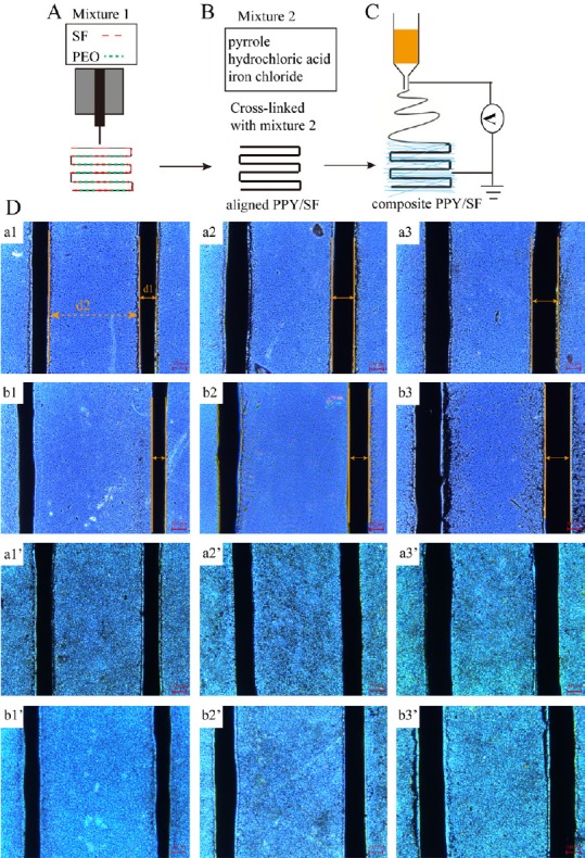
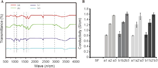
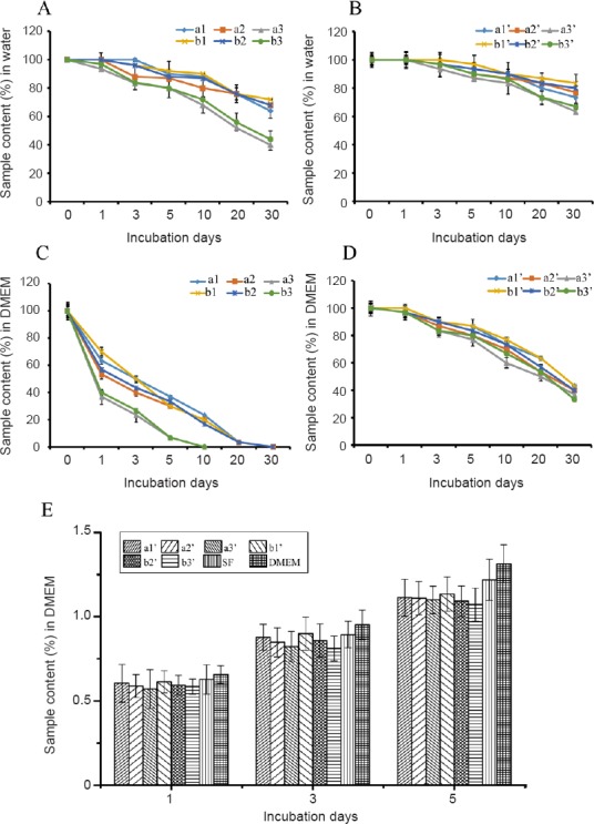
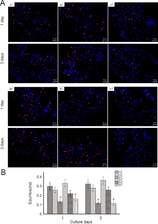
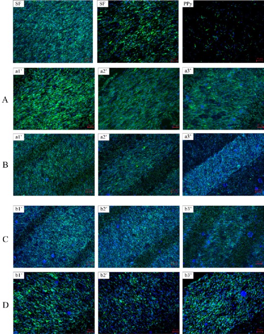
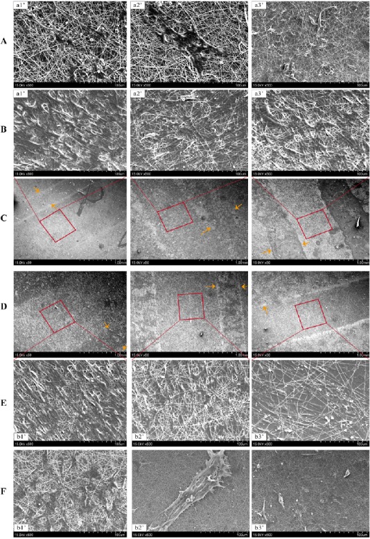
Three dimensional (3D) bioprinting, which involves depositing bioinks (mixed biomaterials) layer by layer to form computer-aided designs, is an ideal method for fabricating complex 3D biological structures. However, it remains challenging to prepare biomaterials with micro-nanostructures that accurately mimic the nanostructural features of natural tissues. A novel nanotechnological tool, electrospinning, permits the processing and modification of proper nanoscale biomaterials to enhance neural cell adhesion, migration, proliferation, differentiation, and subsequent nerve regeneration. The composite scaffold was prepared by combining 3D bioprinting with subsequent electrochemical deposition of polypyrrole and electrospinning of silk fibroin to form a composite polypyrrole/silk fibroin scaffold. Fourier transform infrared spectroscopy was used to analyze scaffold composition. The surface morphology of the scaffold was observed by light microscopy and scanning electron microscopy. A digital multimeter was used to measure the resistivity of prepared scaffolds. Light microscopy was applied to observe the surface morphology of scaffolds immersed in water or Dulbecco's Modified Eagle's Medium at 37°C for 30 days to assess stability. Results showed characteristic peaks of polypyrrole and silk fibroin in the synthesized conductive polypyrrole/silk fibroin scaffold, as well as the structure of the electrospun nanofiber layer on the surface. The electrical conductivity was 1 × 10⁻⁵-1 × 10⁻³ S/cm, while stability was 66.67%. A 3-(4,5-dimethyl-2-thiazolyl)-2,5-diphenyl-2-H-tetrazolium bromide assay was employed to measure scaffold cytotoxicity in vitro. Fluorescence microscopy was used to observe EdU-labeled Schwann cells to quantify cell proliferation. Immunohistochemistry was utilized to detect S100β immunoreactivity, while scanning electron microscopy was applied to observe the morphology of adherent Schwann cells. Results demonstrated that the polypyrrole/silk fibroin scaffold was not cytotoxic and did not affect Schwann cell proliferation. Moreover, filopodia formed on the scaffold and Schwann cells were regularly arranged. Our findings verified that the composite polypyrrole/silk fibroin scaffold has good biocompatibility and may be a suitable material for neural tissue engineering.

Keywords: L929 cells; Schwann cells; biocompatibility; composite nanofiber; conductivity; electrospinning; nerve regeneration; nerve repair; neural regeneration; polypyrrole; scaffold; silk fibroin; three dimensional bioprinting.


Conflict of interest statement

There are no conflicts of interest to declare

Figures

Figure 1
Figure 1
Schematic overview of construct generation. (A) Bioprinting preparation for SF aligned scaffolds. (B) Polymerization of PPy for SF aligned scaffolds. (C) Coating of electrospun SF nanofibers on aligned PPy/SF scaffolds. (D) Surface morphology of aligned PPy/SF scaffolds with varying diameters and distances (a1–b3): a1-80/500, a2-120/500, a3-180/500, b1-80/700, b2-120/700, and b3-180/700. Surface morphology of aligned PPy/SF scaffolds coupled with electrospun SF nanofiber coating with varying diameters and distances (a1’–b3’): a1’-80/500, a2’-120/500, a3’-180/500, b1’-80/700, b2’-120/700, and b3’-180/700. The number in front of the slash represents the diameter of PPy/SF scaffolds, while the number behind the slash represents the distance between channels of PPy/SF scaffolds. Scale bars: 100 μm. SF: Silk fibroin; PPy: polypyrrole; PEO: polyethylene oxide.
Figure 2
Figure 2
Physicochemical properties of scaffolds. (A) Fourier transform infrared spectra of composites: (a) PPy; (b) 3D-printed SF treated with ethanol; (c) PPy-coated 3D-printed SF; (d) Py. Dotted lines represent characteristic peaks. (B) Electrical conductivity of different 3D bioprinted aligned SF scaffolds. Aligned PPy/SF scaffolds of varying diameters and distances (a1–b3): a1-80/500, a2-120/500, a3-180/500, b1-80/700, b2-120/700, and b3-180/700. Composite PPy/SF scaffolds with varying diameters and distances (a1’–b3’): a1’-80/500, a2’-120/500, a3’-180/500, b1’-80/700, b2’-120/700, and b3’-180/700. The number in front of the slash represents the diameter of PPy/SF scaffolds, while the number behind the slash represents the distance between channels of PPy/SF scaffolds. Data represent mean ± SEM. An unpaired two-tailed Student's t-test was used to evaluate comparisons between two groups. For multiple-group comparisons, one-way analysis of variance with Bonferroni's post hoc test was performed to estimate differences among group means. All experiments were carried out in triplicate. SF: Silk fibroin; PPy: polypyrrole; 3D: three-dimensional.
Figure 3
Figure 3
Biological function of scaffolds, as assessed by stability and cytotoxicity. (A) Percentage of retained aligned PPy/SF samples after incubation in water at 37°C. (B) Equalized percentage of retained composite PPy/SF scaffolds in water at 37°C. (C) Percentage of retained aligned PPy/SF samples at 37°C after immersion in DMEM. (D) Equilibrium percentage of retained composite PPy/SF samples in DMEM at 37°C. Aligned PPy/SF scaffolds with varying diameters and distances (a1–b3): a1-80/500, a2-120/500, a3-180/500, b1-80/700, b2-120/700, and b3-180/700. Composite PPy/SF scaffolds with varying diameters and distances (a1’–b3’): a1’-80/500, a2’-120/500, a3’-180/500, b1’-80/700, b2’-120/700, and b3’-180/700. The number in front of the slash represents the diameter of PPy/SF scaffolds, while the number behind the slash represents the distance between channels of PPy/SF scaffolds. The higher the sample content, the more stability the scaffold exhibited. Percentage of retained scaffolds in different groups was assessed after 1 month. (E) Variation in cell viability of L929 cells, as determined by 3-(4,5-dimethyl-2-thiazolyl)-2,5-diphenyl-2-H-tetrazolium bromide assay, following culture in DMEM or differently aligned PPy/SF or composite aligned PPy/SF scaffold extraction fluids for 1, 3, and 5 days. The viability of L929 cells cultured on different scaffolds indicated no significant difference over time. In addition, the amount of L929 cells increased over time. Data represent mean ± SEM. An unpaired two-tailed Student's t-test was used to evaluate comparisons between two groups. For multiple-group comparisons, one-way analysis of variance with Bonferroni's post hoc test was performed to estimate differences among group means. All experiments were carried out in triplicate. *P < 0.05, vs. DMEM group (one-way analysis of variance). SF: Silk fibroin; PPy: polypyrrole; DMEM: Dulbecco's Modified Eagle's Medium; OD: optical density.
Figure 4
Figure 4
Proliferation of Schwann cells seeded on various PPy/SF composite scaffolds as determined by EdU staining and Hoechst 33342 labeling. (A) Proliferation of Schwann cells seeded on PPy/SF composite scaffolds with varying diameters and distances: a1’-80/500, a2’-120/500, a3’-180/500, b1’-80/700, b2’-120/700, and b3’-180/700. The number in front of the slash represents the diameter of PPy/SF scaffolds, while the number behind the slash represents the distance between channels of PPy/SF scaffolds after 1 or 3 days in culture, as determined by EdU staining. Merged image of EdU-positive Schwann cells (red) and Hoechst 33342 labeling of cell nuclei (blue). Scale bars: 50 μm. (B) Proliferation of Schwann cells cultured on a3’ and b3’ was significantly lower than in other groups at all time points. Number of EdU-positive cells on all composite scaffolds increased over time during 3 days of culture. The major trend of proliferation indicated that the proliferation rate decreased as diameter increased. Data represent mean ± SEM. *P < 0.05 vs. other two diameter scaffolds with the same distance (one-way analysis of variance). All experiments were carried out in triplicate. SF: Silk fibroin; PPy: polypyrrole; EdU: 5-ethynyl-20-deoxyuridine.
Figure 5
Figure 5
Immunofluorescence staining of Schwann cells with anti S100β and Hoechst after 3 days of culture. Immunofluorescence staining of Schwann cells with anti S100β and Hoechst after 3 days of culture on various PPy/SF composite scaffolds with varying diameters and distances: a1’-80/500, a2’-120/500, a3’-180/500, b1’-80/700, b2’-120/700, and b3’-180/700. The number in front of the slash represents the diameter of PPy/SF scaffolds, while the number behind the slash represents the distance between channels of PPy/SF scaffolds. Merged image of immunofluorescence staining for S100β (green) and Hoechst 33342 labeling of cell nuclei (blue). (A, D) Magnified image of samples in B, C. Scale bars: 100 μm for B, C; 50 μm for A, D. All composite scaffolds showed good cell attachment and proliferation, and improved problems associated with pure PPy. SF: Silk fibroin; PPy: polypyrrole.
Figure 6
Figure 6
Scanning electron microscopy micrographs of Schwann cells cultured on various PPy/SF composite scaffolds at 3 days in culture. (A, B) Magnification of the space between prints (square frame) and on top of PPy structures (arrows) in C; (E, F) magnification of square frames and arrows in D. Scale bars: 1 mm for C, D; 100 μm for A, B, E, F. After 3 days of culture, Schwann cells exhibited good adhesion and spreading, showing strong cell-cell interactions. The cellular layer is clearly observable on the surface, indicating compatibility of composite PPy/SF scaffolds. Orange arrows indicate the border of aligned scaffolds. SF: Silk fibroin; PPy: polypyrrole.

References

    1. Adams AM, VanDusen KW, Kostrominova TY, Mertens JP, Larkin LM. Scaffoldless tissue-engineered nerve conduit promotes peripheral nerve regeneration and functional recovery after tibial nerve injury in rats. Neural Regen Res. 2017;12:1529–1537. - PMC - PubMed
    1. Ateh DD, Navsaria HA, Vadgama P. Polypyrrole-based conducting polymers and interactions with biological tissues. J R Soc Interface. 2006;3:741–752. - PMC - PubMed
    1. Aznar-Cervantes S, Roca MI, Martinez JG, Meseguer-Olmo L, Cenis JL, Moraleda JM, Otero TF. Fabrication of conductive electrospun silk fibroin scaffolds by coating with polypyrrole for biomedical applications. Bioelectrochemistry. 2012;85:36–43. - PubMed
    1. Baniasadi H, Ramazani SAA, Mashayekhan S. Fabrication and characterization of conductive chitosan/gelatin-based scaffolds for nerve tissue engineering. Int J Biol Macromol. 2015;74:360–366. - PubMed
    1. Belavi PB, Chavan GN, Naik LR, Somashekar R, Kotnala RK. Structural electrical and magnetic properties of cadmium substituted nickel–copper ferrites. Mater Chem Phys. 2012;132:138–144.