. 2018 Oct;18(4):3898-3906.
doi: 10.3892/mmr.2018.9377. Epub 2018 Aug 9.

A natural ent‑kaurane diterpenoid induces antiproliferation in ovarian cancer cells via ERK1/2 regulation and inhibition of cellular migration and invasion

Affiliations

A natural ent‑kaurane diterpenoid induces antiproliferation in ovarian cancer cells via ERK1/2 regulation and inhibition of cellular migration and invasion

Jin Sun Lee et al. Mol Med Rep. 2018 Oct.

Abstract

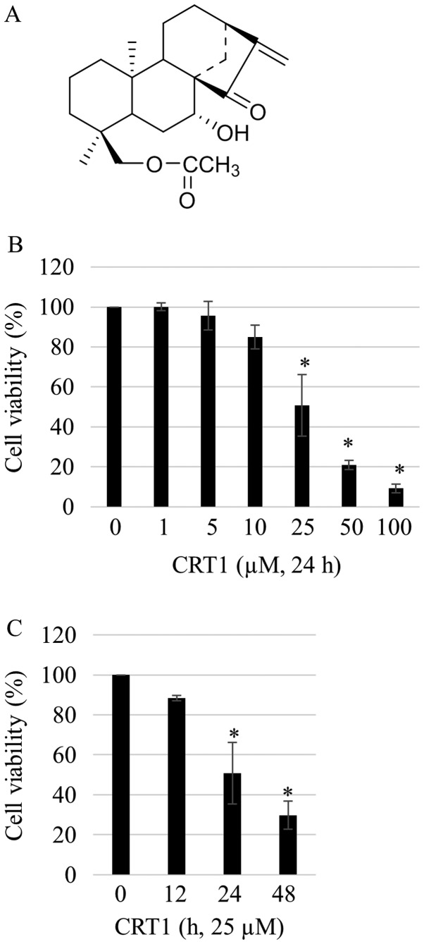
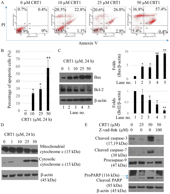
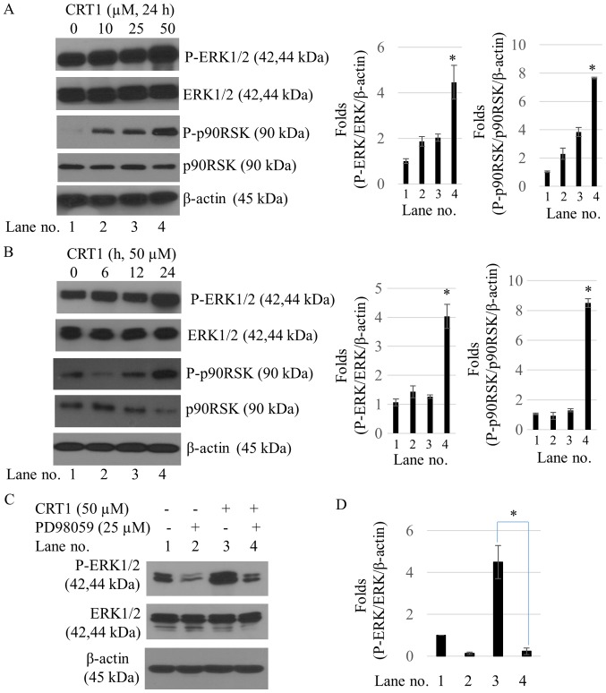
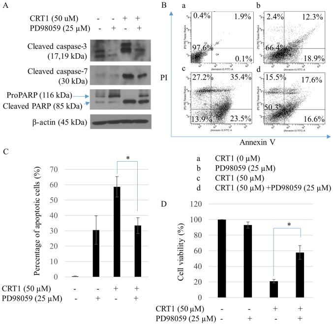
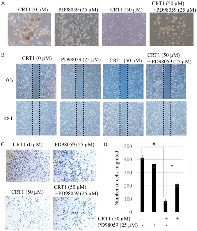
Ovarian cancer is one of the most common causes of female mortalities from gynecological tumors. An ent‑kaurane diterpenoid compound CRT1 (ent‑18‑acetoxy‑7β‑hydroxy kaur‑15‑oxo‑16‑ene), mainly isolated from the Vietnamese herb Croton tonkinesis has been used in folk medicine in Vietnam for cancer treatment. However, the effect of this compound on human ovarian cancer cells has not yet been reported. The objective of the present study was to determine the effect of CRT1 on the cell viability, apoptosis and metastasis of SKOV3 human ovarian cancer cells using a Cell Counting Kit‑8 assay, flow cytometric analysis of Annexin V‑fluorescein isothiocyanate/propidium iodide staining, western blot analysis, soft agar colony forming assay, wound healing assay and Matrigel invasion assay. The results revealed that CRT1 possessed significant anti‑proliferative effects on SKOV3 cells. CRT1 treatment at 25 and 50 µM induced apoptosis, enhanced the percentage of Annexin V‑positive cells, increased the expression of pro‑apoptotic protein B‑cell lymphoma 2 (Bcl‑2)‑associated X protein, cytochrome c release from the mitochondria to the cytosol, cleaved caspase‑3, caspase‑7, caspase‑9, and poly (adenosine diphosphate‑ribose) polymerase. However, it decreased the expression of Bcl‑2 in a dose‑dependent manner. The percentage of necrotic cells increased following CRT1 treatment at <10 µM. CRT1 at 50 µM significantly induced the phosphorylation of extracellular signal‑regulated kinase (ERK). Growth inhibition and the apoptotic effects of CRT1 could be reversed by PD98059, an ERK inhibitor. Additionally, CRT1 inhibited cell migration and invasion via ERK1/2 activation in SKOV3 cells. These results indicated that CRT1, an ent‑kaurane diterpenoid, may be a potential inhibitor of ovarian cancer by the activating ERK1/2/p90 ribosomal S6 kinase signaling pathway.


Figures

Figure 1.
Figure 1.
CRT1 decreases the viability of SKOV3 cells. (A) Chemical structure of CRT1. (B) Cells were treated with CRT1 at the indicated concentrations (1, 5, 10, 25, 50 and 100 µM) for 24 h. The viability of cells was measured by a CCK-8 assay. (C) Following treatment with CRT1 at 25 µM for different time periods (12, 24 and 48 h), cell viability was determined using a CCK-8 assay. Each assay was performed in triplicate. Data presented as the mean ± standard deviation. *P<0.05 vs. control (0 µM or h). CRT1, ent−18-acetoxy-7β-hydroxy kaur-15-oxo-16-ene; CCK-8, Cell Counting Kit-8.
Figure 2.
Figure 2.
CRT1 induces apoptosis in SKOV3 cells. (A) Cells were treated with CRT1 at 25 or 50 µM for 24 h followed by Annexin V-FITC/PI double staining. Apoptotic cells were analyzed by flow cytometry. Data are representatives of three independent experiments. (B) Percentage of apoptotic cells is presented as the mean ± standard deviation. *P<0.05 and **P<0.01 vs. control. (C) Cells were treated with various concentrations of CRT1 for 24 h followed by western blot analysis to measure protein expression levels of Bax and Bcl-2. β-Actin was used as a protein loading control. Protein expression level is presented as the mean ± standard deviation. *P<0.05 and **P<0.01 vs. Lane no. 1. (D) Mitochondria and cytosolic fractions were prepared and western blot analysis was performed to determine cytochrome c levels. (E) Protein levels of caspase-3, −7 and −9 were determined by western blot analysis. Z-VAD-fmk at 100 µM was pretreated for 2 h followed by CRT1 treatment for an additional 24 h. CRT1, ent−18-acetoxy-7β-hydroxy kaur-15-oxo-16-ene; FITC, fluorescein isothiocyanate; PI, propidium iodide; Bcl-2, B-cell lymphoma 2; Bax, Bcl-2-associated X protein; PARP, poly-(adenosine diphosphate-ribose) polymerase.
Figure 3.
Figure 3.
Effect of CRT1 on ERK1/2 phosphorylation in SKOV3 cells. (A) Time-dependent and (B) concentration-dependent effects of CRT1 on the protein expression levels of ERK1/2, p-ERK1/2, p90RSK and p-p90RSK were analyzed by western blotting. Actin was used as a protein loading control. Each experiment was performed in triplicate. All data are presented as the mean ± standard deviation. *P<0.05 vs. the control. (C) ERK inhibitor PD98059 reversed the CRT1-induced phosphorylation of ERK1/2. PD98059 (25 µM) was preincubated for 2 h prior to the addition of CRT1 (50 µM). Phosphorylation levels of ERK1/2 and total ERK, and the expression level of actin were analyzed by western blot analysis with the indicated antibodies. (D) Protein expression level is presented as the mean ± standard deviation. *P<0.05 vs. Lane no. 3. CRT1, ent−18-acetoxy-7β-hydroxy kaur-15-oxo-16-ene; ERK, extracellular signal-regulated kinase; p-, phosphorylated.
Figure 4.
Figure 4.
ERK1/2 regulates CRT1-induced apoptosis and anti-proliferation. (A) ERK inhibitor PD98059 reversed CRT1-induced caspase cleavages. PD98059 (25 µM) was preincubated for 2 h prior to the addition of CRT1 (50 µM). Levels of cleaved caspase-3, caspase-7, PARP and β-actin were analyzed using western blot analysis with the indicated antibodies. (B) Cells were pretreated with PD98059 for 2 h prior to CRT1 (50 µM) treatment. Apoptotic cells were analyzed by Annexin V-FITC/PI double staining assay followed by flow cytometry. Data are representatives of three independent experiments. (C) Percentage of apoptotic cells. (D) Following pre-incubation of PD98059 prior to CRT1 treatment (50 µM), cell viability was determined using Cell Counting Kit-8 assay. Each assay was performed in triplicate. Data are presented as the mean ± standard deviation. *P<0.05 vs. CRT1 single treatment. ERK, extracellular signal-regulated kinase; CRT1, ent−18-acetoxy-7β-hydroxy kaur-15-oxo-16-ene; PARP, poly-(adenosine diphosphate-ribose) polymerase; FITC, fluorescein isothiocyanate; PI, propidium iodide.
Figure 5.
Figure 5.
CRT1 inhibits colony-forming ability, cell migration and invasion of SKOV3 cells by ERK activation. (A) Colony-forming assay for single cell culture of SKOV3 cells treated with CRT1 (50 µM) and/or PD98059 (25 µM). Cells were allowed to culture until visible colonies were evident. Data are presented as photographs: Phase-contrast microscopic examination of the morphology of cells in culture (magnification, ×100). (B) Cell migration was measured by wound healing assay. Cells in culture were wounded and allowed to migrate into the denuded area for 48 h. Five random views were chosen along the scratch wound in each well (magnification, ×100). (C) Transwell invasion assay. CRT1 (50 µM) and/or PD98059 (25 µM)-treated cells were fixed and stained following 48 h as described in the materials and methods. Three randomly selected fields of view were photographed in each well (magnification, ×100). (D) Quantification of the migration of SKOV3 cells. Data represent replicates of three independent experiments and are expressed as the mean ± standard deviation. *P<0.05 vs. CRT1 single treatment; #P<0.05 vs. untreated control. CRT1, ent−18-acetoxy-7β-hydroxy kaur-15-oxo-16-ene; ERK, extracellular signal-regulated kinase.

Similar articles

Cited by

References

    1. Serkies K, Węgrzynowicz E, Jassem J. Paclitaxel and cisplatin chemotherapy for ovarian cancer during pregnancy: Case report and review of the literature. Arch Gynecol Obstet. 2011;283(Suppl 1):S97–S100. doi: 10.1007/s00404-011-1855-4. - DOI - PMC - PubMed
    1. Wen W, Wu J, Liu L, Tian Y, Buettner R, Hsieh MY, Horne D, Dellinger TH, Han ES, Jove R, Yim JH. Synergistic anti-tumor effect of combined inhibition of EGFR and JAK/STAT3 pathways in human ovarian cancer. Mol Cancer. 2015;14:100. doi: 10.1186/s12943-015-0366-5. - DOI - PMC - PubMed
    1. Vaughan S, Coward JI, Bast RC, Jr, Berchuck A, Berek JS, Brenton JD, Coukos G, Crum CC, Drapkin R, Etemadmoghadam D, et al. Rethinking ovarian cancer: Recommendations for improving outcomes. Nat Rev Cancer. 2011;11:719–725. doi: 10.1038/nrc3144. - DOI - PMC - PubMed
    1. Lu Z, Xu S. ERK1/2 MAP kinases in cell survival and apoptosis. IUBMB Life. 2006;58:621–631. doi: 10.1080/15216540600957438. - DOI - PubMed
    1. Cagnol S, Chambard JC. ERK and cell death: Mechanisms of ERK-induced cell death-apoptosis, autophagy and senescence. FEBS J. 2010;277:2–21. doi: 10.1111/j.1742-4658.2009.07366.x. - DOI - PubMed

MeSH terms

Substances