. 2019 Mar;19(3):633-645.
doi: 10.1111/ajt.15072. Epub 2018 Sep 17.

C5aR1 regulates migration of suppressive myeloid cells required for costimulatory blockade-induced murine allograft survival

Affiliations

C5aR1 regulates migration of suppressive myeloid cells required for costimulatory blockade-induced murine allograft survival

Ines Llaudo et al. Am J Transplant. 2019 Mar.

Abstract

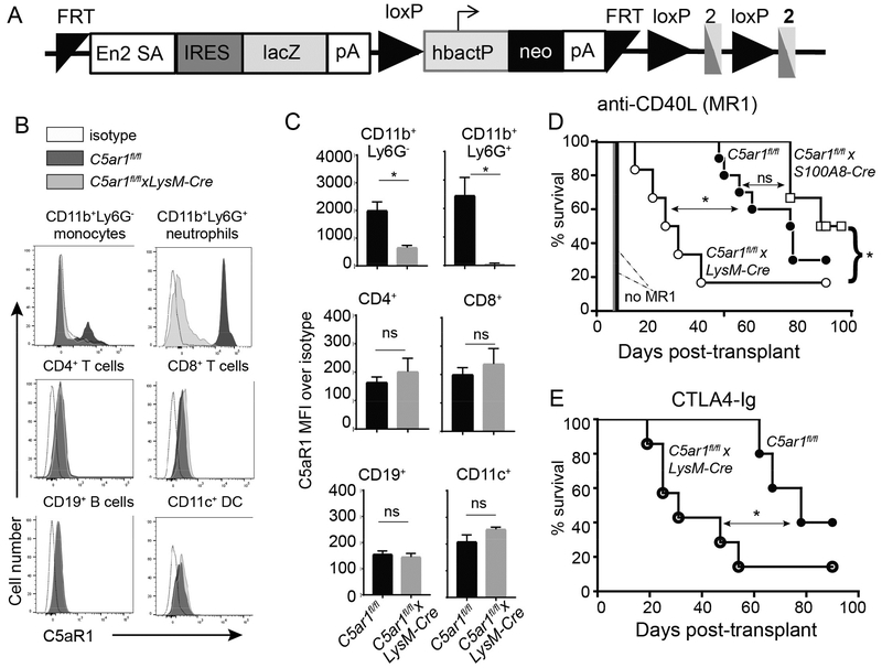
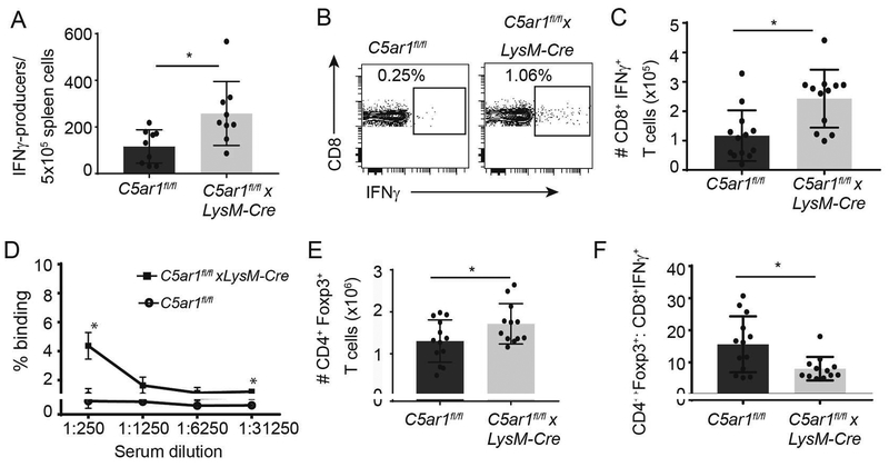
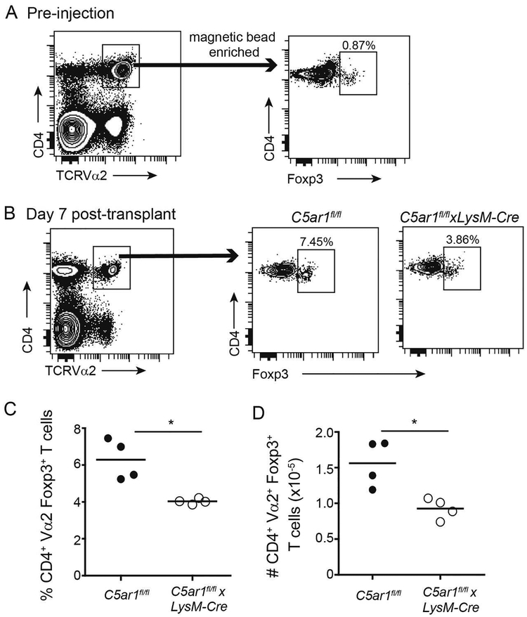
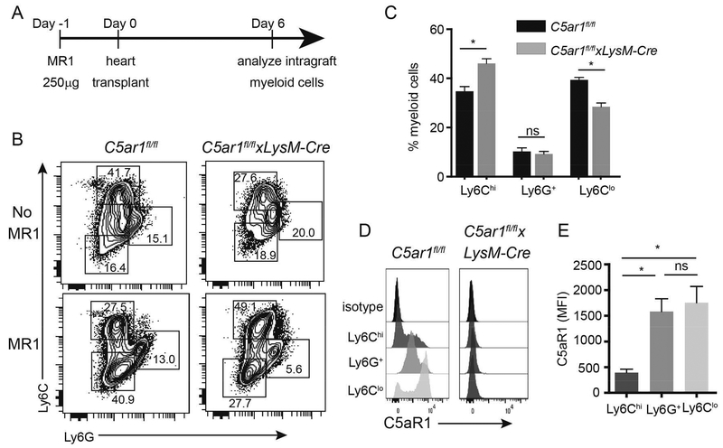
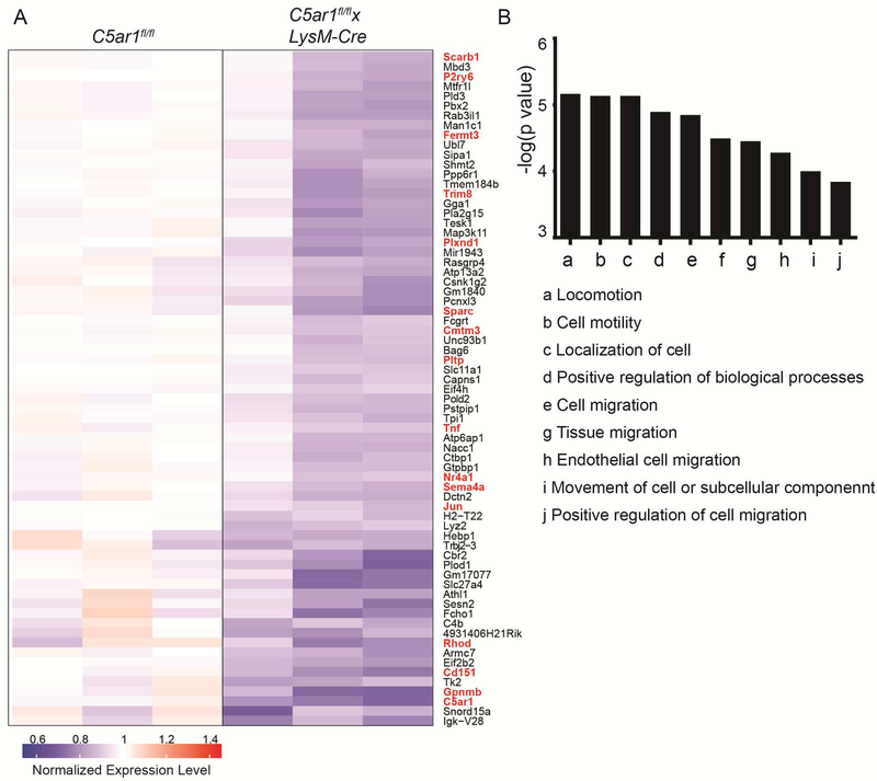
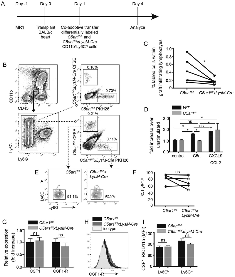
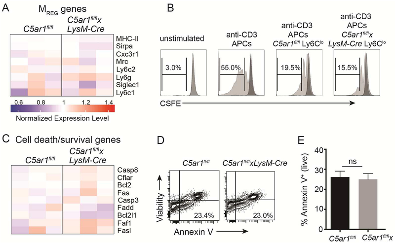
Costimulatory blockade-induced murine cardiac allograft survival requires intragraft accumulation of CD11b+ Ly6Clo Ly6G- regulatory myeloid cells (Mregs) that expand regulatory T cells (Tregs) and suppress effector T cells (Teffs). We previously showed that C5a receptor (C5aR1) signaling on T cells activates Teffs and inhibits Tregs, but whether and/or how C5aR1 affects Mregs required for transplant survival is unknown. Although BALB/c hearts survived >60 days in anti-CD154 (MR1)-treated or cytotoxic T-lymphocyte associated protein 4 (CTLA4)-Ig-treated wild-type (WT) recipients, they were rejected at ~30 days in MR1-treated or CTLA4-Ig-treated recipients selectively deficient in C5aR1 restricted to myeloid cells (C5ar1fl/fl xLysM-Cre). This accelerated rejection was associated with ~2-fold more donor-reactive T cells and ~40% less expansion of donor-reactive Tregs. Analysis of graft-infiltrating mononuclear cells on posttransplant day 6 revealed fewer Ly6Clo monocytes in C5ar1fl/fl xLysM-Cre recipients. Expression profiling of intragraft Ly6Clo monocytes showed that C5aR1 deficiency downregulated genes related to migration/locomotion without changes in genes associated with suppressive function. Cotransfer of C5ar1fl/fl and C5ar1fl/fl xLysM-Cre myeloid cells into MR1-treated allograft recipients resulted in less accumulation of C5ar1-/- cells within the allografts, and in vitro assays confirmed that Ly6Chi myeloid cells migrate to C5a/C5aR1-initiated signals. Together, our results newly link myeloid cell-expressed C5aR1 to intragraft accumulation of myeloid cells required for prolongation of heart transplant survival induced by costimulatory blockade.

Keywords: animal models: murine; basic (laboratory) research/science; immunobiology; immunosuppression/immune modulation; macrophage/monocyte biology; macrophage/monocyte biology: trafficking; tolerance: experimental.


Conflict of interest statement

Disclosure

The authors of this manuscript have no conflicts of interest to disclose as described by the American Journal of Transplantation.

Figures

Figure 1.
Figure 1.
Myeloid cell deficiency of C5ar1 abrogates costimulatory blockade induced prolonged cardiac allograft survival. A. Schematic representation of the targeting insert to conditionally delete C5ar1. Animals transmitting the insert were crossed with flp/flp mice to remove the genes between the 2 FRT sites. The resultant C5ar1fl/fl mice were crossed to a LysM-Cre transgenic to remove a portion of C5ar1 exon 2 from myeloid cells or to an S100A8-Cre transgenic to remove a portion of C5ar1 exon 2 from neutrophils. B-C. Representative flow cytometry plots (B) and quantified surface expression (MFI) of C5aR1 (C) on monocytes and neutrophils (top row) CD4+ and CD8+ T cells (middle row), and B cells and DCs (bottom row). D. Survival of BALB/c hearts transplanted into C5ar1fl/fl, C5arfl/flxLysM-Cre or C5arfl/flxS100A8-Cre recipients treated with anti-CD40L mAb MR1 250μg on day −1 (n=6–10/group). Survival of BALB/c hearts in untreated C5ar1fl/fl (solid black line, no symbol) and C5arfl/flxLysM-Cre (solid gray line, no symbol, n=4/group) recipients. E. Survival of BALB/c hearts transplanted into C5ar1fl/fl or C5arfl/flxLysM-Cre and treated with CTLA4-Ig (n=5–7/group) (E). *p<0.05 by t-test. ns: nonsignificant. *p<0.05, **p<0.05 by survival analysis (Mantel-Cox log rank test). ns=not significant.
Figure 2.
Figure 2.
Absence of myeloid cell expressed C5ar1 augments donor-reactive T cell immune responses in MR1-treated allograft recipients. A. Donor-reactive IFNγ ELISPOTs from C5ar1fl/fl and C5ar1fl/flxLysM-Cre recipients of BALB/c hearts 14 days post-transplant. n=9/group. Frequencies of donor-reactive IFNγ-producers in spleens of untreated C5ar1fl/fl and C5ar1fl/flxLysM-Cre recipients of BALB/c hearts at rejection (day 7) were ~1300/5×105 spleen cells and were not different between groups (not shown). B-C. Representative flow cytometry plots (B) and total numbers per spleen of donor-reactive IFNγ-producing CD8+ T cells (C) from C5ar1fl/fl and C5ar1fl/flxLysM-Cre recipients of BALB/c hearts 14 days post-transplant, n=12/group. D. Serum donor-reactive alloantibodies in C5ar1fl/fl and C5ar1fl/flxLysM-Cre recipients of BALB/c hearts 14 days post-transplant, n=3/group E-F. Total numbers of Foxp3+CD4+ T cells per spleen (E) and calculated ratios of CD4+Foxp3+ cells/donor reactive IFNγ-producers per mouse (TREG:TEFF ratios), n=12/group (F). * p<0.05
Figure 3.
Figure 3.
Absence of myeloid cell-expressed C5ar1 limits in expansion of donor-reactive TREG in MRI-treated allograft recipients. A. Gating strategy for enriching CD4+Vα2+ TEa T cells from spleens of TEa TCR tg mice (left), confirmation of purity of isolated population (right) demonstrating ~0.9% Foxp3+CD4+ T cells within the sorted TEa+ population. The purified cells were adoptively transferred into C5ar1fl/fl and C5ar1fl/flxLysM-Cre and treated with MR1 (day 0), 24 h later transplanted with BALB/c hearts. B-D. Gating strategy, representative flow cytometry plots (B) and quantified results showing percentages (C) and total numbers (D) of adoptively transferred TEa cells analyzed 7 days post-transplant demonstrating fewer Foxp3+ TEa cells in the C5ar1fl/flxLysM-Cre recipients, n=4/group (from 2 separate experiments). p<0.05
Figure 4.
Figure 4.
Myeloid cell-expressed C5ar1 regulates accumulation of Ly6Clo myeloid cells in allografts following recipient treatment with MR1. A. Schematic of experimental design. B. Representative flow cytometry plots of graft-infiltrating anti-Ly6C/Ly6G-stained cells gated on the live CD45+CD11b+ subset from untreated or MR1-treated C5ar1fl/fl and C5ar1fl/flxLysM-Cre recipients of BALB/c hearts. C. Quantified results of MR1 treated recipients (n=8/group). D-E. Representative flow cytometry histograms (D) and quantified MFI results (E) showing absence of C5aR1 expression on graft infiltrating CD11b+ Ly6G/Ly6C subsets from BALB/c allografts transplanted into MR1-treated C5ar1fl/fl and C5ar1fl/flxLysM-Cre (day 6 post-transplant). *p<0.05, ns=not significant.
Figure 5.
Figure 5.
Microarray analysis indicates absence myeloid cell-expressed C5aR1 specifically reduces the expression of genes involved in cell migration and locomotion. Heatmap (A) and enrichment analysis (B) of downregulated genes in microarrays of RNA obtained from flow-sorted intragraft CD11b+Ly6CloLy6G- cells on day 6 after BALB/c hearts were transplanted into MR1-treated C5ar1fl/fl (n=3) and C5ar1fl/flxLysM-Cre (n=4) recipients of BALB/c hearts. The genes that comprise these pathways are depicted in red font in A.
Figure 6.
Figure 6.
Myeloid cell-expressed C5ar1 is required for optimal migration of myeloid cells to allografts in MR1-treated recipients. A. Schematic of experimental design (see text for details). B Representative flow cytometry plots showing gating strategy and revealing lower frequencies of intragraft, adoptively transferred C5ar1fl/flxLysM-Cre Ly6Glo myeloid cells regardless of label with CFSE or PKH26. C. Quantified frequencies of intragraft myeloid cells in day 4 post-transplant allografts. n=6/group, 2 separate experiments. D. Chemotaxis assays of CD11b+Ly6Chi monocyte WT or C5ar1−/− mice in response to C5a (100 ng/ml) and CXCL9 (1μg/mL) + CCL2 (2ng/mL). E-F. Representative flow cytometry histograms (E) and quantified results (F) depicting percentages of Ly6CloLy6G- myeloid cells within the adoptively transferred intragraft populations. G-I. Quantitative RT-PCR for CSF1 and CSF1-R expression (G), and representative flow plot (H) and quantified surface expression (I) of CSF1-R on intragraft myeloid cells from MR1-treated C5ar1fl/fl and C5ar1fl/flxLysM-Cre recipients of BALB/c hearts. *p<0.05, ns=not significant
Figure 7.
Figure 7.
Graft infiltrating C5ar1fl/fl and C5ar1fl/flxLysM-Cre Ly6Clo myeloid cells exhibit equivalent suppressive capacities and rates of apoptosis. A. Heatmap depicting expression levels for MREG-produced genes involved in suppressive function (see Figure 5 for details, n=3–4/group). B. Representative flow cytometry plots of unstimulated or anti-CD3 stimulated WT T cells ± flow sorted CD11b+Ly6CloLy6G- myeloid cells (200,000 T cells, 50,000 CD11c+ APCs and 50,000 myeloid cells) isolated from allografts on day 6 of MR1-treated C5ar1fl/fl (controls) or C5ar1fl/flxLySM-Cre recipients, illustrating suppressive ability of myeloid cells from both genotypes. Representative of results from 2 independent experiments. C. Heatmap depicting expression levels genes involved in cell death/survival (see Figure 5 for details, n=3–4/group). D-E. Representative flow cytometry plot (D) and quantified results (E) depicting frequency of apoptotic (AnnexinV+) CD11b+ Ly6Clo graft-infiltrating cells. *p<0.05, ns=not significant.

Comment in

Similar articles

Cited by

References

    1. Larsen CP, Elwood ET, Alexander DZ, Ritchie SC, Hendrix R, Tucker-Burden C et al. Long-term acceptance of skin and cardiac allografts after blocking CD40 and CD28 pathways. Nature 1996;381(6581):434–438. - PubMed
    1. Garcia MR, Ledgerwood L, Yang Y, Xu J, Lal G, Burrell B et al. Monocytic suppressive cells mediate cardiovascular transplantation tolerance in mice. J Clin Invest 2010;120(7):2486–2496. - PMC - PubMed
    1. Ochando JC, Homma C, Yang Y, Hidalgo A, Garin A, Tacke F et al. Alloantigen-presenting plasmacytoid dendritic cells mediate tolerance to vascularized grafts. Nat Immunol 2006;7(6):652–662. - PubMed
    1. Conde P, Rodriguez M, van der Touw W, Jimenez A, Burns M, Miller J et al. DC-SIGN(+) Macrophages Control the Induction of Transplantation Tolerance. Immunity 2015;42(6):1143–1158. - PMC - PubMed
    1. Pearson TC, Trambley J, Odom K, Anderson DC, Cowan S, Bray R et al. Anti-CD40 therapy extends renal allograft survival in rhesus macaques. Transplantation 2002;74(7):933–940. - PubMed

Publication types

MeSH terms