

Acute noncontrast T1-weighted magnetic resonance imaging predicts chronic radiofrequency ablation lesions
- PMID: 30106244
- PMCID: PMC6235745
- DOI: 10.1111/jce.13709
Acute noncontrast T1-weighted magnetic resonance imaging predicts chronic radiofrequency ablation lesions
Abstract
Background: Magnetic resonance imaging (MRI) has been used to visualize radiofrequency (RF) ablation lesions but the relationship between volumes that enhance in acute MRI and the chronic lesion size is unknown.
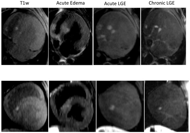
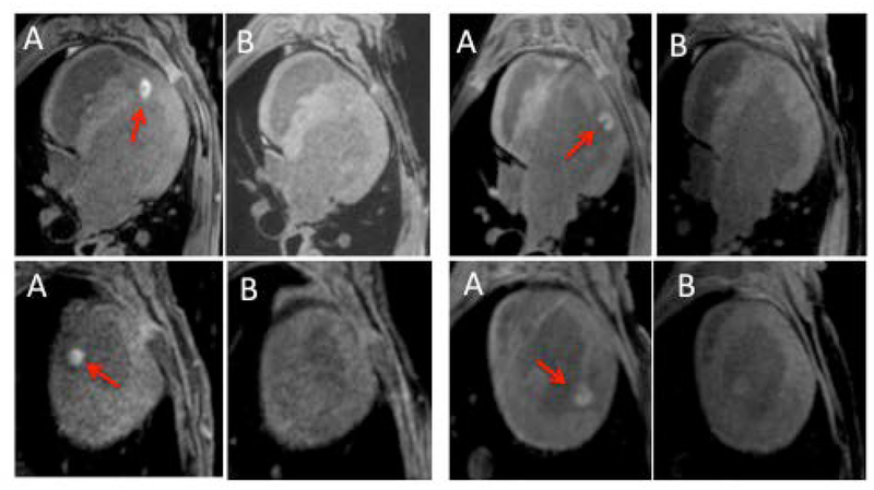
Objectives: The main goal was to use noncontrast (native) T1-weighted (T1w) MRI and late gadolinium enhancement (LGE)-MRI to visualize lesions acutely and chronically and correlate the acute area of enhancement with chronic lesion size in histology.
Materials and methods: In a canine (n = 9) model RF ablation lesions were created in both ventricles. Native T1w MRI and LGE-MRI were acquired acutely after the ablation procedure. After 8 weeks, another set of RF ablations was performed, and the MRI study was repeated. Volume and depth of enhancement in native T1w MRI and LGE-MRI acquired after the initial ablation procedure were correlated with chronic lesion volume and depth in histology.
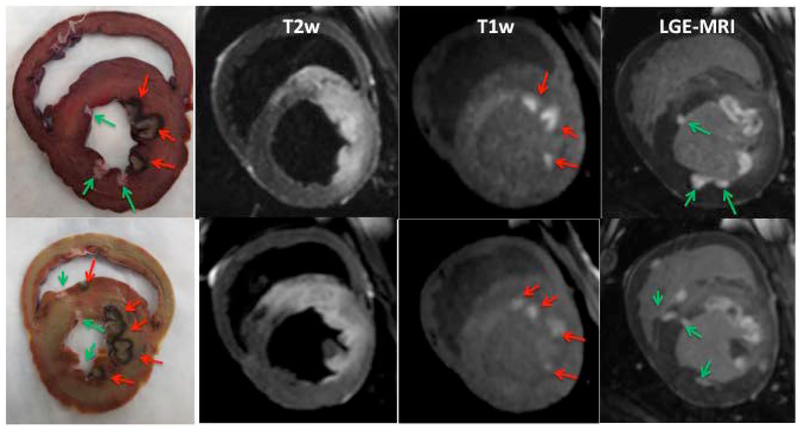
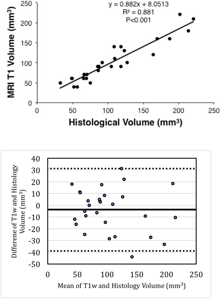
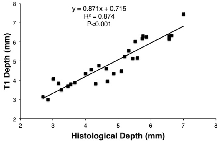
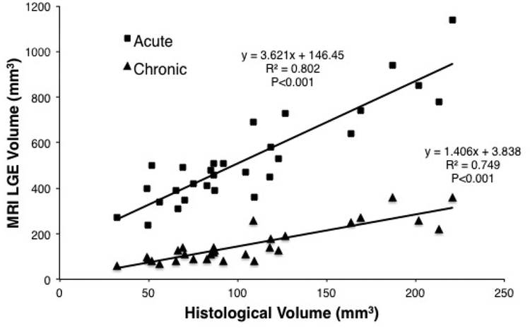
Results: Thirty-three lesions were analyzed. Native T1w MRI visualized the acute lesions but not the chronic lesions. LGE-MRI showed both acute and chronic lesions. Acute native T1w MRI volume (average of 102.1 ± 48.5 mm3 ) and depth (4.9 ± 1.2 mm) correlated well with chronic histological volume (105.9 ± 51.8 mm3 ) and depth (4.8 ± 1.3 mm) with R2 of 0.881 (P < 0.001) and 0.874 (P < 0.001), respectively. Acute LGE-MRI had a significantly higher volume of enhancement of 499.7 ± 214.4 mm3 (P < 0.001) and depth of 7.5 ± 1.8 mm ( P < 0.001) when compared with chronic histological lesion volume and depth.
Conclusions: Native T1w MRI acquired acutely after RF ablation is a good predictor of chronic lesion size. Acute LGE-MRI significantly overestimates the chronic lesion size.
Keywords: LGE-MRI; cardiac magnetic resonance image; catheter ablation; lesion visualization; noncontrast MRI.
© 2018 Wiley Periodicals, Inc.
Conflict of interest statement
Eugene Kholmovski reports equity interest in Marrek Inc; consultant to Marrek Inc.
Figures
References
-
- Parmar BR, Jarrett TR, Kholmovski EG, Hu N, Parker D, MacLeod RS, Marrouche NF, Ranjan R. Poor scar formation after ablation is associated with atrial fibrillation recurrence. Journal of interventional cardiac electrophysiology: an international journal of arrhythmias and pacing, 2015. 44(3): p. 247–256. - PMC - PubMed
-
- Ouyang F, Antz M, Ernst S, Hachiya H, Mavrakis H, Deger FT, Schaumann A, Chun J, Falk P, Hennig D, Liu X, Bänsch D, Kuck KH. Recovered pulmonary vein conduction as a dominant factor for recurrent atrial tachyarrhythmias after complete circular isolation of the pulmonary veins: lessons from double Lasso technique. Circulation, 2005. 111(2): p. 127–135. - PubMed
-
- Cheema A, Dong J, Dalal D, Marine JE, Henrikson CA, Spragg D, Cheng A, Nazarian S, Bilchick KC, Almasry I, Sinha S, Scherr D, Halperin H, Berger R, Calkins H Incidence and time course of early recovery of pulmonary vein conduction after catheter ablation of atrial fibrillation. J Cardiovasc Electrophysiol, 2007. 18(4): p. 387–391. - PubMed
Publication types
MeSH terms
Grants and funding
LinkOut - more resources
Full Text Sources
Other Literature Sources
Medical
