Comparative Study
. 2019 Jun;21(6):566-574.
doi: 10.1177/1098612X18792657. Epub 2018 Aug 14.

Comparison of medical and/or surgical management of 23 cats with intracranial empyema or abscessation

Affiliations
Comparative Study

Comparison of medical and/or surgical management of 23 cats with intracranial empyema or abscessation

Sophie Martin et al. J Feline Med Surg. 2019 Jun.

Abstract

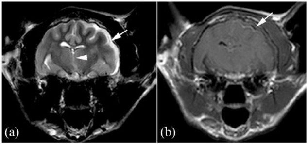
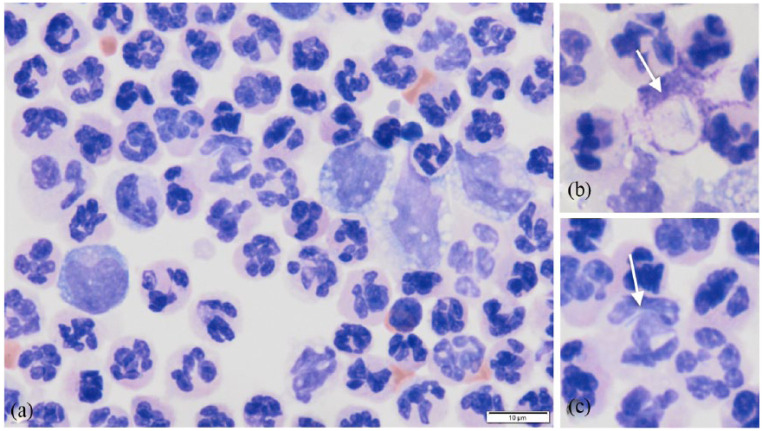
Objectives: Feline intracranial abscessation or empyema is infrequently reported in the veterinary literature. To date, the largest study is based on a population of 19 cats with otogenic infection. The aim of this study was to review a larger population of cats with intracranial empyema from multiple aetiologies and document their signalment, imaging findings, treatment protocols (including medical and/or surgical management) and to compare outcomes.

Methods: Cases presenting to a single referral centre over a 10 year period with compatible history, neurological signs and imaging findings consistent with intracranial abscessation and empyema were reviewed retrospectively.

Results: Twenty-three cats met the inclusion criteria. Advanced imaging (CT and/or MRI) was performed in 22/23 cats; one case was diagnosed via ultrasound. Ten cases underwent medical and surgical management combined, 10 underwent solely medical management and three were euthanased at the time of diagnosis. Short-term outcome showed that 90% of surgically managed and 80% of medically managed cats were alive at 48 h post-diagnosis. Long-term survival showed that surgically managed cases and medically managed cases had a median survival time of 730 days (range 1-3802 days) and 183 days (range 1-1216 days), respectively. No statistical significance in short- or long-term survival ( P >0.05) was found between medically and surgically managed groups.

Conclusions and relevance: Feline intracranial abscessation and empyema are uncommon conditions that have historically been treated with combined surgical and medical management. This study documents that, in some cases, intracranial abscessation and empyema can also be successfully treated with medical management alone.

Keywords: Intracranial empyema; craniectomy; intracranial abscessation; otitis interna; otitis media.


Conflict of interest statement

The authors declared no potential conflicts of interest with respect to the research, authorship, and/or publication of this article.

Figures

Figure 1
Figure 1
Intracranial empyema in a 13-year-old male neutered domestic shorthair cat. (a) T2-weighted (W) and (b) T1W post-gadolinium transverse plane images show the left subarachnoid space is widened by a slightly heterogeneous layer of hyperintensity with mild peripheral contrast enhancement (white arrows). There is also evidence of midline shift (white arrowhead) away from the affected site
Figure 2
Figure 2
(a) T2-weighted (W) magnetic resonance transverse plane image of a 5-year-old female neutered domestic shorthair cat with an intracranial abscess in the frontal and temporal lobes (white arrow) that partially supresses on (b) fluid-attenuated inversion recovery (FLAIR) sequence (white arrow). There was increased T2W and FLAIR signal in the surrounding white matter (black arrows), the musculature overlying the calvarium (white arrowheads) and midline shift of the falx away from the lesion. (c) The T2W sagittal plane image shows signs of increased intracranial pressure with caudal transtentorial (black arrow) and caudal cerebellar herniation (white arrow)
Figure 3
Figure 3
(a) Transverse plane post-intravenous contrast CT image of a 7-year-old male neutered domestic shorthair cat with an intracranial abscess. The lesion is located ventral to the brainstem and shows strong rim enhancement and absent contrast enhancement of the centre of the mass (white arrow). The right middle-ear cavity (black arrow) and the medial portion of the horizontal ear canal (black arrowhead) is filled with peripherally contrast-enhancing soft tissue-attenuating material, suggestive of otitis media and externa. (b) The corresponding T1-weighted (W) magnetic resonance post-gadolinium transverse plane image shows the same lesion location (white arrow) and right ear changes (black arrow and arrowhead in [b] as in [a]), with improved depiction of the soft tissues. (c) The sagittal plane T1W post-gadolinium view shows the extent and cavitated appearance of the lesion (white arrows) ventral to the brainstem
Figure 4
Figure 4
Ultrasound image of a 15-year-old female neutered domestic shorthair cat. Defect within the skull (white arrows), flocculent material communicating with the subcutaneous space and the intracranial cavity
Figure 5
Figure 5
(a) Cytology image of cerebrospinal fluid from a 14-year-old male neutered domestic longhair cat with marked neutrophilic pleocytosis with bacterial sepsis. (b,c) Enlarged images of degenerate neutrophils containing filamentous bacteria (white arrows). Modified Wright’s stain
Figure 6
Figure 6
Kaplan–Meier curve depicting survival of surgically and medically managed cats (purple line) and solely medically treated cats (black line) over time

References

    1. Tsou T-P, Lee P-I, Lu C-Y, et al. . Microbiology and epidemiology of brain abscess and subdural empyema in a medical center: a 10-year experience. J Microbiol Immunol Infect 2009; 42: 405–412. - PubMed
    1. Das AK, Jumani K, Kashyap RC. Subdural empyema: a rare complication of chronic otitis media. Med J Armed Forces India 2005; 61: 281–283. - PMC - PubMed
    1. Costanzo C, Garosi LS, Glass EN, et al. . Brain abscess in seven cats due to a bite wound: MRI findings, surgical management and outcome. J Feline Med Surg 2011; 13: 672–680. - PMC - PubMed
    1. Cardy TJA, Lam R, Peters LM, et al. . Successful medical management of a domestic longhair cat with subdural intracranial empyema and multifocal pneumonia. J Vet Emerg Crit Care 2017; 27: 238–242. - PubMed
    1. Sturges BK, Dickinson PJ, Kortz GD, et al. . Clinical signs, magnetic resonance imaging features, and outcome after surgical and medical treatment of otogenic intracranial infection in 11 cats and 4 dogs. J Vet Intern Med 2006; 20: 648–656. - PubMed

Publication types

MeSH terms

LinkOut - more resources