. 2018 Oct;66(10):737-751.
doi: 10.1369/0022155418793600. Epub 2018 Aug 14.

4',6-Diamidino-2-Phenylindole Distinctly Labels Tau Deposits

Affiliations

4',6-Diamidino-2-Phenylindole Distinctly Labels Tau Deposits

Chengyu Li et al. J Histochem Cytochem. 2018 Oct.

Abstract

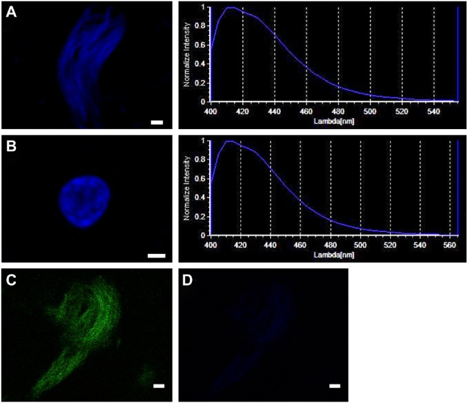
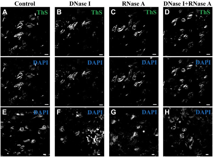
Tau deposits have distinct biochemical characteristics and vary morphologically based on identification with tau antibodies and several chemical dyes. Here, we report 4',6-diamidino-2-phenylindole (DAPI)-positivity of tau deposits. Furthermore, we investigated the cause for this positivity. DAPI was positive in 3R/4R (3-repeat/4-repeat) tau deposits in Alzheimer's disease, myotonic dystrophy, and neurodegeneration with brain iron accumulation, and in 4R tau deposits in corticobasal degeneration, but negative in 4R tau deposits in frontotemporal dementia with parkinsonism-17 and progressive supranuclear palsy. The peak emission wavelength of DAPI after binding to a tau deposit was similar to that after binding to a nucleus. This DAPI-positivity was conspicuous at the optimum concentration of 2 μg/ml. DAPI-positivity was diminished after formic acid treatment, but preserved after nucleic acid elimination and phosphate moiety blocking. Our results suggest that staining with 2 μg/ml DAPI is a common but useful tool to differentially detect tau deposits in various tauopathies.

Keywords: 4′,6-diamidino-2-phenylindole; Phos-tag; neurofibrillary tangle; tauopathies.


Conflict of interest statement

Competing Interests: The author(s) declared no potential conflicts of interest with respect to the research, authorship, and/or publication of this article.

Figures

Figure 1.
Figure 1.
DAPI-positivity of different isoform tau deposits. Tau isoform fluorescence staining is shown in AD (A and B), MyD (C and D), NBIA (E and F), CBD (G), FTDP-17 (H), and PSP (I). The 3R/4R isoform tau deposits were detected in the CA1 of AD, the entorhinal cortex of MyD, and the CA1 of NBIA. The 4R isoform tau deposits were present in the CA1 of CBD, the midbrain of FTDP-17, and the CA1 of PSP. DAPI-positivity was present in AD, MyD, NBIA, and CBD, but not in FTDP-17 and PSP. Images show RD3 (red), RD4 (red), and DAPI (blue). Calibration bars = 10 µm. Abbreviations: DAPI, 4′,6-diamidino-2-phenylindole; AD, Alzheimer’s disease; MyD, myotonic dystrophy; NBIA, neurodegeneration with brain iron accumulation; CBD, corticobasal degeneration; FTDP-17, frontotemporal dementia with parkinsonism-17; PSP, progressive supranuclear palsy.
Figure 2.
Figure 2.
Spectral pattern of DAPI in tau deposits and nuclei. A similar pattern of emission wavelength of DAPI was detected at sites of tau deposits and nuclei. DAPI-positive tau deposit (A, upper left), emission wavelength of DAPI-tau deposit (A, upper right), DAPI-positive nucleus (B, middle left), and emission wavelength of DAPI-nucleus (B, middle right). (C) A tau deposit was identified with ThS (D) and no ThS-tau deposit was detected at the excitation wavelength of DAPI. Calibration bars = 2 µm. Abbreviations: DAPI, 4′,6-diamidino-2-phenylindole; ThS, thioflavin-S.
Figure 3.
Figure 3.
Concentration-dependent specificity of DAPI in tau deposits. Serial dilutions of DAPI (blue) at 1, 2, 6, 12, 24, and 30 µg/ml were used to stain brain sections. In parallel, tau deposits were visualized with ThS (green) to show colocalization of these two stains, as shown in scatter plots. The colocalization was assessed using PCC, MOC, and ICQ values. The optimal concentration of DAPI was 2 µg/ml. Calibration bars = 20 µm. Abbreviations: DAPI, 4′,6-diamidino-2-phenylindole; ThS, thioflavin-S; PCC, Pearson’s correlation coefficient; MOC, Mander’s overlap coefficient; ICQ, intensity correlation quotient.
Figure 4.
Figure 4.
DAPI-positivity of tau deposits is reduced by pretreatment with formic acid. Brain sections were stained with 2 µg/ml DAPI (blue), with or without pretreatment with formic acid, and tau deposits were visualized using ThS (green). (A) Formic acid (–) and (B) formic acid (+). The colocalization of these two stains is shown in scatter plots and was assessed using PCC, MOC, and ICQ values. Calibration bars = 20 µm. Abbreviations: DAPI, 4′,6-diamidino-2-phenylindole; ThS, thioflavin-S; PCC, Pearson’s correlation coefficient; MOC, Mander’s overlap coefficient; ICQ, intensity correlation quotient.
Figure 5.
Figure 5.
DAPI-positivity of tau deposits is preserved even after pretreatment with proteinase K. Proteinase K can be used to eliminate lipofuscin. DAPI-positivity was preserved even after elimination of lipofuscin. The images show AT8 (green) and DAPI (blue) in tau deposits pretreated with proteinase K. (A) Proteinase K (–) and (B) proteinase K (+). The tau deposits (arrows), lipofuscin (arrowhead) and faint outline of nuclei (asterisks) are shown. Calibration bars = 50 µm. Abbreviation: DAPI, 4′,6-diamidino-2-phenylindole.
Figure 6.
Figure 6.
DAPI-positivity of tau deposits is not affected by RNase A and/or DNase I treatments. (A and E) DNase I (–)/RNase A (–); (B and F) DNase I (+)/RNase A (–); (C and G) DNase I (–)/RNase A (+); (D and H) DNase I (+)/RNase A (+). (A–D) All sections were double-stained with ThS (green) and DAPI (blue). (E–H) All sections were stained with DAPI (blue) alone. Calibration bars = 10 µm. Abbreviations: DAPI, 4′,6-diamidino-2-phenylindole; ThS, thioflavin-S.
Figure 7.
Figure 7.
Distribution of Phos-tag and DAPI in NFT-like tau deposits in an NBIA case. Confocal images showing TSA (red) and DAPI (blue). Phos-tag binding was imaged with TSA. (A) No Phos-tag blocking. (B) Phos-tag blocking (thick arrows). Calibration bars = 10 µm. Abbreviations: DAPI, 4′,6-diamidino-2-phenylindole; NFT, neurofibrillary tangle; NBIA, neurodegeneration with brain iron accumulation; TSA, Tyramide Signal Amplification.

Similar articles

References

    1. Ferrer I, López-González I, Carmona M, et al. Glial and neuronal tau pathology in tauopathies: characterization of disease-specific phenotypes and tau pathology progression. J Neuropathol Exp Neurol. 2014;73(1):81–97. - PubMed
    1. Adams SJ, DeTure MA, McBride M, et al. Three repeat isoforms of tau inhibit assembly of four repeat tau filaments. PLoS ONE. 2010;5(5):e10810. - PMC - PubMed
    1. Mackenzie IRA. Neuropathology of atypical parkinsonian disorder. In: Litvan I, editor. Atypical parkinsonian disorders: clinical and research aspects. Current clinical neurology. Totowa, NJ: Humana Press; 2005:47–77.
    1. Ludolph AC, Kassubek J, Landwehrmeyer BG, et al. Tauopathies with parkinsonism: clinical spectrum, neuropathologic basis, biological markers, and treatment options. Eur J Neurol. 2009;16(3):297–309. - PMC - PubMed
    1. Buée L, Delacourte A. Comparative biochemistry of tau in progressive supranuclear palsy, corticobasal degeneration, FTDP-17 and Pick’s disease. Brain Pathol. 1999;9(4):681–93. - PMC - PubMed

Publication types

LinkOut - more resources