. 2018 Nov;48(10):3255-3272.
doi: 10.1111/ejn.14111. Epub 2018 Sep 24.

Prefrontal cortex modulates firing pattern in the nucleus reuniens of the midline thalamus via distinct corticothalamic pathways

Affiliations

Prefrontal cortex modulates firing pattern in the nucleus reuniens of the midline thalamus via distinct corticothalamic pathways

Eric C Zimmerman et al. Eur J Neurosci. 2018 Nov.

Abstract

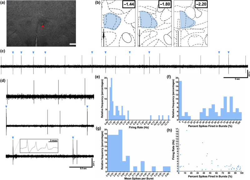
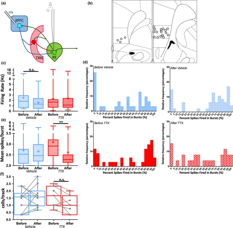
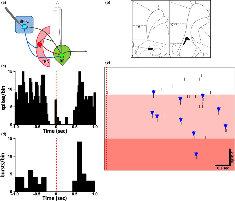
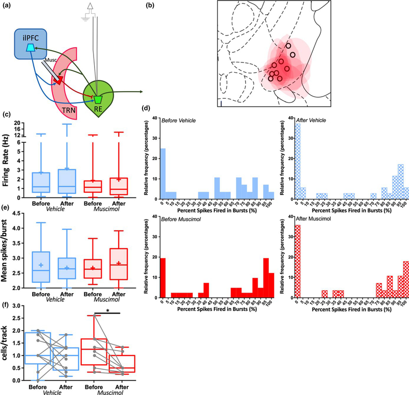
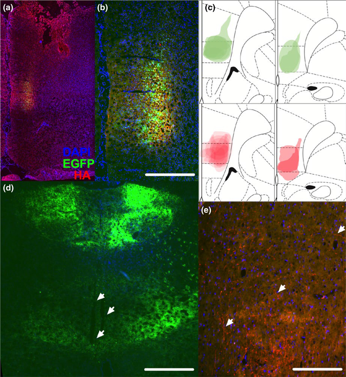
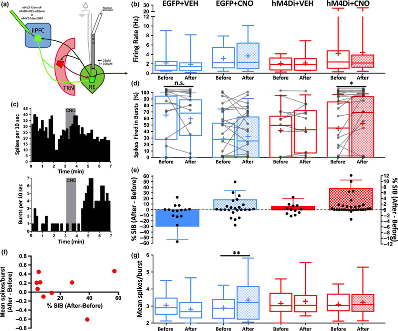
The thalamus has long been recognized for its role in relaying sensory information from the periphery, a function accomplished by its "first-order" nuclei. However, a second category of thalamic nuclei, termed "higher-order" nuclei, have been shown instead to mediate communication between cortical areas. The nucleus reuniens of the midline thalamus (RE) is a higher-order nucleus known to act as a conduit of reciprocal communication between the medial prefrontal cortex (mPFC) and hippocampus. While anatomical and behavioural studies of RE are numerous, circuit-based electrophysiological studies, particularly those examining the impact of cortical input and the thalamic reticular nucleus (TRN) on RE neuron firing, are sparse. To characterize RE neuron firing properties and dissect the circuit dynamics of the infralimbic subdivision of the mPFC (ilPFC), the TRN and RE, we used in vivo, extracellular, single-unit recordings in male Sprague Dawley rats and manipulated neural activity using targeted pharmacological manipulations, electrical stimulation and a projection-specific implementation of designer receptors exclusively activated by designer drugs (DREADDs). We show that ilPFC inhibition reduces multiple burst firing parameters in RE, whereas ilPFC stimulation drives burst firing and dampens tonic firing. In addition, TRN inhibition reduces the number of spontaneously active neurons in RE. Finally, inhibition of ilPFC terminals in RE selectively enhances a subset of burst firing parameters. These findings demonstrate that ilPFC input, both via direct projections and via the TRN, can modulate RE neuron firing pattern in nuanced and complex ways. They also highlight the ilPFC-TRN-RE circuit as a likely critical component of prefrontal-hippocampal interactions.

Keywords: corticothalamic circuits; limbic thalamus; thalamic bursting; thalamic reticular nucleus.


Conflict of interest statement

CONFLICT OF INTEREST

ECZ declares no competing financial interests. AAG reports the following: Johnson & Johnson, Lundbeck, Pfizer, GSK, Merck, Takeda, Dainippon Sumitomo, Otsuka, Lilly, Roche, Asubio, Abbott, Autofony, Janssen, Alkermes, Newron.

Figures

FIGURE 1
FIGURE 1
Baseline firing properties of RE neurons. Extracellular recordings of spontaneously active RE neurons were performed in anesthetized rats under baseline conditions. (a) Representative image of the location of an example neuron recorded in RE. The location of this neuron was marked following recording by iontophoretic application of Chicago Sky Blue dye (dashed arrow). Scale bar = 900 μm. (b) Schematic depiction of the area within RE from which all RE neurons throughout the manuscript were sampled (blue shading). The sampling area comprised a block of tissue including the RE from bregma/dural surface (in mm) AP: −1.4 to −2.2, ML: 0.1–0.5, and DV: −5.5 to −7.5. All neurons included in the analysis were confirmed to be located in the RE by referencing their position to a marked recording site generated via electrophoretic ejection of Chicago Sky Blue from the tip of the recording electrode. (c) Representative trace demonstrating the typical firing pattern of an RE neuron, with mixed tonic and burst (blue triangles) firing. (d) Three representative traces demonstrating the variable amount of burst firing present in the RE neuron population, from completely absent (top), to mixed (middle), to nearly all bursting (bottom). Inset in bottom panel demonstrates typical RE burst architecture. (e–g) Relative frequency histograms of firing rate, the percentage of spikes fired in bursts and the mean spikes per burst for all recorded RE cells. Bin sizes for each column are as follows: e = 0.25 Hz, f = 5%, g = 0.25 spikes/burst. (h) Firing rate plotted against the percentage of spikes fired in bursts. These parameters were not significantly correlated in this group of recorded neurons, although only slowly firing neurons tended to show high levels of bursting. n = 48 cells for panels e–h
FIGURE 2
FIGURE 2
Inhibition of ilPFC reduces burst firing in RE neurons. Spontaneous activity was recorded in multiple RE neurons in multiple animals both before and after acute infusion of TTX (1 μM in 0.5 μl) or dPBS vehicle into ilPFC. (a) Schematic of experimental design. (b) Representation of histological placements of infusion cannulae into ilPFC. (c) The firing rate of RE neurons plotted as “box- and-w hiskers” plots, here and in subsequent figures representing highest and lowest values (highest and lowest horizontal lines), interquartile range (rectangle), mean (“+” symbol) and median (horizontal line in rectangle). Firing rates recorded before vehicle or TTX infusion did not differ from those recorded after infusion. (d) Relative frequency distribution histograms depicting the percentage of spikes fired in bursts in neurons recorded either before or after vehicle (top) or TTX (bottom) infusion into ilPFC. Burst firing propensity was reduced in the population of RE neurons recorded after TTX infusion. Bin size for each column is 5% of spikes fired in bursts. (e) The mean spikes per burst of RE neurons recorded after TTX infusion was decreased compared to those recorded before infusion. (f) The number of spontaneously active RE neurons per electrode track did not differ following vehicle or TTX infusion. **p < 0.01 (Mann–Whitney U test). Before Vehicle n = 31 cells from 12 animals, After Vehicle n = 47 cells, Before TTX n = 34 cells from 9 animals, After TTX n = 26 cells
FIGURE 3
FIGURE 3
Electrical Stimulation of ilPFC induces burst firing and attenuates tonic firing in RE neurons. Spontaneously active RE neurons were recorded both before and during electrical stimulation of ilPFC (0.5 hz, 1 mA) via a concentric bipolar stimulating electrode. Several successive trials were performed in each neuron. (a) Schematic of experimental design. (b) Representation of histological placements of stimulating electrodes in recorded animals. (c) Cumulative perievent histogram of firing rate for a subset of trials in all RE neurons recorded (n = 10 trials/cell) demonstrating inhibition immediately following ilPFC stimulation (red dotted line). (d) Cumulative perievent histogram of burst firing plotted from the same traces as in C demonstrating inhibition immediately following ilPFC stimulation (red dotted line), followed by rebound bursting ~0.6sec poststimulation. (e) Perievent raster of 30 successive trials within a single RE neuron. Electrical stimulation of ilPFC (red dotted line) in successive trials gradually converted the firing pattern of this neuron from tonic to burst firing (white vs. light red shaded areas). After 21 trials, the neuron nearly ceased firing (darker red area). n = 8 cells from three animals
FIGURE 4
FIGURE 4
TRN inhibition decreases the number of spontaneously active RE neurons. Spontaneous activity was recorded in multiple RE neurons in multiple animals both before and after acute infusion of fluorescently tagged muscimol (0.8 μM in 0.2 μl) or dPBS vehicle into TRN (a) Schematic of experimental design. (b) Representation of histological placements of infusion cannulae for vehicle groups (black circles) and maximum extent of fluorescence for muscimol groups (red shading) in TRN. (c) The firing rate of RE neurons recorded before vehicle or muscimol infusion did not differ from those recorded after infusion. (d) Relative frequency distribution histograms depicting the percentage of spikes fired in bursts in neurons recorded either before or after vehicle (top) or muscimol (bottom) infusion into TRN. There were no differences in burst firing propensity before and after vehicle or muscimol infusion. Bin size for each column is 5% of spikes fired in bursts. (e) The mean spikes per burst of RE neurons recorded before vehicle or muscimol infusion did not differ from those recorded after infusion. (f) The number of spontaneously active RE neurons per electrode track recorded after muscimol infusion was decreased compared to those recorded before infusion. *p < 0.05 (Mann– Whitney U test). Before Vehicle n = 28 cells from nine animals, After Vehicle n = 35 cells, Before Muscimol n = 41 cells from eight animals, After Muscimol n = 28 cells
FIGURE 5
FIGURE 5
Histological validation of viral vector expression in ilPFC and RE AAV vector constructs containing the inhibitory DREADD hM4Di (rAAV2-hSyn-HA-hM4Di-IRES-mCitrine) or EGFP only controls (rAAV2-hSyn-EGFP) were infused into ilPFC. Blue = DAPI, Green = EGFP, Red = anti- HA antibody throughout, as in A. (a) Representative coronal section demonstrating expression restricted to ilPFC. (b) Higher magnification image of section picture in A. Scale bar = 300 μm. (c) Superimposed traces of the maximal extent of EGFP expression across EGFP- only animals (green, top) anti- HA immunoreactivity across hM4Di animals (red, bottom) from all animals recorded. (d) Low-m agnification image demonstrating terminal expression throughout the midline thalamus in a EGFP animal with recording electrode tracks present in RE (white arrows). Scale bar = 900 μm. (e) Representative image of HA+ terminal labelling in RE (white arrows). Scale bar = 150 μm
FIGURE 6
FIGURE 6
Inhibition of ilPFC terminals in RE enhances burst firing in RE neurons. Individual RE neurons in rats expressing hM4Di or EGFP only in ilPFC and ilPFC terminals in RE were recorded before, during and after local microinfusion of CNO (60 nl of 100 μM) or dPBS vehicle control (VEH) via a combined glass injection pipette- recording electrode. (a) Schematic diagram demonstrating the virus injection site, relevant circuitry and combined glass injection pipette- recording electrode dimensions. (b) Acute CNO or VEH application had no effect on firing rate in any group. For panels b,d,e,g groups as indicated in panel b. “Before” is the firing parameter measured for 3 m before vehicle or CNO application, and “After” is the firing parameter measured for 3 m after vehicle or CNO application. (c) Cumulative perievent histograms of firing rate (top) and burst firing (bottom) for a single RE neuron in an hM4Di animal before, during (grey box) and after CNO application. (d) The percentage of spikes fired in bursts presented as individual neurons (grey lines) and grouped box- and-w hiskers plots before and after acute CNO application. CNO application enhanced burst firing in RE neurons of animals expressing hM4Di, but this effect was not consistently observed in any other group. (e) Data from D presented as the difference in percentage of spikes fired in bursts (% SIB) before and after vehicle or CNO application. Individual values plotted in black circles on left y- axis and group means + standard error of the mean plotted in bars on right y- axis, for clarity. (f) Data from a subset of neurons in the hM4Di+CNO group that exhibited an increase in burst firing following CNO application, demonstrating inconsistent changes in mean spikes/burst. (g) Acute CNO enhanced the mean number of spikes within a burst in the EGFP + CNO group, but not in any other group. *p < 0.05, **p < 0.01 (Wilcoxon paired signed-r ank test). EGFP + VEH n = 15 neurons from 3 animals, EGFP + CNO n = 18 neurons from four animals, hM4Di+VEH n = 12 neurons from two animals, hM4Di+CNO n = 30 neurons from nine animals

Similar articles

Cited by

References

    1. Alkire MT, Hudetz AG, & Tononi G (2008). Consciousness and anesthesia. Science, 322, 876–880. 10.1126/science.1149213 - DOI - PMC - PubMed
    1. Allen TA, Narayanan NS, Kholodar-Smith DB, Zhao Y, Laubach M, & Brown TH (2008). Imaging the spread of reversible brain inactivations using fluorescent muscimol. Journal of Neuroscience Methods, 171, 30–38. 10.1016/j.jneumeth.2008.01.033 - DOI - PMC - PubMed
    1. Anticevic A, Cole MW, Repovs G, Murray JD, Brumbaugh MS, Winkler AM, … Glahn DC (2014). Characterizing thalamo- cortical disturbances in schizophrenia and bipolar illness. Cerebral Cortex, 24, 3116–3130. 10.1093/cercor/bht165 - DOI - PMC - PubMed
    1. Anticevic A, Haut K, Murray JD, Repovs G, Yang GJ, Diehl C, … Cannon TD (2015). Association of thalamic dysconnectivity and conversion to psychosis in youth and young adults at elevated clinical risk. JAMA Psychiatry, 72, 882–891. 10.1001/jamapsychiatry.2015.0566 - DOI - PMC - PubMed
    1. Barker GRI, & Warburton EC (2018). A critical role for the nucleus reuniens in long-t erm, but not short-term associative recogni- tion memory formation. Journal of Neuroscience, 38, 3208–3217. 10.1523/JNEUROSCI.1802-17.2017 - DOI - PMC - PubMed

Publication types

LinkOut - more resources