. 2018 Oct;12(10):1753-1777.
doi: 10.1002/1878-0261.12372. Epub 2018 Sep 3.

UBXN2A enhances CHIP-mediated proteasomal degradation of oncoprotein mortalin-2 in cancer cells

Affiliations

UBXN2A enhances CHIP-mediated proteasomal degradation of oncoprotein mortalin-2 in cancer cells

Sanam Sane et al. Mol Oncol. 2018 Oct.

Abstract

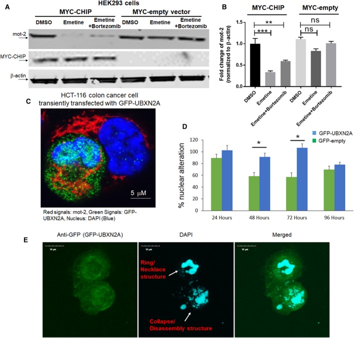
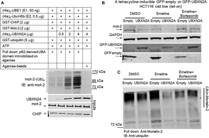
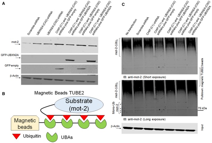
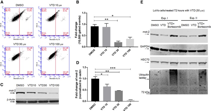
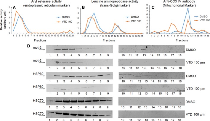
Overexpression of oncoproteins is a major cause of treatment failure using current chemotherapeutic drugs. Drug-induced degradation of oncoproteins is feasible and can improve clinical outcomes in diverse types of cancers. Mortalin-2 (mot-2) is a dominant oncoprotein in several tumors, including colorectal cancer (CRC). In addition to inactivating the p53 tumor suppressor protein, mot-2 enhances tumor cell invasion and migration. Thus, mot-2 is considered a potential therapeutic target in several cancer types. The current study investigated the biological role of a ubiquitin-like protein called UBXN2A in the regulation of mot-2 turnover. An orthogonal ubiquitin transfer technology followed by immunoprecipitation, in vitro ubiquitination, and Magnetic Beads TUBE2 pull-down experiments revealed that UBXN2A promotes carboxyl terminus of the HSP70-interacting protein (CHIP)-dependent ubiquitination of mot-2. We subsequently showed that UBXN2A increases proteasomal degradation of mot-2. A subcellular compartmentalization experiment revealed that induced UBXN2A decreases the level of mot-2 and its chaperone partner, HSP60. Pharmacological upregulation of UBXN2A using a small molecule, veratridine (VTD), decreases the level of mot-2 in cancer cells. Consistent with the in vitro results, UBXN2A+/- mice exhibited selective elevation of mot-2 in colon tissues. An in vitro Anti-K48 TUBE isolation approach showed that recombinant UBXN2A enhances proteasomal degradation of mot-2 in mouse colon tissues. Finally, we observed enhanced association of CHIP with the UBXN2A-mot-2 complex in tumors in an azoxymethane/dextran sulfate sodium-induced mouse CRC model. The existence of a multiprotein complex containing UBXN2A, CHIP, and mot-2 suggests a synergistic tumor suppressor activity of UBXN2A and CHIP in mot-2-enriched tumors. This finding validates the UBXN2A-CHIP axis as a novel and potential therapeutic target in CRC.

Keywords: CHIP E3 ligase; UBXN2A; colorectal cancer; mortalin-2; mouse; veratridine.


Figures

Figure 1
Figure 1
UBXN2A and CHIP proteins form a ternary complex with mot‐2. (A) Schematic drawings of the protein structures of mot‐2 and the CHIP E3 ubiquitin ligase. Mot‐2 has two major domains, an N‐terminal ATPase domain (ATP) and a C‐terminal substrate‐binding domain (SBD). These two domains are reciprocally controlled by the presence of ATP/ADP on the ATP domain and a client protein bound to the SBD (Dores‐Silva et al., 2015). The CHIP E3 ubiquitin ligase has three major domains: a tetratricopeptide repeat (TPR) domain located at the N terminus, a U‐box domain at its C terminus, and a mixed‐charge domain located in the middle of the protein (McDonough and Patterson, 2003). (B) HeLa cell lysates were subjected to IP using anti‐mot‐2 antibodies. IP experiments showed mot‐2 can pull down CHIP. (C) WB analysis was used to verify the lack of the CHIP protein in CHIP knockout mice using small intestine (SI) and large intestine (LI) tissue lysates. To verify the mot‐2‐CHIP complex in vivo, colon tissue (LI) lysates from C57Bl/6 WT (CHIP+/+) or CHIP knockout (CHIP−/−) were subjected to IP using anti‐CHIP antibodies immobilized on magnetic IgA. WB showed mot‐2 protein can be pulled down only from WT colon lysates where CHIP proteins are present (HC, heavy chain of immunoglobulins). (D) HCT‐116 cells were exposed to stress using the genotoxic stress agent etoposide for 24 h followed by IP experiments with anti‐UBXN2A antibodies immobilized on magnetic beads coupled with protein A. Control groups received DMSO treatment. WB analysis showed interaction of CHIP and UBXN2A takes place in the cytoplasm and not the nucleus due to the absence of CHIP in the nucleus. More importantly, we observed genotoxic stress induces nucleo‐cytoplasmic translocation of UBXN2A, which leads to an increase in UBXN2A binding to CHIP in cytoplasmic fraction. (E) In another set of experiments, DMSO‐ and etoposide (Eto)‐treated cells were subjected to IP using anti‐CHIP antibodies. WB results show the existence of a triple complex containing mot‐2, CHIP, and UBXN2A. More importantly, E indicates etoposide enhances the association of CHIP and mot‐2 protein alongside an elevation of UBXN2A in the cytoplasm, as previously described. Collectively, the experiments conducted in this figure indicate UBXN2A, mot‐2, and CHIP proteins can be present simultaneously in a multiprotein complex.
Figure 2
Figure 2
Overexpression of CHIP and UBXN2A decreases mot‐2 protein levels in cells. (A) HEK293 cells were transiently transfected with MYC‐tag CHIP or MYC‐tag empty vectors. After 24 h, cells were treated with DMSO, emetine, or a combination of emetine and bortezomib for another 24 h, followed by WB. (B) Measurement of protein bands revealed overexpression of MYC‐CHIP but not MYC‐empty significantly decreases mot‐2 while in the presence of bortezomib, an inhibitor of the 26S proteasome, downregulation of mot‐2 was blocked in the presence of emetine in cells expressing MYC‐CHIP (n = 3, **P < 0.01, ***P < 0.001‐Tukey's multiple comparison test, mean ± SE). (C) HCT‐116 cells were transiently transfected with GFP‐UBXN2A. After 48 h, cells were stained with anti‐mot‐2 followed by fluorescent secondary antibody. (C) z‐stack confocal maximum intensity projection images of mot‐2 (red signals), GFP‐UBXN2A (green signals), and DAPI (blue signals‐nucleus). Scale bar is 5 μm. (D, E) Morphological examination revealed a developing nuclear condensation and fragmentation (Ring/necklace as well as collapse/disassembly structures) in HCT‐116 cells expressing GFP‐UBXN2A in comparison with cells expressing GFP alone in the first 72 h after transient transfection. The number of apoptotic cells started to reduce after 72 h due to formation of resistant cells (n = 150 cells per group, *P < 0.05‐ Tukey's multiple comparison test, mean ± SE). Scale bar is 10 μm.
Figure 3
Figure 3
Stable induction of UBXN2A decreases mot‐2 and slows migration in cancer cells developing resistance to apoptosis. We generated a Tet‐on‐inducible HCT‐116 cell line capable of stably expressing GFP‐empty or GFP‐UBXN2A proteins upon DOX treatments. Cells were induced with DOX for 48 h, then fixed and stained for mot‐2 proteins (red signals). (A, B) Representative confocal images of mot‐2 in cells expressing GFP‐empty (A) and GFP‐UBXN2A (B). (C) Quantitative analysis of mot‐2 proteins measured by imagej software (NIH, Bethesda, MD, USA). The mean pixel intensity shows induction of UBXNA leads to significant reduction in mot‐2 by 25% (n = 100–150 cells per group, ***P < 0.001 t‐test for two groups, mean ± SE). Scale bar is 5 μm. (D) In a similar set of experiments, DOX‐treated cells were simultaneously treated with 5‐fluorouracil (5‐FU), a common antitumor drug for the treatment of human CRC (n = 100–150 cells per group, ***P < 0.001 t‐test for two groups, mean ± SE). Quantitation of signals indicates that the combination of DOX and 5‐FU has no synergistic suppressive effect on mot‐2 protein levels. (E, F) Tet‐on HCT‐116 metastatic cells were seeded on each side of the Ibidi culture‐insert and treated with DOX for 72 h. Dead/residual cells generated by UBXN2A overexpression were washed out at day 3, and the culture‐insert was detached in order to form a cell‐free gap in the cell monolayer (upper panel). Remaining cells were allowed to migrate for another 24 h. Wound images were obtained using a Leica inverted microscope equipped with a camera. Representative images show more significant closure of the cell‐free gap by cells expressing GFP‐empty than by cells expressing GFP‐UBXN2A after 24 h (F). Scale bar is 200 μm.
Figure 4
Figure 4
The UBXN2A/CHIP axis promotes ubiquitination and 26S proteasomal degradation of mot‐2 protein. (A) In vitro ubiquitination assay containing E1, E2, and CHIP as the E3 ubiquitin ligase, which received recombinant mot‐2 and different concentrations of recombinant UBXN2A. Reactions were subjected to a p62‐UBA pull‐down assay. (A) The typical accumulation of a polyubiquitinated ladder of mot‐2, which was enhanced in the presence of UBXN2A in a dose‐dependent manner. The P62‐UBA strategy followed by immunoblot with anti‐mot‐2 antibodies allowed us to specifically observe ubiquitinated mot‐2 generated in reactions. (B) A set of Tet‐on HCT‐116 cells stably expressing GFP‐empty or GFP‐UBXN2A were treated with DMSO (vehicle), emetine, or a combination of emetine and bortezomib. WB showed UBXN2A decreases mot‐2 protein level while mot‐2 remains intact in the presence of bortezomib regardless of the absence or present of UBXN2A. (C) Cell lysates used in B were subjected to IP using anti‐mot‐2 antibodies immobilized on magnetic beads. WB analysis with FK2 anti‐ubiquitin antibodies revealed UBXN2A decreases the ubiquitinated level of mot‐2 while bortezomib reverses UBXN2A's effect on mot‐2, indicating UBXN2A promotes proteasomal degradation of ubiquitinated mot‐2. Experiments in this figure were repeated two times with similar results.
Figure 5
Figure 5
UBXN2A–CHIP axis is essential for ubiquitination of mot‐2. (A, B) HCT‐116 cells were transiently transfected with lentiviral shRNA against CHIP and UBXN2A as well as GFP‐empty or GFP‐UBXN2A plasmids. (A) Western blot analysis indicates increased mot‐2 protein in cell expressing shRNA against UBXN2A alone and an even further increase when both UBXN2A and CHIP shRNA are present (lanes 2, 3, 4, and 6 versus lane 1). However, re‐expression of GFP‐UBXN2A and not GFP‐empty in UBXN2A‐silenced cells led to a reduction in mot‐2 protein (lanes 5 and 7 versus lanes 4 and 6). (B, C) Cell lysates were incubated with Magnetic Beads TUBE2 followed by WB analysis. WB showed a combination of UBXN2A and CHIP shRNAs decreases the ubiquitinated mot‐2 proteins (lanes 4 and 6 versus lane 1) while re‐expression of GFP‐UBXN2A and not GFP‐empty increases the ubiquitinated mot‐2 proteins (lanes 5 and 7 versus lanes 4 and 6). Longer exposure showed no major changes in mono‐ and multi‐monoubiquitination mot‐2 proteins expressing shRNAs and GFP‐empty or GFP‐UBXN2A plasmids. Bortezomib was added 4 h before preparing cell lysates. An aliquot of total cell lysates (20% of IP) was used as the input control.
Figure 6
Figure 6
LoVo cells treated with VTD show a dose‐dependent reduction in mot‐2 levels. (A, B) LoVo colon cancer cells were treated with the indicated concentrations of VTD or vehicle (DMSO) for 72 h followed by staining with anti‐mot‐2/Alexa Fluor 546. Cells were examined using flow cytometry, and data were normalized with DMSO. Data are shown as mean ± SEM of three data readings (n = 3) where *P < 0.05 and **P < 0.01 using Tukey's modified Student's t‐test. (C, D) In a similar set of experiments, LoVo cells were treated with DMSO or different concentrations of VTD for 72 h. Cell lysates were subjected to WB experiments. Statistical analysis of mot‐2 protein bands normalized by β‐actin shows VTD significantly deceases mot‐2 proteins in a dose‐dependent manner (n = 3, *P < 0.05, **P < 0.01, ***P < 0.001 Tukey's modified student's t‐test, mean ± SEM). (E) Three groups of LoVo cells were treated with DMSO, VTD (30 μm), or VTD (30 μm) plus bortezomib for 72 h, followed by cell lysate preparation. Bortezomib was added 24 h before preparing cell lysates. WB analysis showed VTD‐dependent reduction in mot‐2 can be reversed by bortezomib, while VTD showed no effects on HSC70. Lower Panel E shows the efficient accumulation of ubiquitinated proteins in the presence of bortezomib. Experiments in Panel E were repeated two times with similar results.
Figure 7
Figure 7
VTD selectively decreases protein levels of mot‐2 and its chaperone partner, HSP60, in both cytoplasmic and mitochondrial compartments. LoVo cells were treated with 100 μm VTD for 72 h followed by iodixanol gradient ultracentrifuge fractionations. (A–C) ER, Golgi, and mitochondrial markers determined the locations of these three membrane compartments. (D) Collected fractions (1–18) were probed with anti‐mot‐2, anti‐HSP60, and anti‐HSC70 antibodies. Results show a dramatic reduction in mot‐2 and HSP60 in both cytosolic‐ and mitochondrial‐enriched fractions, whereas HSC70 levels slightly decreased in the presence of VTD.
Figure 8
Figure 8
UBXN2A/CHIP is associated with mot‐2 in colorectal tissues and targets endogenous mot‐2 for proteasomal degradation in colorectal tumors. Whole tissue homogenates contain all enzymatic reactions for proper function of the endogenous ubiquitin–proteasome pathway, and therefore, they can be an ideal starting material for examining the in vivo proteasomal degradation of mot‐2 in the presence of recombinant UBXN2A proteins. (A) Tissue homogenates in the presence and the absence of GST‐UBXN2A were subjected to pull‐down experiments using K48‐TUBE followed by WB using anti‐mot‐2 antibodies. In addition, a set of tissue homogenates were additionally treated with bortezomib, a potent 26S proteasome inhibitor. (B) Normal adjacent tissues and colorectal tumors were dissected from the AOM/DSS colon cancer mouse model. Normal adjacent and tumor tissues were pooled from three animals for IP experiments. (C) Tissue lysates were subjected to IP using anti‐UBXN2A antibodies immobilized on IgA magnetic beads. Pulled‐down proteins were subjected to WB experiments using anti‐mot‐2, anti‐CHIP, and anti‐UBXN2A antibodies [I and II. Adjacent tissues (colon Distal); III. Adjacent tissues (rectum); IV. Tumors (colon Distal); V. Tumors (rectum)]. Results indicate association of UBXN2A with mot‐2 and the CHIP E3 ubiquitin ligase, particularly in tumors originated in the colon area. Experiments illustrated in A and C were repeated three times with similar results (HC: heavy chain and LC: light chain). (D) Schematic diagram of UBXN2Atm1a(KOMP)Mbp allele. The UBXN2A tm1a allele was initially a nonexpressive form due to the trapping cassette (LacZ/Neo), disrupting the UBXN2A transcript. Lower panel in D shows an agarose gel image of PCR products for WT (301 bp) and heterozygous UBXN2A (+/−, 301 bp and 514 bp) mice (n = 3). The left lane (MW) shows DNA molecular weight markers. (E, F) Western blotting of PFC, heart, and proximal and distal colon tissues dissected from WT and UBXN2A heterozygote (UBXN2A+/−) mice revealed haploinsufficiency of UBXN2A (E), (n = 4 per group error bars display SE to an observed mean in +/− mice group) can lead to elevation of mot‐2, particularly in distal colon tissues. Numbers shown below mot‐2 protein bands are relative intensities of the bands with the level in WT tissues as 1.0. Western blots shown are representative of three independent experiments (F). UBXN2A haploinsufficient mice display a moderate reduction in UBXN2A's protein partner, p97 as well. (G) UBXN2A facilitates ubiquitination of mot‐2 oncoprotein by the CHIP E3 ubiquitin ligase in cancer cells.

Similar articles

Cited by

References

    1. Abdullah A, Sane S, Branick KA, Freeling JL, Wang H, Zhang D and Rezvani K (2015a) A plant alkaloid, veratridine, potentiates cancer chemosensitivity by UBXN2A‐dependent inhibition of an oncoprotein, mortalin‐2. Oncotarget 16, 23561–23581. - PMC - PubMed
    1. Abdullah A, Sane S, Freeling JL, Wang H, Zhang D and Rezvani K (2015b) Nucleocytoplasmic translocation of UBXN2A is required for apoptosis during DNA damage stresses in colon cancer cells. J Cancer 6, 1066–1078. - PMC - PubMed
    1. Agsteribbe E, Huckriede A, Veenhuis M, Ruiters MH, Niezen‐Koning KE, Skjeldal OH, Skullerud K, Gupta RS, Hallberg R, van Diggelen OP et al (1993) A fatal, systemic mitochondrial disease with decreased mitochondrial enzyme activities, abnormal ultrastructure of the mitochondria and deficiency of heat shock protein 60. Biochem Biophys Res Commun 193, 146–154. - PubMed
    1. Alexandru G, Graumann J, Smith GT, Kolawa NJ, Fang R and Deshaies RJ (2008) UBXD7 binds multiple ubiquitin ligases and implicates p97 in HIF1alpha turnover. Cell 134, 804–816. - PMC - PubMed
    1. Ando K, Oki E, Zhao Y, Ikawa‐Yoshida A, Kitao H, Saeki H, Kimura Y, Ida S, Morita M, Kusumoto T et al (2013) Mortalin is a prognostic factor of gastric cancer with normal p53 function. Gastric Cancer 17, 255–262. - PubMed

Publication types

MeSH terms