. 2018 Nov:74:106-120.
doi: 10.1016/j.bbi.2018.08.008. Epub 2018 Aug 11.

Female mice are protected from space radiation-induced maladaptive responses

Affiliations

Female mice are protected from space radiation-induced maladaptive responses

Karen Krukowski et al. Brain Behav Immun. 2018 Nov.

Abstract

Interplanetary exploration will be humankind's most ambitious expedition and the journey required to do so, is as intimidating as it is intrepid. One major obstacle for successful deep space travel is the possible negative effects of galactic cosmic radiation (GCR) exposure. Here, we investigate for the first time how combined GCR impacts long-term behavioral and cellular responses in male and female mice. We find that a single exposure to simulated GCR induces long-term cognitive and behavioral deficits only in the male cohorts. GCR exposed male animals have diminished social interaction, increased anxiety-like phenotype and impaired recognition memory. Remarkably, we find that the female cohorts did not display any cognitive or behavioral deficits after GCR exposure. Mechanistically, the maladaptive behavioral responses observed only in the male cohorts correspond with microglia activation and synaptic loss in the hippocampus, a brain region involved in the cognitive domains reported here. Furthermore, we measured reductions in AMPA expressing synaptic terminals in the hippocampus. No changes in any of the molecular markers measured here are observed in the females. Taken together these findings suggest that GCR exposure can regulate microglia activity and alter synaptic architecture, which in turn leads to a range of cognitive alterations in a sex dependent manner. These results identify sex-dependent differences in behavioral and cognitive domains revealing promising cellular and molecular intervention targets to reduce GCR-induced chronic cognitive deficits thereby boosting chances of success for humans in deep space missions such as the upcoming Mars voyage.

Keywords: Galactic cosmic ray; Microglia; Radiation; Sex-differences; Synapse.


Conflict of interest statement

Conflict of interest

The authors report no conflict of interest.

Figures

Fig. 1.
Fig. 1.
Experimental Design. Animals were exposed to mixed ion radiation, galactic cosmic ray (proton, helium, oxygen) at two doses (15 and 50 cGy) on day 0. Behavioral analysis are shown relative to radiation exposure. EPM = Elevated Plus Maze; Social = three-chamber social task; OF = Open Field; NOR = Novel Object Recognition.
Fig. 2.
Fig. 2.
Anxiety-like behavior at 45 days post GCR exposure. Anxiety-like behavior was measured by time spent in the open arms (including center space) in the elevated plus maze. No differences in exploration times were observed in male (A) or female (B) cohorts. Exploration times standardized to sex-matched 0 cGy group. A one-way ANOVA did not reveal any significant differences between groups. Individual animal scores represented in dots, lines depict group mean and SEM. Males n = 11–13; Females n = 13–15.
Fig. 3.
Fig. 3.
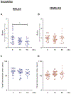
Measurement of sociability after GCR exposure. Beginning at 45 days post exposure (GCR – 0, 15, 50 cGy) animals were tested for sociability deficits by the three-chamber social approach task. In the sociability part of this task the test animal is exposed to a mouse enclosed in a cage (non-aggressive, age-sex matched) or an empty cage for ten minutes. Sociability is measured by a preference to spend time with the mouse over the empty cage standardized to sex-matched 0 cGy group in the first 5 min of testing. (A) Male mice exposed to 50 cGy of GCR had significant impairments in sociability when compared to the 0 cGy group (F = 3.96; p = 0.02). (B) GCR exposure did not impact sociability in the female cohorts. One-way ANOVA did not reveal any significant differences between groups. Interaction times standardized to sex-matched 0 cGy group. (C) Males- Total interaction time with both the mouse and the empty cage. One-way ANOVA did not reveal any significant differences between groups. (D) Females- Total interaction time with both the mouse and the empty cage. One-way ANOVA did not reveal any significant differences between groups. Individual animal scores represented in dots, lines depict group mean and SEM. *p < 0.05. Males n = 13–16; Females n = 15–17.
Fig. 4.
Fig. 4.
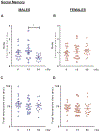
Measurement of social memory after GCR exposure. Beginning at 45 days post exposure (GCR - 0, 15, 50 cGy) animals were tested for social memory deficits by the three-chamber social approach task. In the social memory part of this task the test animal is exposed to a familiar mouse enclosed in a cage (from sociability part) or a novel mouse enclosed in a cage (non-aggressive, age-sex matched) for five minutes. Social memory is measured by a preference to spend time with the novel mouse over the familiar mouse standardized to sex-matched 0 cGy group. (A) Male mice exposed to 50 cGy of GCR had significant impairments in social memory when compared to the 15 cGy group (F = 3.99; p = 0.02). (B) GCR exposure did not impact sociability in the female cohorts. One-way ANOVA did not reveal any significant differences between groups. Interaction times standardized to sex-matched 0 cGy group. (C) Males- Total interaction time with both the novel and familiar mouse. One-way ANOVA did not reveal any significant differences between groups. (D) Females- Total interaction time with both the novel and familiar mouse. One-way ANOVA did not reveal any significant differences between groups. Individual animal scores represented in dots, lines depict group mean and SEM. *p < 0.05. Males n = 14–15; Females n = 16–17.
Fig. 5.
Fig. 5.
High doses of GCR exposure induce anxiety-like behavior in male only. Beginning at 80 days post exposure, anxiety-like behavior was measured in the open field task. Animals are individually allowed to explore of 30 × 30 cm arena and time spent in the center (17 × 17 cm) was calculated. Decreased time in the center is indicative of anxiety-like behavior. (A) Male mice exposed to 50 cGy of GCR spent significantly less time in the center of the field when compared to 0c Gy groups (F = 4.05; p = 0.02). (B) No differences were measured in center time between the female cohorts. Exploration times standardized to sex-matched 0 cGy group. Individual animal scores represented in dots, lines depict group mean and SEM. *p < 0.05. Males n = 14–15; Females n = 16–18.
Fig. 6.
Fig. 6.
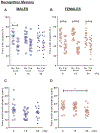
Radiation-induced recognition memory impairments in male cohorts only. Animals were exposed to GCR radiation (0, 15, 50 cGy). Beginning 80 days later, animals were tested for memory deficits. Memory deficits were measured by novel object recognition. Animals were exposed to two identical objects, 24 hrs later the animals are exposed to one familiar object and one novel object. Memory deficits are calculated by a deficit in distinguishing the new (novel) object. Nv = novel. Fm = Familiar. (A) Male cohorts exposed to either 15 or 50 cGy of GCR were unable to distinguish the objects, denoting memory impairment. Two-way ANOVA found significant differences in object distinction (p = 0.001). Bonferroni post-hoc analysis. (B) All female cohorts distinguished the novel from familiar objects. Two-way ANOVA found significant differences in object distinction (p = 0.0001). Bonferroni post-hoc analysis. (C) Males- Total exploration time with both objects is depicted per group. (D) Females- Total exploration time with both objects is depicted per group. A significant decrease in interaction time in the 50 cGy group was measured when compared to the 0 cGy cohort (F = 4.18, p = 0.02). Bonferroni post-doc analysis denotation shown = *p < 0.05, **p < 0.01. ***p < 0.001 Individual animal scores represented in dots, lines depict group mean and SEM. Males n = 14–15; Females n = 15–18.
Fig. 7.
Fig. 7.
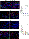
GCR induced changes in microglia phenotypes in male cohorts only. Microglia levels were measured by Iba-1 staining in the dorsal hippocampus. Representative images from MALE cohorts- 0 cGy DG (A), 50 cGy DG (B), 0 cGy CA2/3 (C) 50 cGy CA2/3 (D). (E) Males exposed to 50 cGy of triple ion had increased Iba-1 expression in the DG, CA1 and CA2/3. Two-way ANOVA revealed a significant region effect (p < 0.01), group effect (p < 0.001) and interaction (p < 0.01). Bonferroni post-hoc analysis reveals differences between groups. Representative images from FEMALE cohorts- 0 cGy DG (F), 50 cGy DG (G), 0 cGy CA2/3 (H) 50 cGy CA2/3 (I). (J) No differences in Iba-1 expression was measured. Two-way ANOVA revealed a significant region effect (p < 0.05) and interaction (p < 0.05), however Bonferroni post-hoc analysis did not reveal any differences between the groups. No reactivity was observed in secondary alone. 200x magnification. **p < 0.01. ***p < 0.001 Individual animal scores represented in dots, lines depict group mean and SEM. Scale bar = 100 μm denoted by white line in (A). Males n = 3–4; Females n = 4.
Fig. 8.
Fig. 8.
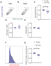
Alterations in synapse composition after GCR exposure in the male hippocampi. Hippocampi were collected and synaptosomes were isolated by sucrose gradient. (A, B) Representative plots used to determine synaptosome levels first by size calibration beads, then co-expression of pre and post synaptic markers. Pre-synaptic marker-Synapsin-1 and post synaptic markers- PSD-95 and GluR1. (C) Significant decreases in total synaptosome numbers in the 50 cGy group when compared to 0 cGy group. Student t-test. (D, E) No differences were measured in total synapsin-1 expression (D) or PSD95 expression (E). (F,G) Decreases in surface GluR1 expression was measured in the 50 cGy group when compared to the 0 cGy group. (F) Representative histogram plot with 0 cGy group in grey and 50 cGy group in blue. (G) Quantification of total GluR1 expressing terminals. Events were collected on an LSRII and analyzed in FlowJo. **p < 0.01. ***p < 0.001 Individual animal scores represented in dots, lines depict group mean and SEM. n = 6–10.
Fig. 9.
Fig. 9.
No change in synapse levels measured in female cohorts. Hippocampi were collected and synaptosomes were isolated by sucrose gradient. (A) No differences were observed in total synaptosome (co-staining with synapsin-1 and PSD95) numbers in the female 50 cGy group when compared to female 0 cGy group. Student t-test. (B, C) No differences were measured in total synapsin-1 expression (B) or PSD95 expression (C). (D) No differences in surface GluR1 expression was measured in the female 50 cGy group when compared to the female 0 cGy group. Quantification of total GluR1 expressing terminals. Events were collected on an LSRII and analyzed in FlowJo. Individual animal scores represented in dots, lines depict group mean and SEM. n = 7–10.

Comment in

  • Perhaps woman are better astronauts?
    Rummel C. Rummel C. Brain Behav Immun. 2018 Nov;74:47-48. doi: 10.1016/j.bbi.2018.09.021. Epub 2018 Sep 23. Brain Behav Immun. 2018. PMID: 30257190 No abstract available.

References

    1. Cucinotta FA, Kim MH, Willingham V, George KA, 2008. Physical and biological organ dosimetry analysis for international space station astronauts. Radiat. Res 170, 127–138. 10.1667/RR1330.1. - DOI - PubMed
    1. Cucinotta FA, Nikjoo H, Goodhead DT, 1998. The effects of delta rays on the number of particle-track traversals per cell in laboratory and space exposures. Radiat. Res 150, 115–119. - PubMed
    1. Zeitlin C, et al., 2013. Measurements of energetic particle radiation in transit to Mars on the Mars Science Laboratory. Science 340, 1080–1084. 10.1126/science.1235989. - DOI - PubMed
    1. Nelson GA, 2016. Space Radiation and Human Exposures. A Primer Radiat. Res 185, 349–358. 10.1667/RR14311.1. - DOI - PubMed
    1. Cucinotta FA, 2014. Space radiation risks for astronauts on multiple International Space Station missions. PLoS ONE 9, e96099. 10.1371/journal.pone.0096099. - DOI - PMC - PubMed

Publication types