. 2018 Oct:19:62-73.
doi: 10.1016/j.redox.2018.07.021. Epub 2018 Aug 3.

The Parkinson's disease gene product DJ-1 modulates miR-221 to promote neuronal survival against oxidative stress

Affiliations

The Parkinson's disease gene product DJ-1 modulates miR-221 to promote neuronal survival against oxidative stress

Stephanie E Oh et al. Redox Biol. 2018 Oct.

Abstract

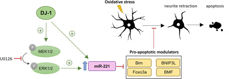
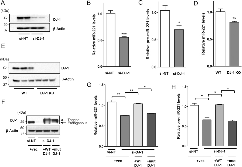
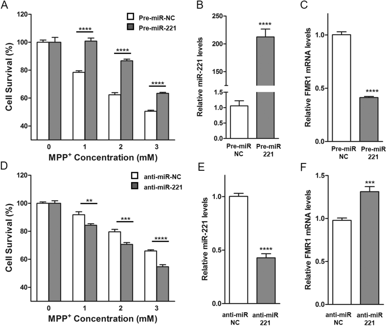
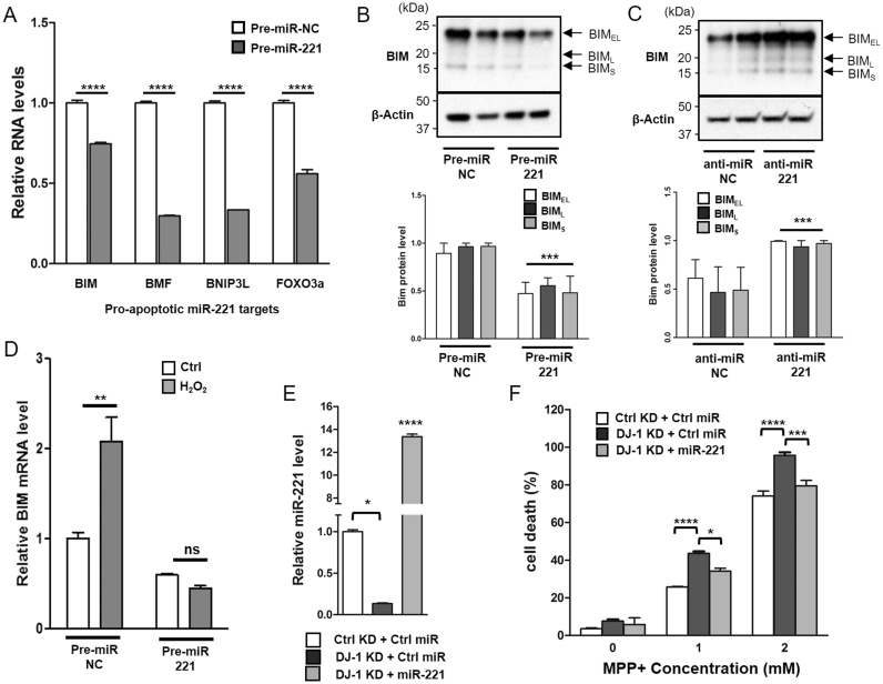
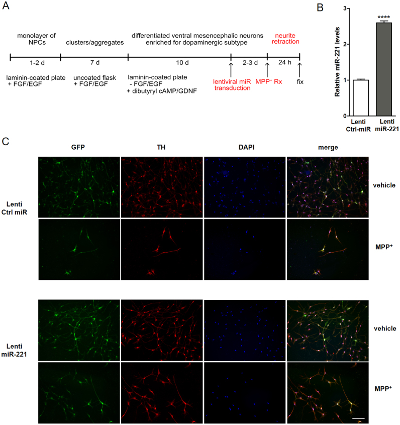
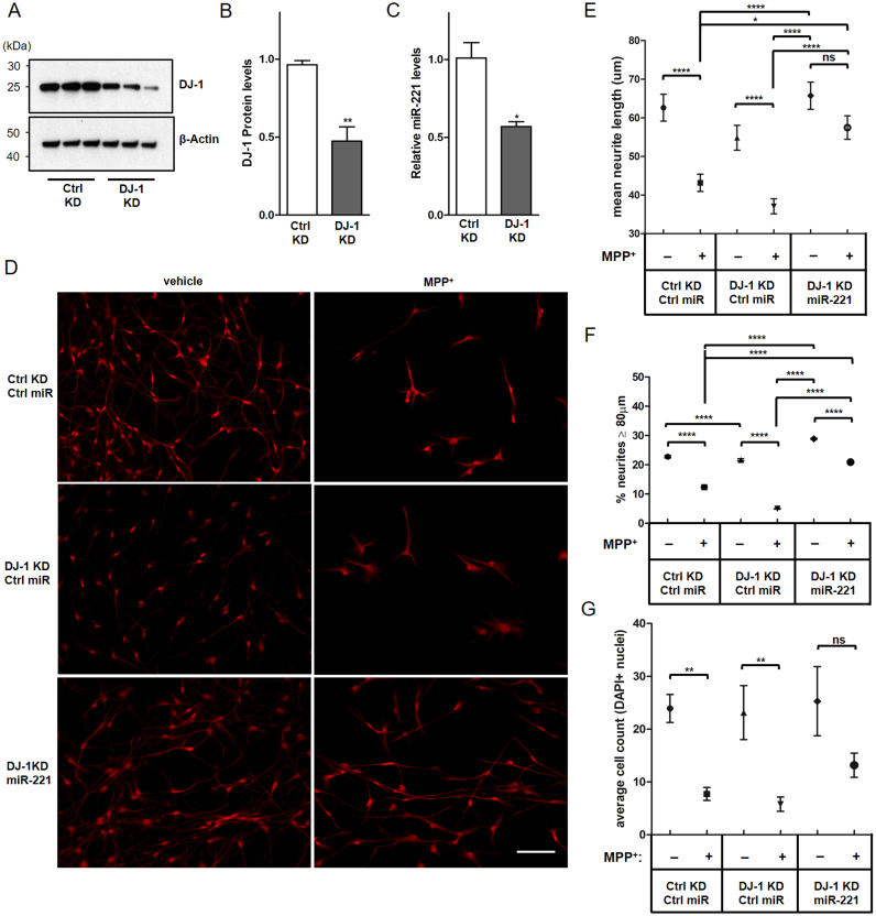
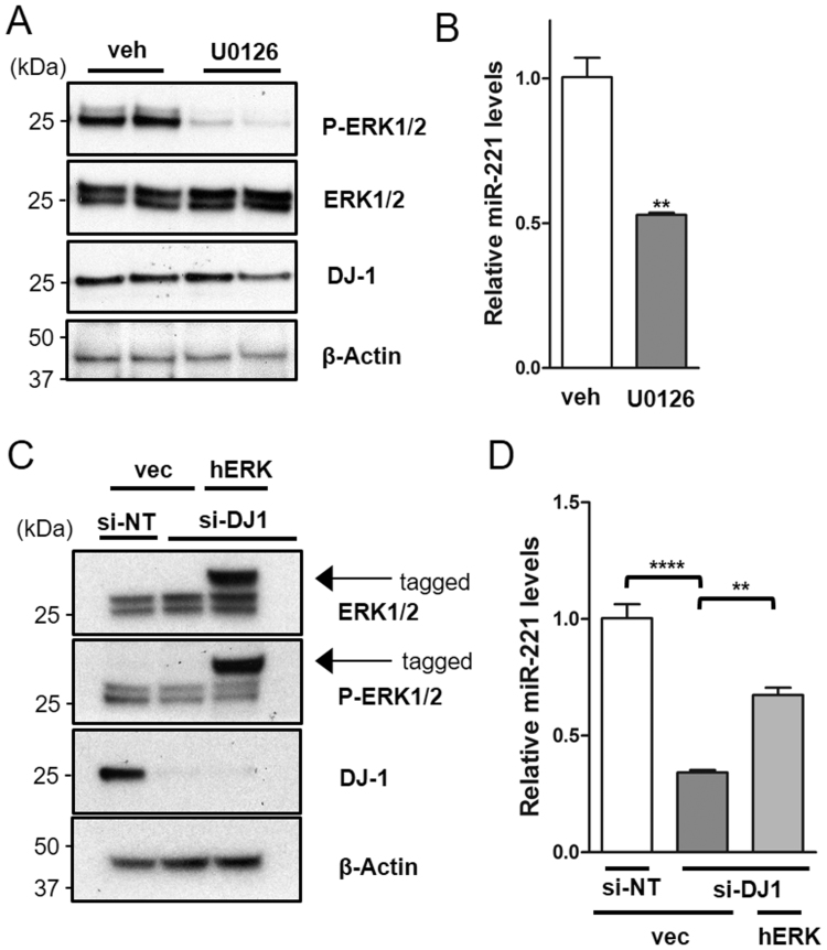
DJ-1 is a highly conserved protein that protects neurons against oxidative stress and whose loss of function mutations are linked to recessively inherited Parkinson's disease (PD). While a number of signaling pathways have been shown to be regulated by DJ-1, its role in controlling cell survival through non-coding RNAs remains poorly understood. Here, using a microarray screen, we found that knocking down DJ-1 in human neuroblastoma cells results in down-regulation of microRNA-221 (miR-221). This is one of the most abundant miRNAs in the human brain and promotes neurite outgrowth and neuronal differentiation. Yet the molecular mechanism linking miR-221 to genetic forms of PD has not been studied. Consistent with the microarray data, miR-221 expression is also decreased in DJ-1-/- mouse brains. Re-introduction of wild-type DJ-1, but not its PD-linked pathogenic M26I mutant, restores miR-221 expression. Notably, over-expression of miR-221 is protective against 1-methyl-4-phenylpyridinium (MPP+)-induced cell death, while inhibition of endogenous miR-221 sensitizes cells to this toxin. Additionally, miR-221 down-regulates the expression of several pro-apoptotic proteins at basal conditions and prevents oxidative stress-induced up-regulation of bcl-2-like protein 11 (BIM). Accordingly, miR-221 protects differentiated DJ-1 knock-down ReNcell VM human dopaminergic neuronal cells from MPP+-induced neurite retraction and cell death. DJ-1 is a known activator of the mitogen-activated protein kinase (MAPK)/extracellular-regulated kinase (ERK) pathway and may modulate miR-221 levels in part through this pathway. We found that inhibiting ERK1/2 decreases miR-221 levels, whereas over-expressing ERK1 in DJ-1 knock-down cells increases miR-221 levels. These findings point to a new cytoprotective mechanism by which DJ-1 may increase miR-221 expression through the MAPK/ERK pathway, subsequently leading to repression of apoptotic molecules. The inability of a pathogenic DJ-1 mutant to modulate miR-221 further supports the relevance of this mechanism in neuronal health and its failure in DJ-1-linked PD.

Keywords: Autosomal recessive; DJ-1; Oxidative stress; PARK7; Parkinson's disease; miR-221; microRNA (miRNA).


Figures

fx1
Graphical abstract
Fig. 1
Fig. 1
miR-221 expression is modulated by wild-type DJ-1 but not its pathogenic mutant. SH-SY5Y cells were transfected with either pooled DJ-1 siRNA (si-DJ-1) or non-targeting control siRNA (si-NT). (A) Immunoblotting shows that DJ-1 is knocked down effectively by pooled DJ-1 siRNA. (B) DJ-1 knock-down (KD) decreases mature miR-221 and (C) precursor pre-miR-221. (D) DJ-1 knockout mouse brains have lower levels of mature miR-221 compared to wild-type mice. (E) DJ-1 knockout mice lack DJ-1 expression. (F) SH-SY5Y cells were transfected with either si-NT or si-DJ-1 for 24 h, followed by transfection with either empty control vector, FLAG-tagged wild-type DJ-1, or FLAG-tagged pathogenic M26I mutant DJ-1. Immunoblotting shows expression of FLAG-tagged wild-type and mutant DJ-1 protein, and the downregulation of endogenous DJ-1 protein in DJ-1 KD cells. (G) Transfection of wild-type DJ-1 but not its pathogenic M26I mutant in DJ-1 KD cells rescued mature miR-221 expression and (H) precursor pre-mir-221 levels. Data are presented as means ± S.E.M. Asterisks denote statistically significant differences (*p ≤ 0.05, **p ≤ 0.01, ***p ≤ 0.001) relative to control. (B–D) analyzed using two-tailed student's t-test. (G, H) analyzed using one-way ANOVA with Bonferroni post-hoc multiple comparisons test.
Fig. 2
Fig. 2
miR-221 is cytoprotective under MPP+insult. (A) SH-SY5Y cells were transfected with either pre-miR negative control (pre-miR-NC) or pre-miR-221. Transfected cells were treated with indicated concentrations of MPP+ for 24 h before assessing cell survival using MTS assay. Over-expression of miR-221 resulted in significantly higher cell viability following MPP+ treatment. (B) Transfected cells showed robust increase in mature miR-221 levels and (C) down-regulation of known miR-221 mRNA target, fragile X mental retardation 1 transcript (FMR1). (D) SH-SY5Y cells were transfected with either anti-miR negative control (anti-miR-NC) or anti-miR-221 that binds to endogenous mature miR-221. Cell survival of transfected cells was assessed after MPP+ treatment at the indicated concentrations for 24 h, and showed that inhibition of endogenous miR-221 resulted in a significant decrease of cell viability. (E) Anti-miR-221 transfected cells showed a decrease in mature miR-221 levels and (F) up-regulation of FMR1. Data are presented as means ± S.E.M. Asterisks denote statistically significant differences (**p ≤ 0.01, ***p ≤ 0.001, ****p ≤ 0.0001) relative to control. (B, C, E, F) analyzed using two-tailed student's t-test. (A, D) analyzed using two-way ANOVA with Bonferroni post-hoc multiple comparisons test.
Fig. 3
Fig. 3
miR-221 Down-Regulates transcripts involved in apoptosis, including Pro-Apoptotic protein BIM, and protects DJ-1 knock-down cells from cell death. (A) Compared to cells transfected with negative control pre-miR (pre-miR-NC), SH-SY5Y cells transfected with pre-miR-221 showed a decrease of mRNA transcripts implicated in apoptosis, including BIM, BMF, BNIP3L, and FOXO3A. (B) All three isoforms of BIM protein (BIMEL, BIML, BIMS) were decreased by over-expression of miR-221 and (C) increased by the inhibition of miR-221 using anti-miR-221. (D) Pre-miR-221 transfected cells were treated with 250 uM H2O2 for 24 h. Compared to control, over-expression of miR-221 prevented the oxidative stress mediated induction of pro-apoptotic BIM transcript. (E) Cells were made to stably express non-targeting control (Ctrl) short hairpin RNA (shRNA) or pooled DJ-1 shRNA. Cells were then transduced with either lentiviral control miR (Ctrl miR) or miR-221 for 24 h. Stable DJ-1 KD decreases the levels of mature miR-221, while transduction with lentiviral miR-221 robustly increases its levels. (F) Cells were then treated with the indicated concentrations of MPP+ for 24 h, and cell death was assessed using LDH assay. Compared to control KD cells, stable DJ-1 KD leads to increased cell death, which is rescued when miR-221 is over-expressed. Data are presented as means ± S.E.M. Asterisks denote statistically significant differences (*p ≤ 0.05, **p ≤ 0.01, ***p ≤ 0.001, ****p ≤ 0.0001) relative to control. ns = not significant. (A, D, F) analyzed using two-way ANOVA with Bonferroni post-hoc multiple comparisons test. (B, C) analyzed using two-tailed student's t-test. (E) Analyzed using one-way ANOVA with Bonferroni post-hoc multiple comparisons test.
Fig. 4
Fig. 4
miR-221 mitigates the loss of neuron morphology in terminally differentiated, human ReNcell VM dopaminergic neurons treated with MPP+. To investigate the role of miR-221 in terminally differentiated human neuronal cells, (A) neural progenitor cells (NPCs) derived from the ventral mesencephalon (ReNcell VM) were terminally differentiated to dopaminergic neurons using an established pre-aggregation protocol. (B) Differentiated ReNcell VM cells were transduced with lentivirus containing control (lenti-Ctrl miR) or miR-221 (lenti-miR-221). Cells transduced with lenti-miR-221 showed robust up-regulation of mature miR-221. (C) These cells were treated with 0.5 mM MPP+ or vehicle (DMEM) for 24 h. Differentiated ReNcell VM cells exhibited GFP expression from viral transduction, stained positively for the dopaminergic neuron marker tyrosine hydroxylase (TH). Following MPP+ exposure, cells exhibited loss of neurite morphology, which was mitigated by miR-221 over-expression (Scale bar: 100 µm). Data are presented as means ± S.E.M. Asterisks denote statistically significant differences (****p ≤ 0.0001) relative to control. (B) analyzed using two-tailed student's t-test.
Fig. 5
Fig. 5
miR-221 prevents MPP+-induced neurite retraction and cell loss in DJ-1 KD ReNcell VM dopaminergic neurons. ReNcell NPCs were made to stably express either control non-targeting shRNA or pooled DJ-1 shRNA, then differentiated into post-mitotic dopaminergic neurons. (A) Immunoblotting shows that DJ-1 is knocked down effectively by DJ-1 shRNA. (B) Quantification of (A). (C) As seen previously in SH-SY5Y cells, DJ-1 KD decreases miR-221 levels in differentiated ReNcell VM neurons. (D) Control KD (Ctrl KD) or DJ-1 KD cells were transduced with either lenti-Ctrl miR or lenti-miR-221 and exposed to 0.5 mM MPP+ or vehicle (DMEM) for 24 h. MPP+ exposure resulted in cell loss and neuritic retraction (Scale bar: 100 µm). (E) Measurements of neurite length indicated that miR-221 protects DJ-1 KD neurites from MPP+. (F) Compared to control KD cells, long neurites (≥ 80 µm, longer than 75th percentile of all neurites) were less preserved among DJ-1 KD cells treated with MPP+. This effect was reversed when DJ-1 KD cells were made to over-express miR-221. (G) MPP+ treatment significantly decreased the cell count in Ctrl miR transduced neurons, whereas miR-221 transduced neurons showed a smaller, non-significant decrease in cell count. For (E, G), n = 450 neurites per group, representative of length measurements taken from 15 microscopic fields per experimental group across three biological replicates. For (F), mean values of percent neurites ≥ 80 µm long were generated with bootstrap resampling, where each sample run contained n = 100 neurites per group. Resampling runs were repeated 500 times to generate confidence intervals for mean values of percent neurites ≥ 80 µm. For (G), Average cell count was assessed by counting DAPI-positive nuclei in these fields. For (B, C, E, G), data are presented as means ± S.E.M. For (F), data are presented as means ± Confidence Interval. Asterisks denote statistically significant differences (*p ≤ 0.05, **p ≤ 0.01, ***p ≤ 0.001, ****p ≤ 0.0001) relative to control. ns = not significant. (B, C) analyzed using two-tailed student's t-test. (E, F, G) Analyzed using one-way ANOVA with Bonferroni post-hoc multiple comparisons test.
Fig. 6
Fig. 6
DJ-1 modulates miR-221 expression through the MAPK/ERK1/2 pathway. To investigate whether DJ-1 could modulate miR-221 levels through its effect on the MAPK/ERK pathway, (A) SH-SY5Y cells were treated with MEK1/2 inhibitor, U0126 (50 µM), or vehicle (DMSO) for 24 h. This led to a decrease in levels of active, phosphorylated ERK1/2 without impacting the levels of total ERK1/2 or DJ-1 protein. (B) The inhibition of ERK1/2 phosphorylation led to down-regulation of miR-221 levels. (C) DJ-1 KD cells were transfected with FLAG-tagged hERK1 or vector-only control for 48 h. hERK1 overexpression increased phosphorylation of ERK1 in DJ-1 KD cells (D) and partially rescued the decrease in miR-221 levels. Data are presented as means ± S.E.M. Asterisks denote statistically significant differences (**p ≤ 0.01, ****p ≤ 0.0001) relative to control. (B) analyzed using two-tailed student's t-test. (D) analyzed using one-way ANOVA with Bonferroni post-hoc multiple comparisons test. Veh = vehicle. Vec = vector-only control.

Similar articles

Cited by

References

    1. Bonifati V., Rizzu P., van Baren M.J., Schaap O., Breedveld G.J., Krieger E., Dekker M.C., Squitieri F., Ibanez P., Joosse M., van Dongen J.W., Vanacore N., van Swieten J.C., Brice A., Meco G., van Duijn C.M., Oostra B.A., Heutink P. Mutations in the DJ-1 gene associated with autosomal recessive early-onset parkinsonism. Science. 2003;299:256–259. - PubMed
    1. Choi J., Sullards M.C., Olzmann J.A., Rees H.D., Weintraub S.T., Bostwick D.E., Gearing M., Levey A.I., Chin L.S., Li L. Oxidative damage of DJ-1 is linked to sporadic Parkinson and Alzheimer diseases. J. Biol. Chem. 2006;281:10816–10824. - PMC - PubMed
    1. Bandopadhyay R., Kingsbury A.E., Cookson M.R., Reid A.R., Evans I.M., Hope A.D., Pittman A.M., Lashley T., Canet-Aviles R., Miller D.W., McLendon C., Strand C., Leonard A.J., Abou-Sleiman P.M., Healy D.G., Ariga H., Wood N.W., de Silva R., Revesz T., Hardy J.A., Lees A.J. The expression of DJ-1 (PARK7) in normal human CNS and idiopathic Parkinson's disease. Brain: J. Neurol. 2004;127:420–430. - PubMed
    1. Taira T., Saito Y., Niki T., Iguchi-Ariga S.M.M., Takahashi K., Ariga H. DJ-1 has a role in antioxidative stress to prevent cell death. EMBO Rep. 2004;5:213–218. - PMC - PubMed
    1. Mullett S.J., Di Maio R., Greenamyre J.T., Hinkle D.A. DJ-1 expression modulates astrocyte-mediated protection against neuronal oxidative stress. J. Mol. Neurosci. 2013;49:507–511. - PMC - PubMed

Publication types