Case Reports
. 2018 Sep 1;141(9):2576-2591.
doi: 10.1093/brain/awy209.

SYT1-associated neurodevelopmental disorder: a case series

Affiliations
Case Reports

SYT1-associated neurodevelopmental disorder: a case series

Kate Baker et al. Brain. .

Abstract

Synaptotagmin 1 (SYT1) is a critical mediator of fast, synchronous, calcium-dependent neurotransmitter release and also modulates synaptic vesicle endocytosis. This paper describes 11 patients with de novo heterozygous missense mutations in SYT1. All mutations alter highly conserved residues, and cluster in two regions of the SYT1 C2B domain at positions Met303 (M303K), Asp304 (D304G), Asp366 (D366E), Ile368 (I368T) and Asn371 (N371K). Phenotypic features include infantile hypotonia, congenital ophthalmic abnormalities, childhood-onset hyperkinetic movement disorders, motor stereotypies, and developmental delay varying in severity from moderate to profound. Behavioural characteristics include sleep disturbance and episodic agitation. Absence of epileptic seizures and normal orbitofrontal head circumference are important negative features. Structural MRI is unremarkable but EEG disturbance is universal, characterized by intermittent low frequency high amplitude oscillations. The functional impact of these five de novo SYT1 mutations has been assessed by expressing rat SYT1 protein containing the equivalent human variants in wild-type mouse primary hippocampal cultures. All mutant forms of SYT1 were expressed at levels approximately equal to endogenous wild-type protein, and correctly localized to nerve terminals at rest, except for SYT1M303K, which was expressed at a lower level and failed to localize at nerve terminals. Following stimulation, SYT1I368T and SYT1N371K relocalized to nerve terminals at least as efficiently as wild-type SYT1. However, SYT1D304G and SYT1D366E failed to relocalize to nerve terminals following stimulation, indicative of impairments in endocytic retrieval and trafficking of SYT1. In addition, the presence of SYT1 variants at nerve terminals induced a slowing of exocytic rate following sustained action potential stimulation. The extent of disturbance to synaptic vesicle kinetics is mirrored by the severity of the affected individuals' phenotypes, suggesting that the efficiency of SYT1-mediated neurotransmitter release is critical to cognitive development. In summary, de novo dominant SYT1 missense mutations are associated with a recognizable neurodevelopmental syndrome, and further cases can now be diagnosed based on clinical features, electrophysiological signature and mutation characteristics. Variation in phenotype severity may reflect mutation-specific impact on the diverse physiological functions of SYT1.


Figures
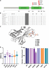
Figure 1
Figure 1
SYT1 de novo mutations cluster in the C2B domain. (A) 2D cartoon of SYT1 domain structure depicting position of patients’ mutations (Jay and Brouwer, 2016). (B) Evolutionary conservation at mutation sites. (C) Structure of the C2B domain of SYT1 in ribbon representation coloured grey (McNicholas et al., 2011). Mutated residues are shown in ball and stick representation coloured in atom colour (carbons orange) and Ca2+ ions in cyan. The side chains of Ile374 and Lys301, which pack against Met303, are shown in grey ball and stick representation. (D and E) Simulations (1.3 µs) were performed on C2B models derived from the calcium-bound soluble NMR structure (PDB 1k5w). Models of the mutant C2B domains were generated using Molsoft ICM Pro. (D) The average RMSD of the backbone atoms of each protein, compared to the starting structures, across all frames in the simulations. Data are mean ± standard error of the mean (SEM). (E) The Ca2+-binding ability of the C2B domains was analysed by tracking the distances between the bound calcium ions and the gamma carbon of Asp363 (equivalent to human Asp364) throughout the trajectories. Data are percentage occupancies ± SEM at the calcium 1 (filled) and calcium 2 (striped) sites for each protein over the simulation time; calcium ions were considered bound if the distances between the ions and the gamma carbon of Asp363 were <6 Å. *P < 0.01, **P < 0.0001 versus wild-type, two-way ANOVA with Dunnett’s multiple comparisons tests.
Figure 2
Figure 2
SYT1 de novo mutations are associated with low frequency oscillation bursts on EEG. Clinical EEG acquired for 6 of 11 patients with SYT1 mutations during early childhood. EEG for three patients with recurrent mutation SYT1 I368T: (A) Patient 2, age 8 months. (B) Patient 1, age 2 years. (C) Patient 10, age 3 years. EEG for patients with other mutations: (D) Patient 4, mutation SYT1 D366E, age 2 years. (E) Patient 5, mutation SYT1 N371K, age 2 years. (F) Patient 7, SYT1 N371K, age 3 years. Scale: x = 1 s, y = 600 µV.
Figure 3
Figure 3
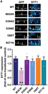
SYT1 mutants, except M303K, are expressed as efficiently as wild-type protein. Cultured hippocampal neurons were transfected with SYT1 variants. (A) Representative images of neurons transfected with SYT1 variants (tagged with pHluorin, a variant of GFP), fixed at rest and immunolabelled for GFP and SYT1. Greyscale panels (left) highlight transfected neurons (GFP), and false colour panels (right) display SYT1 immunofluorescence staining, with warmer colours indicating more intense staining. Arrowheads highlight transfected (filled) and non-transfected (open) nerve terminals. Scale bar = 5 μm. (B) Bar graph shows SYT1 immunofluorescence intensity in transfected neurons relative to non-transfected neurons in the same field of view. Data displayed as mean ± SEM, n = 3–4. **P < 0.01 compared to wild-type, one-way ANOVA with Dunnett’s multiple comparison test.
Figure 4
Figure 4
SYT1 variants display mutation-specific defects in trafficking. Cultured hippocampal neurons transfected with SYT1 variants were fixed at rest (basal; B), or immediately after 30 s incubation with 50 mM KCl (KCl; K), or after 2.5 min recovery in standard saline buffer following 30 s depolarization with 50 mM KCl (recover; R). All steps were performed at 37°C. (A) Diagram showing the localization of synaptic vesicle proteins (dark blue) in a presynaptic terminal at rest (left, basal), following stimulation (middle, KCl), and after recovery (right, recover). Colour intensity of background in presynaptic terminal represents fluorescence intensity of labelled proteins. Arrows (pink) indicate direction of change in protein localization and fluorescence signal. (B) Representative images of neurons transfected with SYT1-pHluorin variants, fixed and immunolabelled for GFP. Scale bar = 5 μm. (C) The distribution of fluorescence intensity along neurites determined by CV analysis, where a high CV equates to a punctate localization, indicative of targeting to presynaptic terminals. Data is mean CV ± SEM, n = 5–9. #P < 0.05, ##P < 0.01 compared to wild-type within same condition, two-way ANOVA with Dunnett’s multiple comparison test. *P < 0.05, **P < 0.01, ***P < 0.001 compared to basal; +P < 0.05, ++P < 0.01 compared to KCl; all by two-way ANOVA with Tukey’s multiple comparison test.
Figure 5
Figure 5
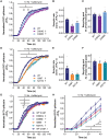
SYT1 mutants slow evoked exocytosis. Hippocampal neurons transfected with SYT1-pHluorin variants were stimulated with a train of 1200 action potentials at 10 Hz, in the presence of 1 µM bafilomycin A1 to block vesicle reacidification. (A and D) Time course of mean ΔF/F0 of SYT1-pHluorin variants normalized to stimulation peak. (B and E) Mean initial rate of exocytosis calculated from linear fit of ΔF/F0 per second over the first 20 s of stimulation (normalized to stimulation peak). (C and F) Total vesicle pool mobilized by 1200 action potentials at 10 Hz, normalized to NH4Cl peak. (A) P < 0.05 for SYT1D304G-pH and *P < 0.05 for SYT1D366E-pH against SYT1WT-pH over time indicated by bar [wild-type (WT) n = 6, D304G n = 7, D366E n = 6, repeated measures ANOVA with Dunnett’s multiple comparisons test]. (B) **P < 0.01 (n as in A, one-way ANOVA versus wild-type with Dunnett’s multiple comparisons test). (C) Not significant by one-way ANOVA, n as in A. (D) *P < 0.05 for SYT1I368T-pH and P < 0.05 for SYT1N371K-pH against SYT1WT-pH over time indicated by bar (wild-type n = 7, I368T n = 5, N371K n = 6, repeated measures ANOVA with Dunnett’s multiple comparisons test). (E) *P < 0.05 (n as in D, one-way ANOVA versus wild-type with Dunnett’s multiple comparison test). (F) Not significant by one-way ANOVA, n as in D. (G and H) Hippocampal neurons transfected with SYT1-pHluorin variants were perfused with high (4 mM) Ca2+ buffer (wild-type 4 mM, D304G, D366E, I368T, N371K) or normal Ca2+ buffer (wild-type 2 mM), and were stimulated with a train of 1200 action potentials at 10 Hz, in the presence of 1 µM bafilomycin A1 to block synaptic vesicle re-acidification. (G) Time course of mean ΔF/F0 of SYT1-pHluorin variants normalized to stimulation peak. P < 0.05 for SYT1D304G #P < 0.05 for SYT1I368T-pH, *P < 0.05 for SYT1D366E-pH, and P < 0.05 for SYT1N371K-pH, against 4 mM SYT1WT-pH over time indicated by bar (n = 8 for 4 mM wild-type, D366E, I368T; n = 7 for 2 mM wild-type, D304G, N371K, repeated measures ANOVA with Tukey’s multiple comparisons test). (H) Same data as in G, but cut-off at 40 s for clarity. All data represented as mean ± SEM.

References

    1. Baker K, Gordon SL, Grozeva D, van Kogelenberg M, Roberts NY, Pike M et al. . Identification of a human synaptotagmin-1 mutation that perturbs synaptic vesicle cycling. J Clin Invest 2015; 125: 1670–8. - PMC - PubMed
    1. Bellus GA, Hefferon TW, Ortiz de Luna RI, Hecht JT, Horton WA, Machado M et al. . Achondroplasia is defined by recurrent G380R mutations of FGFR3. Am J Hum Genet 1995; 56: 368–73. - PMC - PubMed
    1. Blanchard MG, Willemsen MH, Walker JB, Dib-Hajj SD, Waxman SG, Jongmans MC et al. . De novo gain-of-function and loss-of-function mutations of SCN8A in patients with intellectual disabilities and epilepsy. J Med Genet 2015; 52: 330–7. - PMC - PubMed
    1. Cafiero C, Marangi G, Orteschi D, Ali M, Asaro A, Ponzi E et al. . Novel de novo heterozygous loss-of-function variants in MED13L and further delineation of the MED13L haploinsufficiency syndrome. Eur J Hum Genet 2015; 23: 1499–504. - PMC - PubMed
    1. Ebrahimi-Fakhari D, Saffari A, Westenberger A, Klein C. The evolving spectrum of PRRT2-associated paroxysmal diseases. Brain 2015; 138 (Pt 12): 3476–95. - PubMed

Publication types

MeSH terms