Multicenter Study
. 2018 Sep 1;77(9):793-802.
doi: 10.1093/jnen/nly056.

Immunohistochemical Method and Histopathology Judging for the Systemic Synuclein Sampling Study (S4)

Collaborators, Affiliations
Multicenter Study

Immunohistochemical Method and Histopathology Judging for the Systemic Synuclein Sampling Study (S4)

Thomas G Beach et al. J Neuropathol Exp Neurol. .

Abstract

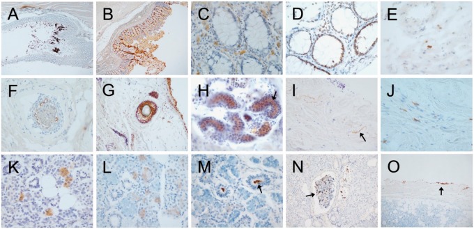
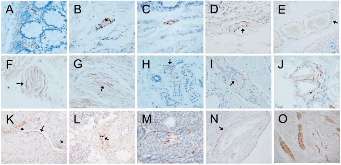
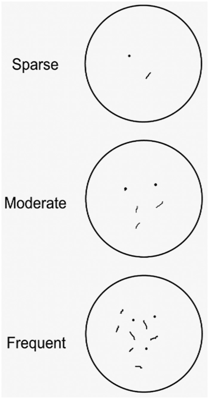
Immunohistochemical (IHC) α-synuclein (Asyn) pathology in peripheral biopsies may be a biomarker of Parkinson disease (PD). The multi-center Systemic Synuclein Sampling Study (S4) is evaluating IHC Asyn pathology within skin, colon and submandibular gland biopsies from 60 PD and 20 control subjects. Asyn pathology is being evaluated by a blinded panel of specially trained neuropathologists. Preliminary work assessed 2 candidate immunoperoxidase methods using a set of PD and control autopsy-derived sections from formalin-fixed, paraffin-embedded blocks of the 3 tissues. Both methods had 100% specificity; one, utilizing the 5C12 monoclonal antibody, was more sensitive in skin (67% vs 33%), and was chosen for further use in S4. Four trainee neuropathologists were trained to perform S4 histopathology readings; in subsequent testing, their scoring was compared to that of the trainer neuropathologist on both glass slides and digital images. Specificity and sensitivity were both close to 100% with all readers in all tissue types on both glass slides and digital images except for skin, where sensitivity averaged 75% with digital images and 83.5% with glass slides. Semiquantitative (0-3) density score agreement between trainees and trainer averaged 67% for glass slides and 62% for digital images.


Figures

FIGURE 1.
FIGURE 1.
Photomicrographs of autopsy sections immunohistochemically stained for pathological Asyn by the 2 compared methods illustrating common types of non-specific staining. Specific versus non-specific staining morphologies have been defined by their neuronal or non-neuronal appearance and empirically by their differential presence in normal control subjects versus subjects with autopsy-confirmed PD or other Lewy body diseases. Immunoperoxidase reaction product is brown, the counterstain is blue. Panels (A–E) are of sigmoid colon, (F–J) of skin from scalp, and (K–O) of submandibular gland. Most staining features were seen with both staining methods. (A, B) Sigmoid colon stained with the 5C12 method (A) and the Nantes method (B), showing non-specific DAB deposition on lumenal contents and within surface epithelial cells. (C) Mucosa of sigmoid colon, depicting frequent melanin-containing macrophages (melanosis coli) within the lamina propria, a relatively common non-specific finding (5C12 method). (D) Mucosa showing non-specific staining of epithelial cell nuclei; melanin-containing macrophages are also present in the lamina propria (5C12 method). (E) Macrophages non-specifically stained within the submucosa (5C12 method). (F, G) Non-specific staining of hair follicles ([F] with the 5C12 method, [G] with the Nantes method). Also seen in (G) are non-specifically stained melanocytes in the epidermis. (H) Sweat glands with non-specific staining of lumenal surfaces (arrow points to example; 5C12 method). (I) Collagen fibers (arrow points to an example) in the dermis, non-specifically taking up DAB (Nantes method). (J) Macrophages in the dermis with non-specific staining of cytoplasmic contents (5C12 method). (K, L) Diffuse, non-specific staining of cytoplasm of serous epithelial cells of submandibular gland ([K] with Nantes method, [L] with 5C12 method). (M) Non-specific staining of lumenal contents (arrow) of a duct within submandibular gland (5C12 method). (N) Non-specific DAB deposition on secretion (arrow) within a submandibular gland duct (5C12 method). (O) Non-specific DAB deposition along the edge of a submandibular gland biopsy (5C12 method).
FIGURE 2.
FIGURE 2.
Photomicrographs of autopsy sections immunohistochemically stained for pathological Asyn by the 2 compared methods illustrating common types of presumptively specific staining. Specific versus non-specific staining morphologies have been defined by their neuronal or non-neuronal appearance and empirically by their differential presence in normal control subjects versus subjects with autopsy-confirmed PD or other Lewy body diseases. Immunoperoxidase reaction product is brown, the counterstain is blue. Panels (A–E) are of sigmoid colon, (F–J) of skin from scalp, and (K–O) of submandibular gland. Most staining features were seen with both staining methods. (A) Several “beaded” fibers, consistent with axons, within the lamina propria (5C12 method). (B, C) Short fibers and puncta, the former consistent with axons, the latter consistent with presynaptic terminals or dystrophic neurites, within a ganglion of the submucosal plexus ([B] with the 5C12 method, [C] with the Nantes method). (D) Short perivascular fibers (arrow) and puncta, consistent with axons, within the submucosa (5C12 method). (E) Longer fiber, consistent with an axon, closely applied to the abluminal surface of a blood vessel within the submucosa (arrow; 5C12 method). (F, G) Fibers consistent with axons (arrows point to examples), within small nerve fascicles in the dermis ([F] with Nantes method, [G] with 5C12 method). (H) Fiber consistent with axon (arrow) applied to the ablumenal surface of a small blood vessel within a cluster of dermal sweat glands (5C12 method). (I) Fine fibers consistent with axons (arrow points to an example) on the surface of an arrector pili muscle in the dermis (5C12 method). (J) Multiple fibers applied to the ablumenal surfaces of dermal blood vessels; fibers running in parallel adjacent to 1 vessel are within a small nerve fascicle (5C12 method). (K) Long fibers (arrow) applied to the ablumenal surface of a blood vessel within the submandibular gland (Nantes method). Also seen are fibers running in parallel within a small nerve fascicle (arrowhead on left) and puncta presumptively representing cross-sectioned axons within a nerve fascicle (arrowhead on right). (L) Fibers running in parallel within a small submandibular gland nerve fascicle (Nantes method); 2 fibers have focal enlargements, consistent with dystrophic change (arrow). (M) Several fibers consistent with axons amongst serous epithelial cells (5C12 method). (N) Long fiber(s) closely applied to the ablumenal surface (arrow) of an arteriole within the submandibular gland (5C12 method). (O) Several large nerve fascicles within the stroma of the submandibular gland, each with numerous immunoperoxidase-stained fibers consistent with axons (5C12 method).
FIGURE 3.
FIGURE 3.
Template used by judges to grade the density of presumptively specific fibers and puncta representing axons and presynaptic terminals. Depicted is the appearance with a 40× microscope objective and 10× ocular magnification.

References

    1. Atik A, Stewart T, Zhang J.. Alpha-synuclein as a biomarker for Parkinson’s disease. Brain Pathol 2016;26:410–8 - PMC - PubMed
    1. Simonsen AH, Kuiperij B, El-Agnaf OM, et al. The utility of alpha-synuclein as biofluid marker in neurodegenerative diseases: A systematic review of the literature. Biomarkers Med 2016;10:19–34 - PubMed
    1. Mollenhauer B, Parnetti L, Rektorova I, et al. Biological confounders for the values of cerebrospinal fluid proteins in Parkinson’s disease and related disorders. J Neurochem 2016;139 (Suppl. 1):290–317 - PubMed
    1. Catafau AM, Bullich S.. Non-amyloid PET imaging biomarkers for neurodegeneration: Focus on tau, alpha-synuclein and neuroinflamation. Curr Alzheimer Res 2016;14:169–77 - PubMed
    1. Rajput AH, Rozdilsky B, Rajput A.. Accuracy of clinical diagnosis in parkinsonism: a prospective study. Can J Neurol Sci 1991;18:275–8 - PubMed

Publication types

MeSH terms