

Methylomic Analysis of Ovarian Cancers Identifies Tumor-Specific Alterations Readily Detectable in Early Precursor Lesions
- PMID: 30108103
- PMCID: PMC6295225
- DOI: 10.1158/1078-0432.CCR-18-1199
Methylomic Analysis of Ovarian Cancers Identifies Tumor-Specific Alterations Readily Detectable in Early Precursor Lesions
Abstract
Purpose: High-grade serous ovarian carcinoma (HGSOC) typically remains undiagnosed until advanced stages when peritoneal dissemination has already occurred. Here, we sought to identify HGSOC-specific alterations in DNA methylation and assess their potential to provide sensitive and specific detection of HGSOC at its earliest stages.
Experimental design: MethylationEPIC genome-wide methylation analysis was performed on a discovery cohort comprising 23 HGSOC, 37 non-HGSOC malignant, and 36 histologically unremarkable gynecologic tissue samples. The resulting data were processed using selective bioinformatic criteria to identify regions of high-confidence HGSOC-specific differential methylation. Quantitative methylation-specific real-time PCR (qMSP) assays were then developed for 8 of the top-performing regions and analytically validated in a cohort of 90 tissue samples. Lastly, qMSP assays were used to assess and compare methylation in 30 laser-capture microdissected (LCM) fallopian tube epithelia samples obtained from cancer-free and serous tubal intraepithelial carcinoma (STIC) positive women.
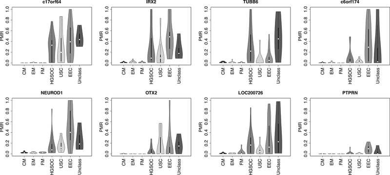
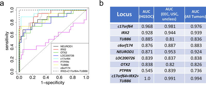
Results: Bioinformatic selection identified 91 regions of robust, HGSOC-specific hypermethylation, 23 of which exhibited an area under the receiver-operator curve (AUC) value ≥ 0.9 in the discovery cohort. Seven of 8 top-performing regions demonstrated AUC values between 0.838 and 0.968 when analytically validated by qMSP in a 90-patient cohort. A panel of the 3 top-performing genes (c17orf64, IRX2, and TUBB6) was able to perfectly discriminate HGSOC (AUC 1.0). Hypermethylation within these loci was found exclusively in LCM fallopian tube epithelia from women with STIC lesions, but not in cancer-free fallopian tubes.
Conclusions: A panel of methylation biomarkers can be used to accurately identify HGSOC, even at precursor stages of the disease.
©2018 American Association for Cancer Research.
Conflict of interest statement
The authors declare no potential conflicts of interest.
Figures


Similar articles
-
Methylomic Landscapes of Ovarian Cancer Precursor Lesions.Clin Cancer Res. 2020 Dec 1;26(23):6310-6320. doi: 10.1158/1078-0432.CCR-20-0270. Epub 2020 Aug 17. Clin Cancer Res. 2020. PMID: 32817081 Free PMC article.
-
Characterization of MicroRNA-200 pathway in ovarian cancer and serous intraepithelial carcinoma of fallopian tube.BMC Cancer. 2017 Jun 17;17(1):422. doi: 10.1186/s12885-017-3417-z. BMC Cancer. 2017. PMID: 28623900 Free PMC article.
-
Differential DNA methylation in high-grade serous ovarian cancer (HGSOC) is associated with tumor behavior.Sci Rep. 2019 Nov 29;9(1):17996. doi: 10.1038/s41598-019-54401-w. Sci Rep. 2019. PMID: 31784612 Free PMC article.
-
Extracellular matrix in high-grade serous ovarian cancer: Advances in understanding of carcinogenesis and cancer biology.Matrix Biol. 2023 Apr;118:16-46. doi: 10.1016/j.matbio.2023.02.004. Epub 2023 Feb 11. Matrix Biol. 2023. PMID: 36781087 Review.
-
Exploring the Role of Fallopian Ciliated Cells in the Pathogenesis of High-Grade Serous Ovarian Cancer.Int J Mol Sci. 2018 Aug 24;19(9):2512. doi: 10.3390/ijms19092512. Int J Mol Sci. 2018. PMID: 30149579 Free PMC article. Review.
Cited by
-
Genome-wide methylation profiling identified novel differentially hypermethylated biomarker MPPED2 in colorectal cancer.Clin Epigenetics. 2019 Mar 7;11(1):41. doi: 10.1186/s13148-019-0628-y. Clin Epigenetics. 2019. PMID: 30846004 Free PMC article.
-
The Mechanism of Xiaoyao San in the Treatment of Ovarian Cancer by Network Pharmacology and the Effect of Stigmasterol on the PI3K/Akt Pathway.Dis Markers. 2021 Jun 29;2021:4304507. doi: 10.1155/2021/4304507. eCollection 2021. Dis Markers. 2021. Retraction in: Dis Markers. 2023 Jul 12;2023:9791279. doi: 10.1155/2023/9791279. PMID: 34306252 Free PMC article. Retracted.
-
Role of DNA methylation and non‑coding RNAs expression in pathogenesis, detection, prognosis, and therapy‑resistant ovarian carcinoma (Review).Mol Med Rep. 2025 Jun;31(6):144. doi: 10.3892/mmr.2025.13509. Epub 2025 Apr 4. Mol Med Rep. 2025. PMID: 40183399 Free PMC article. Review.
-
Up-regulation of circ_LARP4 suppresses cell proliferation and migration in ovarian cancer by regulating miR-513b-5p/LARP4 axis.Cancer Cell Int. 2020 Jan 6;20:5. doi: 10.1186/s12935-019-1071-z. eCollection 2020. Cancer Cell Int. 2020. PMID: 31911757 Free PMC article.
-
Multimodal Spatial Profiling Reveals Immune Suppression and Microenvironment Remodeling in Fallopian Tube Precursors to High-Grade Serous Ovarian Carcinoma.bioRxiv [Preprint]. 2024 Sep 27:2024.09.25.615007. doi: 10.1101/2024.09.25.615007. bioRxiv. 2024. Update in: Cancer Discov. 2025 Jun 3;15(6):1180-1202. doi: 10.1158/2159-8290.CD-24-1366. PMID: 39386723 Free PMC article. Updated. Preprint.
References
-
- Society AC. Cancer Facts & Figures 2017. Atlanta: American Cancer Society; 2017.
-
- Force USPST. Screening for ovarian cancer: Us preventive services task force recommendation statement. JAMA 2018;319(6):588–94 doi 10.1001/jama.2017.21926. - DOI - PubMed
-
- Wittenberger T, Sleigh S, Reisel D, Zikan M, Wahl B, Alunni-Fabbroni M, et al. DNA methylation markers for early detection of women’s cancer: promise and challenges. Epigenomics 2014;6(3):311–27 doi 10.2217/epi.14.20. - DOI - PubMed
-
- Baylin SB, Jones PA. Epigenetic Determinants of Cancer. Cold Spring Harbor Perspect Biol 2016;8(9):35 doi 10.1101/cshperspect.a019505. - DOI - PMC - PubMed
-
- Timp W, Feinberg AP. Cancer as a dysregulated epigenome allowing cellular growth advantage at the expense of the host. Nature Reviews Cancer 2013;13:497 doi 10.1038/nrc3486. - DOI - PMC - PubMed
Publication types
MeSH terms
Substances
Grants and funding
LinkOut - more resources
Full Text Sources
Other Literature Sources
Medical
