

Synchrotron-Based X-Ray Fluorescence Microscopy as a Technique for Imaging of Elements in Plants
- PMID: 30108140
- PMCID: PMC6181034
- DOI: 10.1104/pp.18.00759
Synchrotron-Based X-Ray Fluorescence Microscopy as a Technique for Imaging of Elements in Plants
Abstract
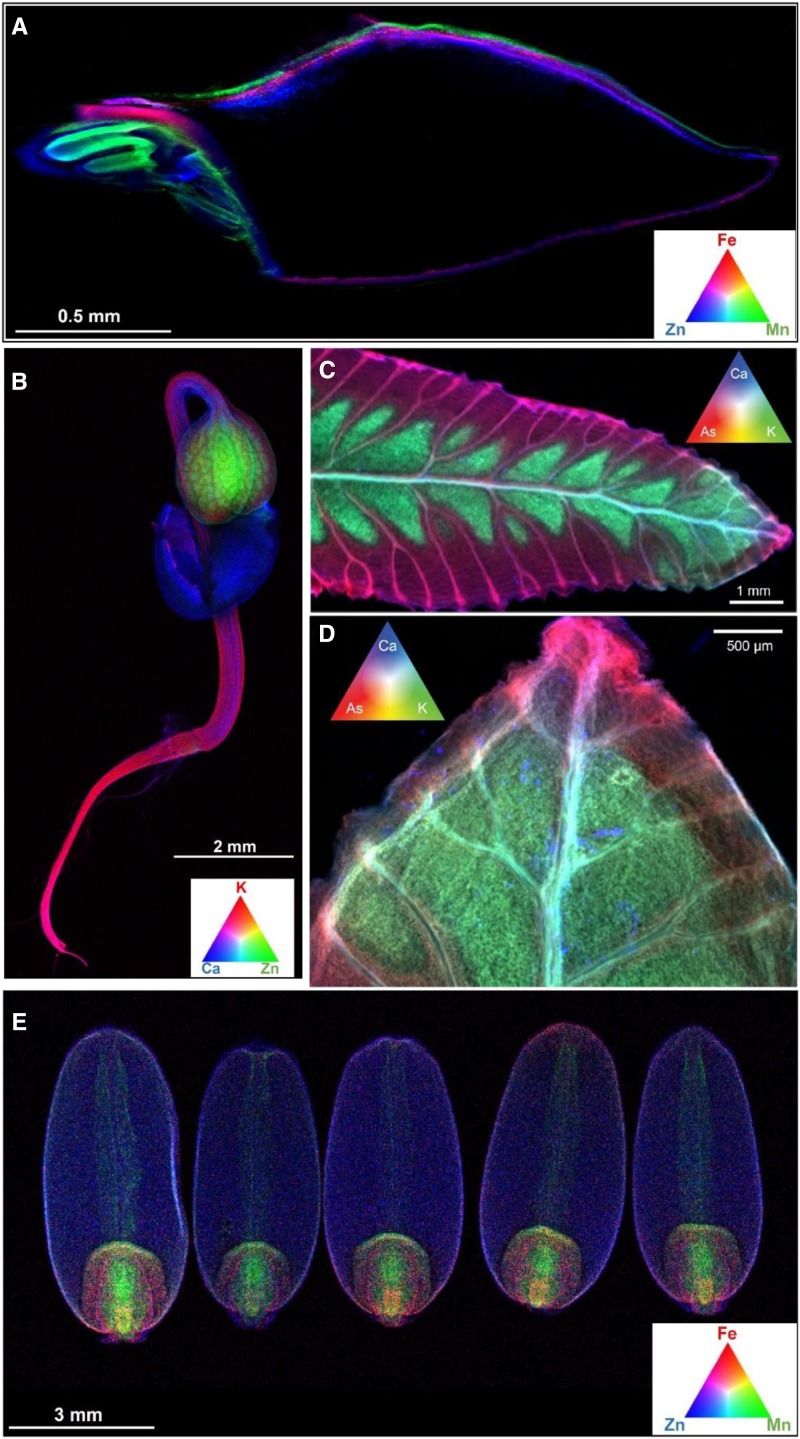
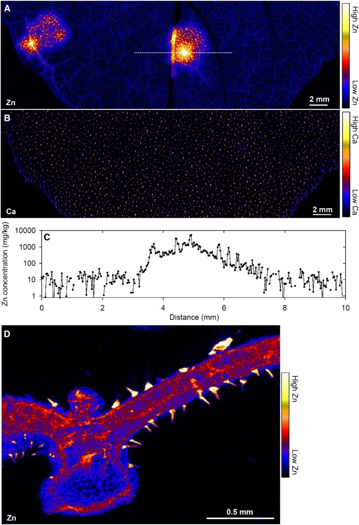
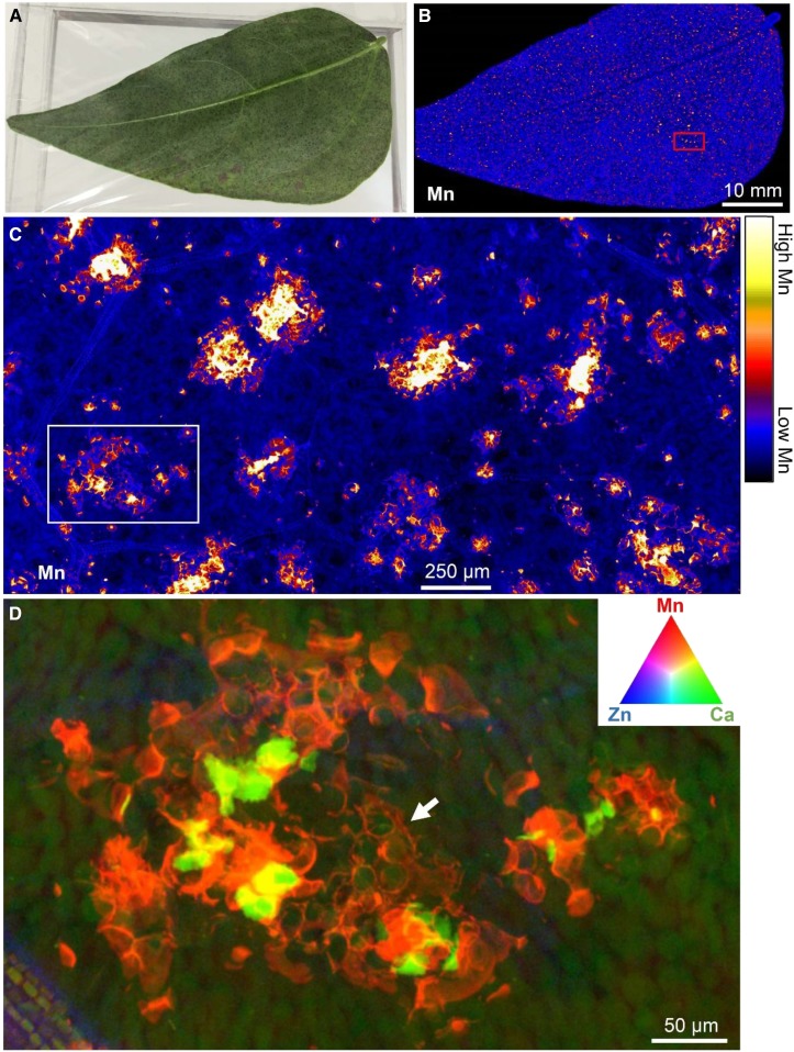
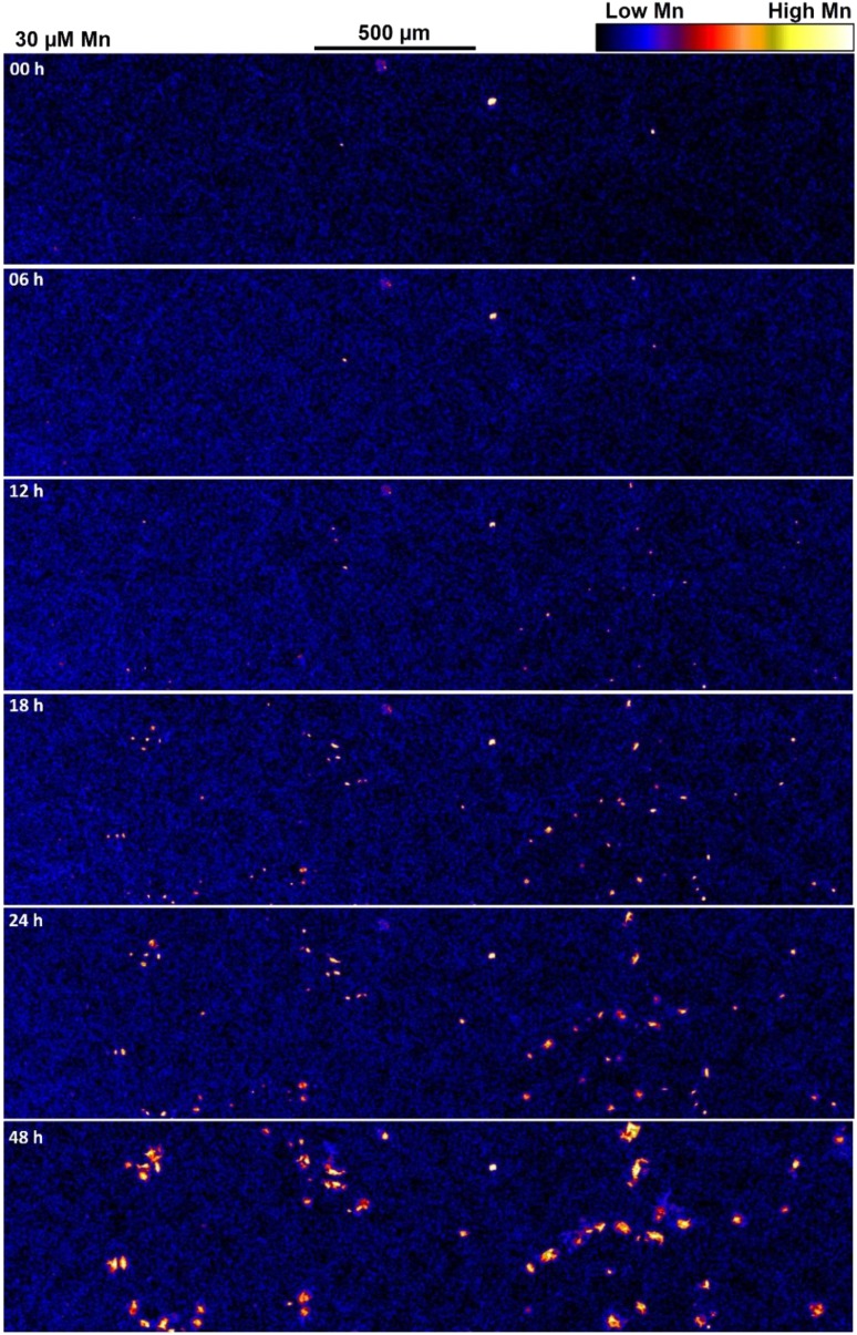
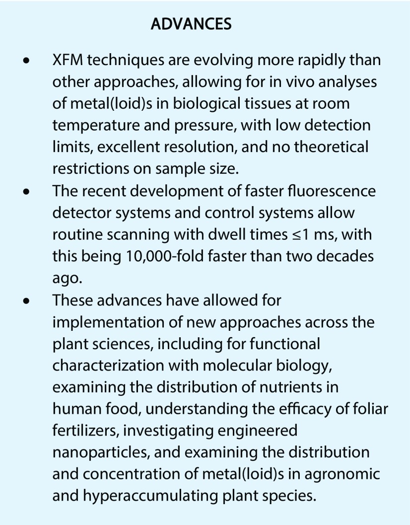
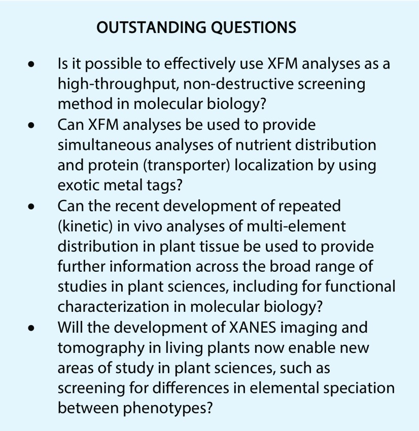
Understanding the distribution of elements within plant tissues is important across a range of fields in plant science. In this review, we examine synchrotron-based x-ray fluorescence microscopy (XFM) as an elemental imaging technique in plant sciences, considering both its historical and current uses as well as discussing emerging approaches. XFM offers several unique capabilities of interest to plant scientists, including in vivo analyses at room temperature and pressure, good detection limits (approximately 1-100 mg kg-1), and excellent resolution (down to 50 nm). This has permitted its use in a range of studies, including for functional characterization in molecular biology, examining the distribution of nutrients in food products, understanding the movement of foliar fertilizers, investigating the behavior of engineered nanoparticles, elucidating the toxic effects of metal(loid)s in agronomic plant species, and studying the unique properties of hyperaccumulating plants. We anticipate that continuing technological advances at XFM beamlines also will provide new opportunities moving into the future, such as for high-throughput screening in molecular biology, the use of exotic metal tags for protein localization, and enabling time-resolved, in vivo analyses of living plants. By examining current and potential future applications, we hope to encourage further XFM studies in plant sciences by highlighting the versatility of this approach.
© 2018 American Society of Plant Biologists. All rights reserved.
Figures
Similar articles
-
Synchrotron XFM tomography for elucidating metals and metalloids in hyperaccumulator plants.Metallomics. 2022 Nov 23;14(11):mfac069. doi: 10.1093/mtomcs/mfac069. Metallomics. 2022. PMID: 36099903 Free PMC article.
-
Visualizing Metal Distribution in Plants Using Synchrotron X-Ray Fluorescence Microscopy Techniques.Methods Mol Biol. 2023;2665:177-189. doi: 10.1007/978-1-0716-3183-6_14. Methods Mol Biol. 2023. PMID: 37166601
-
X-ray elemental mapping techniques for elucidating the ecophysiology of hyperaccumulator plants.New Phytol. 2018 Apr;218(2):432-452. doi: 10.1111/nph.14810. Epub 2017 Oct 10. New Phytol. 2018. PMID: 28994153 Review.
-
Practical review on the use of synchrotron based micro- and nano- X-ray fluorescence mapping and X-ray absorption spectroscopy to investigate the interactions between plants and engineered nanomaterials.Plant Physiol Biochem. 2017 Jan;110:13-32. doi: 10.1016/j.plaphy.2016.07.018. Epub 2016 Jul 20. Plant Physiol Biochem. 2017. PMID: 27475903 Review.
-
Synchrotron-based X-Ray Approaches for Examining Toxic Trace Metal(loid)s in Soil-Plant Systems.J Environ Qual. 2017 Nov;46(6):1175-1189. doi: 10.2134/jeq2016.09.0361. J Environ Qual. 2017. PMID: 29293828 Review.
Cited by
-
Arabidopsis thaliana zinc accumulation in leaf trichomes is correlated with zinc concentration in leaves.Sci Rep. 2021 Mar 5;11(1):5278. doi: 10.1038/s41598-021-84508-y. Sci Rep. 2021. PMID: 33674630 Free PMC article.
-
Genetically Modified Plants: Nutritious, Sustainable, yet Underrated.J Nutr. 2020 Oct 12;150(10):2628-2634. doi: 10.1093/jn/nxaa220. J Nutr. 2020. PMID: 32725215 Free PMC article.
-
Visualization and Quantitative Evaluation of Functional Structures of Soybean Root Nodules via Synchrotron X-ray Imaging.Plant Phenomics. 2024 Jul 17;6:0203. doi: 10.34133/plantphenomics.0203. eCollection 2024. Plant Phenomics. 2024. PMID: 39021394 Free PMC article.
-
Assessing radiation dose limits for X-ray fluorescence microscopy analysis of plant specimens.Ann Bot. 2020 Mar 29;125(4):599-610. doi: 10.1093/aob/mcz195. Ann Bot. 2020. PMID: 31777920 Free PMC article.
-
Disease Ionomics: Understanding the Role of Ions in Complex Disease.Int J Mol Sci. 2020 Nov 17;21(22):8646. doi: 10.3390/ijms21228646. Int J Mol Sci. 2020. PMID: 33212764 Free PMC article. Review.
References
-
- Ajiboye B, Cakmak I, Paterson D, de Jonge MD, Howard DL, Stacey SP, Torun AA, Aydin N, McLaughlin MJ (2015) X-ray fluorescence microscopy of zinc localization in wheat grains biofortified through foliar zinc applications at different growth stages under field conditions. Plant Soil 392: 357–370
-
- Beetz T, Jacobsen C (2003) Soft X-ray radiation-damage studies in PMMA using a cryo-STXM. J Synchrotron Radiat 10: 280–283 - PubMed
-
- Berglund A, Brelid H, Rindby A, Engström P (1999) Spatial distribution of metal ions in spruce wood by synchrotron radiation microbeam X-ray fluorescence analysis. Holzforschung 53: 474
-
- Bertsch PM, Hunter DB (2001) Applications of synchrotron-based X-ray microprobes. Chem Rev 101: 1809–1842 - PubMed
Publication types
MeSH terms
LinkOut - more resources
Full Text Sources
Other Literature Sources
