. 2018 Aug 28;115(35):E8201-E8210.
doi: 10.1073/pnas.1810719115. Epub 2018 Aug 14.

Structure of the mammalian TRPM7, a magnesium channel required during embryonic development

Affiliations

Structure of the mammalian TRPM7, a magnesium channel required during embryonic development

Jingjing Duan et al. Proc Natl Acad Sci U S A. .

Abstract

The transient receptor potential ion channel subfamily M, member 7 (TRPM7), is a ubiquitously expressed protein that is required for mouse embryonic development. TRPM7 contains both an ion channel and an α-kinase. The channel domain comprises a nonselective cation channel with notable permeability to Mg2+ and Zn2+ Here, we report the closed state structures of the mouse TRPM7 channel domain in three different ionic conditions to overall resolutions of 3.3, 3.7, and 4.1 Å. The structures reveal key residues for an ion binding site in the selectivity filter, with proposed partially hydrated Mg2+ ions occupying the center of the conduction pore. In high [Mg2+], a prominent external disulfide bond is found in the pore helix, which is essential for ion channel function. Our results provide a structural framework for understanding the TRPM1/3/6/7 subfamily and extend the knowledge base upon which to study the diversity and evolution of TRP channels.

Keywords: TRP channel; chanzyme; ion channel; kinase; zinc.


Conflict of interest statement

The authors declare no conflict of interest.

Figures
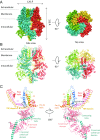
Fig. 1.
Fig. 1.
Overall structure of TRPM7-Mg2+. (A) Cryo-EM reconstruction density map of TRPM7-Mg2+ at 3.7-Å overall resolution. Each channel subunit is color-coded. (B) Ribbon diagram of the mouse TRPM7 model. (C) Ribbon diagrams depicting two side views of a single subunit. Red boxes: hydrophobic helix (almost alawys 759–769) anchored to inner leaflet of the plasma membrane. Black boxes: S2–S3 helix.
Fig. 2.
Fig. 2.
Ion conduction pathway. (A) Ion conduction pathway along the pore of TRPM7-DVF (Top), TRPM7-Mg2+ (Middle), and TRPM7-EDTA (Bottom), shown as dots and mapped using HOLE. Three continuous residues (Glu1047, Gly1046, and Phe1045) in the pore helix form the funnel-shaped selectivity filter (1045FGE1047). The lower gate, Asn1097, is located before the TRP domain. (B) Pore radius along the central axis (calculated with HOLE). Glu1047-Phe1045 and Asn1097 form narrow constrictions at the selectivity filter and lower gate, respectively.
Fig. 3.
Fig. 3.
Mg2+ binding site. (A) Top and side views of the tetrameric TRPM7-Mg2+ structure with bound Mg2+. (B) Side view of the TRPM7-Mg2+ pore, with residues important for cation binding shown in stick representation. (C) Side view of the TRPM7-EDTA pore, with residues in the selectivity filter shown in stick representation. (D) Enlarged side view of the TRPM7-Mg2+ pore. Residues that contribute to cation-binding sites are shown as sticks, and the putative Mg2+ is the red sphere. (E) Top view of the TRPM7-Mg2+ pore.
Fig. 4.
Fig. 4.
Assembly and domain interactions. (A) One TRPM7 subunit from the plane of the membrane with regions of interest depicted by boxes. (B) Expanded view of A showing interactions between the N terminus (pink) and the TRP domain (blue). (C) Expanded view of A showing interactions between N (pink) and C (green) termini. Residues at domain interfaces are labeled, with potential hydrogen bonds and electrostatic interactions shown as dashed lines.
Fig. 5.
Fig. 5.
Structural comparison of TRPM7 and TRPM4. (A) Overlaid monomer of TRPM7 (blue, pink, and cyan) and TRPM4 (gray). (B) Enlarged view of key pore loop disulfide bond between Cys1056 and Cys1066. (C) Aligned TRPM7 (blue) and TRPM4 (gray) dimer pores. Residues of interest are labeled and shown as sticks. (D) Whole-cell I-V relationships of HEK293T TRPM6/7 double KO cells expressing MBP-tagged truncated mouse TRPM7 (blue), with 4 mM Mg2+(gold), mTRPM7 in 4 mM Mg2+, or truncated mouse E1047Q-TRPM7 (black). Bar charts represent the mean currents recorded at +100 mV and −100 mV; E1047Q (n = 18, 103 ± 10 pA/pF and −82 ± 1 pA/pF), mTRPM7 (n = 10, 111 ± 10 pA/pF and −12 ±00.3 pA/pF), and mTRPM7+4 mM Mg2+ (mTRPM7+Mg2+, n = 10, 5 ± 1 pA/pF and −2 ± 0.1 pA/pF). (E) Side and bottom views of superimposed cytosolic domains of TRPM7 (blue and pink) and TRPM4 (gray); boxes highlight extracellular regions containing cysteines.
Fig. 6.
Fig. 6.
Comparison of TRPM7-Mg2+ and TRPM7-EDTA structures. (A) Superimposition of the transmembrane module from TRPM7-Mg2+ (blue) and TRPM7-EDTA structures (pink). (B) Site of the broken disulfide bond between Cys1056 and Cys1066 in the pore domain. Key discernible residues, Cys1056 and Cys1066, are labeled. Shown is superimposition of the disulfide bond between S5 and S6 in the TRPM7-Mg2+ (blue) and TRPM7-EDTA (pink) structures. (C) Whole-cell I-V relationship of HEK293T TRPM6/7 double KO cells expressing MBP-tagged truncated mouse TRPM7 (mTRPM7; orange), C1056S (red), C1065S (green), and C1056S/C1066S (violet). (D) Mean currents of mTRPM7 and mTRPM7 cysteine(s) mutant measured at +100 mV and −100 mV; nontransfected (n = 10, 2.3 ± 0.5 and −1.6 ± 0.1), mTRPM7 (Time0) (n = 12, 5.1 ± 0.1 and −2.1 ± 0.1 pA/pF), C1056S/C1066S (n = 12, 55 ± 3 and −22 ± 3 pA/pF), C1056S (n = 12, 124 ± 5 and −56 ± 2 pA/pF), C1066S (n = 10, 90 ±10 and −25 ± 4 pA/pF), and mTRPM7 (n = 10, 111 ± 10 and −12 ± 1 pA/pF).

Comment in

  • TRPM7 reflected in Cryo-EMirror.
    Chubanov V, Mittermeier L, Gudermann T. Chubanov V, et al. Cell Calcium. 2018 Dec;76:129-131. doi: 10.1016/j.ceca.2018.11.004. Epub 2018 Nov 15. Cell Calcium. 2018. PMID: 30470536

Similar articles

Cited by

References

    1. Clapham DE. TRP channels as cellular sensors. Nature. 2003;426:517–524. - PubMed
    1. Ramsey IS, Delling M, Clapham DE. An introduction to TRP channels. Annu Rev Physiol. 2006;68:619–647. - PubMed
    1. Runnels LW. TRPM6 and TRPM7: A Mul-TRP-PLIK-cation of channel functions. Curr Pharm Biotechnol. 2011;12:42–53. - PMC - PubMed
    1. Jin J, et al. The channel kinase, TRPM7, is required for early embryonic development. Proc Natl Acad Sci USA. 2012;109:E225–E233. - PMC - PubMed
    1. Jin J, et al. Deletion of Trpm7 disrupts embryonic development and thymopoiesis without altering Mg2+ homeostasis. Science. 2008;322:756–760. - PMC - PubMed

Publication types