

Adhesion of Staphylococcus aureus to Corneocytes from Atopic Dermatitis Patients Is Controlled by Natural Moisturizing Factor Levels
- PMID: 30108169
- PMCID: PMC6094479
- DOI: 10.1128/mBio.01184-18
Adhesion of Staphylococcus aureus to Corneocytes from Atopic Dermatitis Patients Is Controlled by Natural Moisturizing Factor Levels
Abstract
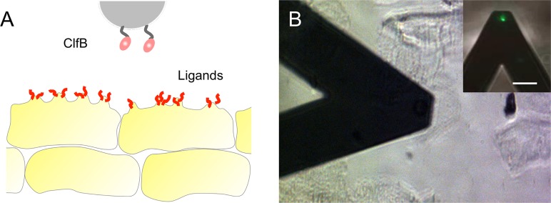
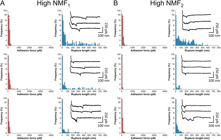
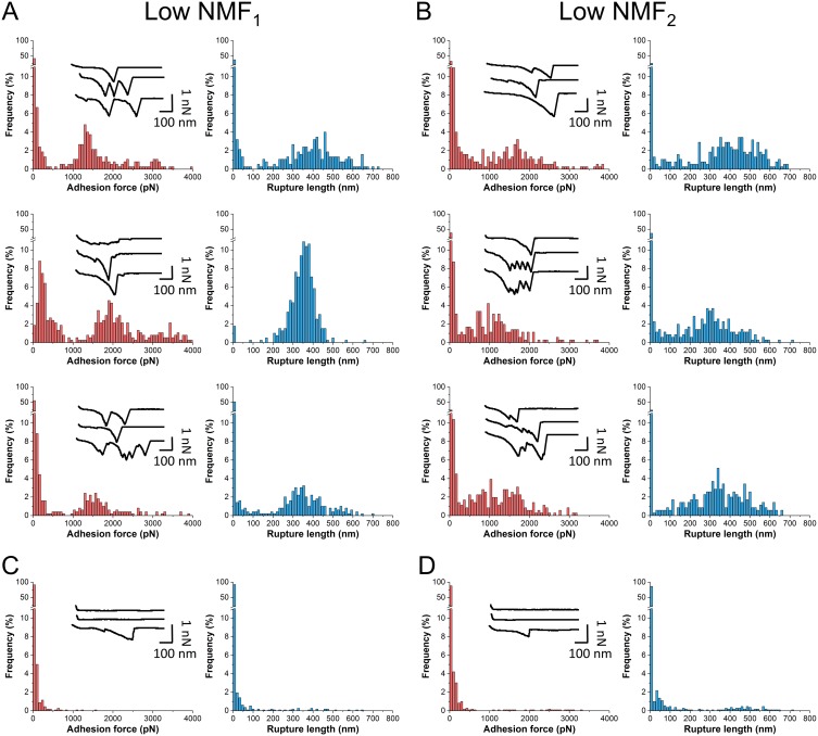
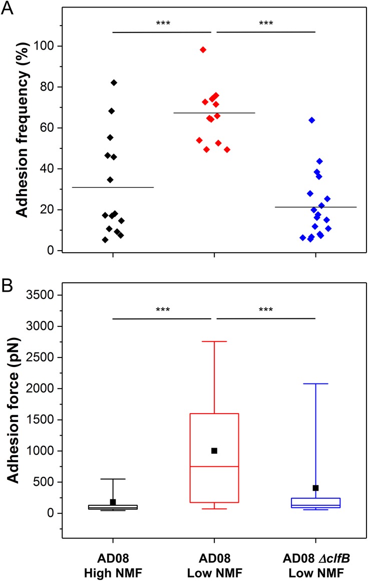
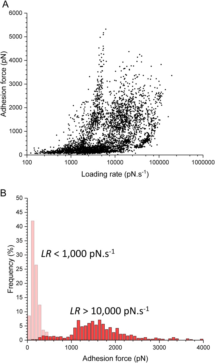
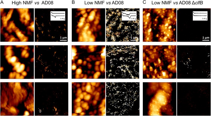
The bacterial pathogen Staphylococcus aureus plays an important role in atopic dermatitis (AD), a chronic disorder that mostly affects children. Colonization of the skin of AD patients by S. aureus exacerbates the disease, but the molecular determinants of the bacterium-skin adhesive interactions are poorly understood. Specifically, reduced levels of natural moisturizing factor (NMF) in the stratum corneum have been shown to be associated with more severe AD symptoms, but whether this is directly related to S. aureus adhesion is still an open question. Here, we demonstrate a novel relationship between NMF expression in AD skin and strength of bacterial adhesion. Low-NMF corneocytes, unlike high-NMF ones, are covered by a dense layer of nanoscale villus protrusions. S. aureus bacteria isolated from AD skin bind much more strongly to corneocytes when the NMF level is reduced. Strong binding forces originate from a specific interaction between the bacterial adhesion clumping factor B (ClfB) and skin ligands. Remarkably, mechanical tension dramatically strengthens ClfB-mediated adhesion, as observed with catch bonds, demonstrating that physical stress plays a role in promoting colonization of AD skin by S. aureus Collectively, our findings demonstrate that patient NMF levels regulate the strength of S. aureus-corneocyte adhesion, the first step in skin colonization, and suggest that the ClfB binding mechanism could represent a potential target for new therapeutic treatments.IMPORTANCE Bacterium-skin interactions play important roles in skin disorders, yet their molecular details are poorly understood. In this study, we decipher the molecular forces at play during adhesion of Staphylococcus aureus to skin corneocytes in the clinically important context of atopic dermatitis (AD), also known as eczema. We identify a unique relationship between the level of natural moisturizing factor (NMF) in the skin and the strength of bacterium-corneocyte adhesion. Bacterial adhesion is primarily mediated by the surface protein clumping factor B (ClfB) and is enhanced by physical stress, highlighting the role of protein mechanobiology in skin colonization. Similar to a catch bond behavior, this mechanism represents a promising target for the development of novel antistaphylococcal agents.
Keywords: Staphylococcus aureus; adhesion; atopic dermatitis; biofilms; skin.
Copyright © 2018 Feuillie et al.
Figures

Similar articles
-
Clumping Factor B Promotes Adherence of Staphylococcus aureus to Corneocytes in Atopic Dermatitis.Infect Immun. 2017 May 23;85(6):e00994-16. doi: 10.1128/IAI.00994-16. Print 2017 Jun. Infect Immun. 2017. PMID: 28373353 Free PMC article.
-
Staphylococcus aureus binds to the N-terminal region of corneodesmosin to adhere to the stratum corneum in atopic dermatitis.Proc Natl Acad Sci U S A. 2021 Jan 5;118(1):e2014444118. doi: 10.1073/pnas.2014444118. Proc Natl Acad Sci U S A. 2021. PMID: 33361150 Free PMC article.
-
Staphylococcal Corneocyte Adhesion: Assay Optimization and Roles of Aap and SasG Adhesins in the Establishment of Healthy Skin Colonization.Microbiol Spectr. 2022 Dec 21;10(6):e0246922. doi: 10.1128/spectrum.02469-22. Epub 2022 Oct 11. Microbiol Spectr. 2022. PMID: 36219106 Free PMC article.
-
Staphylococcal Biofilms in Atopic Dermatitis.Curr Allergy Asthma Rep. 2017 Oct 23;17(12):81. doi: 10.1007/s11882-017-0750-x. Curr Allergy Asthma Rep. 2017. PMID: 29063212 Free PMC article. Review.
-
Staphylococcus aureus and Atopic Dermatitis: A Complex and Evolving Relationship.Trends Microbiol. 2018 Jun;26(6):484-497. doi: 10.1016/j.tim.2017.11.008. Epub 2017 Dec 9. Trends Microbiol. 2018. PMID: 29233606 Review.
Cited by
-
Binding Strength of Gram-Positive Bacterial Adhesins.Front Microbiol. 2020 Jun 25;11:1457. doi: 10.3389/fmicb.2020.01457. eCollection 2020. Front Microbiol. 2020. PMID: 32670256 Free PMC article. Review.
-
Epidermis as a Platform for Bacterial Transmission.Front Immunol. 2021 Dec 1;12:774018. doi: 10.3389/fimmu.2021.774018. eCollection 2021. Front Immunol. 2021. PMID: 34925344 Free PMC article. Review.
-
Skin barrier dysfunction and filaggrin.Arch Pharm Res. 2021 Jan;44(1):36-48. doi: 10.1007/s12272-021-01305-x. Epub 2021 Jan 18. Arch Pharm Res. 2021. PMID: 33462753 Review.
-
Interaction between the microbiota and the skin barrier in aging skin: a comprehensive review.Front Physiol. 2024 Jan 19;15:1322205. doi: 10.3389/fphys.2024.1322205. eCollection 2024. Front Physiol. 2024. PMID: 38312314 Free PMC article. Review.
-
How Microbes Use Force To Control Adhesion.J Bacteriol. 2020 May 27;202(12):e00125-20. doi: 10.1128/JB.00125-20. Print 2020 May 27. J Bacteriol. 2020. PMID: 32253344 Free PMC article. Review.
References
Publication types
MeSH terms
Substances
LinkOut - more resources
Full Text Sources
Other Literature Sources
Medical
