. 2018 Oct 5;293(40):15397-15418.
doi: 10.1074/jbc.RA118.003063. Epub 2018 Aug 14.

HGF-induced formation of the MET-AXL-ELMO2-DOCK180 complex promotes RAC1 activation, receptor clustering, and cancer cell migration and invasion

Affiliations

HGF-induced formation of the MET-AXL-ELMO2-DOCK180 complex promotes RAC1 activation, receptor clustering, and cancer cell migration and invasion

Wenjing Li et al. J Biol Chem. .

Abstract

The MET proto-oncogene-encoded receptor tyrosine kinase (MET) and AXL receptor tyrosine kinase (AXL) are independently operating receptor tyrosine kinases (RTKs) that are functionally associated with aggressive and invasive cancer cell growth. However, how MET and AXL regulate the migratory properties of cancer cells remains largely unclear. We report here that the addition of hepatocyte growth factor (HGF), the natural ligand of MET, to serum-starved human glioblastoma cells induces the rapid activation of both MET and AXL and formation of highly polarized MET-AXL clusters on the plasma membrane. HGF also promoted the formation of the MET and AXL protein complexes and phosphorylation of AXL, independent of AXL's ligand, growth arrest-specific 6 (GAS6). The HGF-induced MET-AXL complex stimulated rapid and dynamic cytoskeleton reorganization by activating the small GTPase RAC1, a process requiring both MET and AXL kinase activities. We further found that HGF also promotes the recruitment of ELMO2 and DOCK180, a bipartite guanine nucleotide exchange factor for RAC1, to the MET-AXL complex and thereby stimulates the RAC1-dependent cytoskeleton reorganization. We also demonstrated that the MET-AXL-ELMO2-DOCK180 complex is critical for HGF-induced cell migration and invasion in glioblastoma or other cancer cells. Our findings uncover a critical HGF-dependent signaling pathway that involves the assembly of a large protein complex consisting of MET, AXL, ELMO2, and DOCK180 on the plasma membrane, leading to RAC1-dependent cell migration and invasion in various cancer cells.

Keywords: Ras-related C3 botulinum toxin substrate 1 (Rac1); cancer; cell invasion; cell migration; cell signaling; glioblastoma; guanine nucleotide exchange factor (GEF); receptor tyrosine kinase.


Conflict of interest statement

The authors declare that they have no conflicts of interest with the contents of this article

Figures
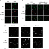
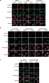
Figure 1.
Figure 1.
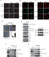
HGF binding to MET induces receptor clustering and patch formation on the plasma membrane. A, U373-MG cells were serum-starved and then stimulated with or without HGF (50 ng/ml) for 7 min. Cells were immunostained with an antibody against p-MET (Tyr-1234/1235–phosphorylated), followed with a secondary antibody conjugated to Alexa Fluor 647 (red). Cells were further immunostained with an anti-total MET antibody preconjugated to Alexa Fluor 488 (green). Arrows indicate the clusters of receptors on the plasma membrane. B, the MET clustering is sensitive to the MET inhibitor crizotinib. Cells were treated similarly as in A, except that cells were serum-starved and treated with 1 μm crizotinib or control (DMSO) for 3 h before the addition of HGF or left untreated. C, time course for MET cluster formation on the plasma membrane. Serum-starved cells were stimulated with HGF for the indicated time, fixed, and immunostained with the anti-p-MET (Tyr-1234/1235–phosphorylated) antibody. Scale bars in A–C, 20 μm.
Figure 2.
Figure 2.
HGF induces the activation and co-clustering of AXL and MET on plasma membrane. A, serum-starved U373-MG cells were left untreated (0) or treated with HGF (50 ng/ml) or GAS6 (400 ng/ml) for 7 min. The cells were processed for immunostaining with antibodies for either p-MET (Tyr-1234/1235–phosphorylated), total AXL (AXL), or p-AXL (Tyr-779–phosphorylated). Arrows indicate the clusters of receptors on the plasma membrane. B, co-clustering of the Tyr-779–phosphorylated AXL and MET on the plasma membrane. Serum-starved U373-MG cells were treated with HGF as in A and doubly immunostained with antibodies for p-AXL-Tyr-779 (red) and total MET (green) antibodies. Scale bar, 20 μm. C, co-clustering of AXL and MET on the plasma membrane. Cells were treated with HGF as in B but stained with antibodies for AXL (red) and MET (green). Cells were then examined by confocal microscopy under a higher-magnification objective (×40 oil objective, NA 1.3). Scale bar, 10 μm. D, HGF induces the direct interaction between MET and AXL on the cell surface. Serum-starved U373-MG cells were left untreated or treated with HGF (50 ng/ml) for 7 min. The cells were fixed and processed for the PLA using mouse anti-MET and rabbit anti-AXL antibodies. The white arrow indicates the PLA spots detected on the plasma membrane. Scale bar, 10 μm. The numbers of average spots per cells were quantified after counting 50 cells under each condition (means with S.D. are shown; ***, p < 0.001). E, serum-starved U373-MG cells were treated as in A. Western blot analysis was conducted using antibodies specific for the indicated proteins with actin as a loading control. Longer exposures were shown for the p140 kinase-dead (p140KD) and p120 kinase-dead (p120KD) forms of AXL, which are differentially phosphorylated in the HGF- or GAS6-treated samples. F, cells were treated similarly as in B with HGF, and cell lysates were subjected to immunoprecipitation with the mouse anti-phosphotyrosine mAb (p-Tyr-100)-conjugated resins, followed by Western blot analysis with antibodies specific for AXL (total AXL), p-AXL (Tyr-779–phosphorylated), MET (total MET), and p-MET (Tyr-1234/1235–phosphorylated) as indicated. G, same as F except that cell lysates were subjected to immunoprecipitation (IP) with control IgG and anti-MET antibodies. The immunocomplexes were Western blotted with antibodies specific for AXL (total AXL), p-AXL (Tyr-779–phosphorylated), MET (total MET), and p-MET (Tyr-1234/1235–phosphorylated) as indicated.
Figure 3.
Figure 3.
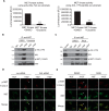
Regulation of MET and AXL RTK activities in the MET–AXL complexes. A, in vitro MET kinase activity assay using either poly(Glu4, Tyr1) (left) or AXL Tyr-779 peptide (right) as substrates. Reactions were carried out in the presence of DMSO (vehicle control) or the MET inhibitor crizotinib (2 μm). Phosphorylation products were measured using the ADP-Glo system and displayed as luminescence units. Assays were carried out in duplicates (shown are means with S.D. (error bars); ***, p < 0.001). B, MET kinase activity is required for the MET–AXL complex formation. Serum-starved U373-MG cells were pretreated with DMSO or 1 μm MET kinase inhibitor crizotinib for 3 h and then treated with or without HGF for 7 min. Cell lysates were used for immunoprecipitation (IP) with anti-MET antibodies and Western blotted with antibodies for AXL, p-AXL (Tyr-779–phosphorylated), MET, and p-MET (Tyr-1234/1235–phosphorylated) as indicated. C, the same as B except that the cells were pretreated with 1 μm AXL kinase inhibitor R428. D and E, AXL is required for MET clustering and activation. U373-MG cells were transfected with control (luciferase; Luc) or AXL siRNAs for 72 h, followed by serum starvation for 3 h (D). Alternatively, U373-MG cells were simultaneously serum-starved and treated with 1 μm AXL inhibitor R428 or DMSO control for 3 h (E). Cells were then stimulated with or without HGF for 7 min, followed by doubly immunostaining with antibodies for MET (total Met) and p-MET (Tyr-1234/1235–phosphorylated). Scale bars, 20 μm.
Figure 4.
Figure 4.
HGF induces clustering of MET and AXL in the presence of GAS6. A, top row, serum-starved U373-MG cells were treated with either HGF (50 ng/ml), GAS6 (400 ng/ml), or HGF (50 ng/ml) and GAS6 (400 ng/ml) together for 7 min. Bottom row, serum-starved cells were pretreated with various indicated concentrations of GAS6 for 7 min. HGF (50 ng/ml) was then added and incubated with the cells for an additional 7 min. The cells were processed for clustering analysis using anti-p-MET (Tyr-1234/1235–phosphorylated) antibodies. B, the serum-starved cells were treated with either HGF (50 ng/ml), GAS6 (400 ng/ml), or HGF (50 ng/ml) and GAS6 (400 ng/ml) together or with PBS as control for 7 min. The proteins were immunoprecipitated (IP) with anti-MET antibodies and Western blotted with anti-AXL, p-MET (Tyr-1234/1235–phosphorylated) and MET antibodies. C, serum-starved U373-MG cells were treated with HGF (50 ng/ml), GAS6 (400 ng/ml), or simultaneously with HGF and GAS6 together at the various concentrations as indicated for 7 min and then processed for p-MET clustering analysis as in A. D, the serum-starved cells were treated with GAS6 (400 ng/ml) or simultaneously GAS6 together with the indicated concentrations of HGF for 7 min. The proteins were analyzed by Western blotting. E, the same as in D, except that cells were treated simultaneously with HGF (50 ng/ml) together with the various concentrations of GAS6 for 7 min as indicated. F, starved cells were pretreated with various concentrations of GAS6 for 10 min, and then HGF (50 ng/ml) was subsequently added as indicated for a further 7 min. The cells were processed for Western blotting with the indicated antibodies.
Figure 5.
Figure 5.
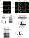
The HGF-activated MET–AXL RTK complex regulates cytoskeleton organization and RAC1 activation. A, U373-MG cells were serum-starved and treated with or without HGF for 7 min. Cells were immunostained with antibodies for p-MET (Tyr-1234/1235–phosphorylated) (red) and phalloidin (green) as indicated. B, U373-MG cells were transfected with control (luciferase; Luc) or RAC1 siRNAs for 72 h. The cells were then serum-starved for 3 h, subsequently stimulated with or without HGF, and immunostained with anti-MET (total MET) antibody (green) and phalloidin (magenta). C, HGF induces an increase of RAC1-GTP levels. Serum-starved U373-MG cells were treated with HGF for 7 min or left untreated, cell lysates were assayed for RAC1-GTP using the pulldown assay, and the starting material was also examined for total RAC1 by Western blotting. RAC1-GTP pulldown assays were conducted in duplicates, and the blots were scanned and quantified (means with S.D. (error bars) are shown; ***, p < 0.001). Cell lysates were also analyzed by Western blotting with the indicated antibodies (right). D, serum-starved U373-MG cells were pretreated with DMSO (control), 1 μm R428, or 1 μm crizotinib for 3 h and then processed for RAC1-GTP pulldown assay as in C. Quantification was conducted and normalized to the without-HGF (−HGF) sample in each pair. Statistical analysis was conducted for the indicated samples (***, p < 0.001; *, p < 0.05). E, U373-MG cells were transfected with control (luciferase) or AXL siRNAs for 72 h. Cells were then processed and assayed for the RAC1-GTP levels and the protein phosphorylation status as in C. Statistical analysis was conducted for each pair of samples (***, p < 0.001; **, p < 0.01).
Figure 6.
Figure 6.
ELMO2 is co-clustered with the activated MET in response to HGF. A, serum-starved U373-MG cells were treated with or without HGF for 7 min, and the cells were processed for double-immunostaining with antibodies for p-MET (Tyr-1234/1235–phosphorylated) (red) and ELMO2 (green) as indicated. B, serum-starved U373-MG cells were pretreated with DMSO or 1 μm MET kinase inhibitor crizotinib for 3 h and then treated with or without HGF. Cells were immunostained as in A. C, association of ELMO2 with AXL in the HGF-stimulated cells. U373-MG cells were treated as in A. Cell lysates were used for immunoprecipitation (IP) with anti-AXL antibodies or control IgG. The immunocomplexes were blotted with anti-MET or anti-ELMO2. The blot was then stripped and reblotted with anti-AXL antibodies. D, same as in C except that cell lysates were immunoprecipitated with anti-MET antibody and then blotted with anti-ELMO2 or anti-p-AXL (Tyr-779–phosphorylated) antibodies. The blots were then sequentially stripped and reblotted with anti-AXL, anti-p-MET (Tyr-1234/1235–phosphorylated), and anti-MET antibodies as indicated. E, same as in C except cell lysates were immunoprecipitated with anti-ELMO2 antibody and blotted with anti-MET and anti-ELMO2 antibodies as indicated. The blot was then stripped and reblotted with anti-AXL antibodies. F, serum-starved U373-MG cells were pretreated with DMSO or 1 μm MET kinase inhibitor crizotinib as in B. Cells were co-immunostained with anti-AXL (total AXL) (red) and anti-ELMO2 (green) antibodies. G, cells were treated as in F except for pretreatment with 1 μm AXL kinase inhibitor R428 and immunostained with anti-p-MET (Tyr-1234/1235–phosphorylated) (red) and anti-ELMO2 (green) antibodies as indicated. H, cells were transfected with control (luciferase; Luc) or AXL siRNAs for 72 h. Cells were serum-starved and then treated with or without HGF for 7 min. Cells were immunostained with anti-AXL (total AXL) (red) and anti-ELMO2 (green) antibodies as indicated.
Figure 7.
Figure 7.
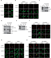
DOCK180 is recruited into the MET–AXL–ELMO2 complex in response to HGF. A, DOCK180 is associated with ELMO2 in HGF-stimulated cells. Serum-starved U373-MG cells were treated with or without HGF for 7 min. Cell lysates were used for immunoprecipitation (IP) with anti-ELMO2 antibodies. The immunocomplexes were blotted with anti-DOCK180 or anti-ELMO2 antibodies. Blots were then sequentially stripped and reblotted with MET and then AXL antibodies. B, serum-starved U373-MG cells were pretreated with DMSO or 1 μm crizotinib for 3 h and then treated with or without HGF for 7 min. Cell lysates were processed for immunoprecipitation with anti-ELMO2 antibody and blotted with the indicated antibodies. C, RAC1-GTP assay. U373-MG cells were transfected with control (luciferase; Luc), ELMO2, or DOCK180 siRNAs for 72 h; cells were starved for 3 h; and then cells were stimulated with or without HGF for 7 min. Cell lysates were assayed for RAC1-GTP using the pulldown assay as described in the legend to Fig. 5C, and results are quantified. Statistical analysis was conducted for each pair of samples as indicated (***, p < 0.001; **, p < 0.01; *, p < 0.05). Error bars, S.D. D, serum-starved U373-MG cells were pretreated with DMSO or 1 μm crizotinib for 3 h and then treated with or without HGF for 7 min. Cell lysates were processed for immunoprecipitation with anti-DOCK180 antibody and blotted with the indicated antibodies. E and F, same as in D except that cells were pretreated with 1 μm R428. Cell lysates were subjected to immunoprecipitation with anti-ELMO2 antibody (E) or anti-DOCK180 antibody (F) and Western blotted with anti-AXL, anti-ELMO2, anti-MET, and anti-DOCK180 antibodies as indicated. G and H, U373-MG cells were transfected with control (Luc) or AXL siRNAs for 72 h. Cells were serum-starved for 3 h and then stimulated with or without HGF for 7 min. Cell lysates were immunoprecipitated with anti-MET antibody (G) or anti-DOCK180 antibody (H). Immunoprecipitates were then examined by Western blot analysis with anti-ELMO2, anti-p-AXL (Tyr-779–phosphorylated) and anti-AXL antibodies as indicated.
Figure 8.
Figure 8.
Regulation of actin cytoskeleton by ELMO2 and DOCK180. A, MET and AXL kinase activities are required for the HGF-induced cytoskeleton reorganization. Serum-starved U373-MG cells were pretreated with DMSO, 1 μm crizotinib or 1 μm R428 for 3 h and then treated with or without HGF for 7 min. Cells were processed for co-immunostaining with anti-p-MET (Tyr-1234/1235–phosphorylated) (red) and anti-ELMO2 (green) antibodies as indicated. Cells were also counterstained with phalloidin (magenta). B, ELMO2, but not ELMO1, regulates cytoskeleton reorganization in response to HGF. U373-MG cells were transfected with control (luciferase; Luc), ELMO2, or ELMO1 siRNAs for 72 h. Cells were then serum-starved and stimulated with HGF as in A. Cells were co-immunostained with antibodies for p-MET (Tyr-1234/1235–phosphorylated) (red) and MET (total Met) (green) and then counterstained with phalloidin (magenta) as indicated. C, cells were transfected with control (luciferase) or DOCK180 siRNAs for 72 h and processed as in B. Cells were co-immunostained with antibodies for p-MET (Tyr-1234/1235–phosphorylated) (red) and ELMO2 (green) and then counterstained with phalloidin (magenta) as indicated.
Figure 9.
Figure 9.
AXL, ELMO2, DOCK180, and RAC1 are each required for HGF-induced cells migration and invasion. A, cell migration assay. Serum-starved U373-MG cells were pretreated with 1 μm crizotinib or 1 μm R428 for 3 h. An equal number of cells were then seeded in the Oris cell migration apparatus for 16 h before removal of the stopper. Cells were then treated with HGF (50 ng/ml) or left untreated for another 24 h. Cells were subsequently fixed, stained with DAPI, and imaged with a Nikon fluorescence microscope, and the numbers of cells that migrated into in the central circular areas were quantified using the ImageJ software. HGF-dependent cell migration was quantified (see “Experimental procedures” for details) and shown on the right (shown are the means with S.D. (error bars)). Statistical analysis was conducted for each of the drug-treated samples versus the DMSO control (***, p < 0.001). B, cells were transfected with siRNAs against MET, AXL, ELMO2, DOCK180, and RAC1 for 24 h and then used for a cell migration assay as described in A. HGF-dependent cell migration was quantified and shown. Statistical analysis was conducted for each condition versus luciferase siRNA (Luc) control (***, p < 0.001). Knockdown efficiency for each gene was verified by Western blot analysis with the corresponding antibodies. C, invasion assay using a Boyden chamber precoated with Matrigel. Cells were pretreated with either 1 μm Crizotinib or 1 μm R428 or specific siRNAs as in A and B. An equal number of cells were then seeded to the top chamber of the Boyden chamber, and the bottom chamber contained HGF (50 ng/ml). After 24 h, cells that migrated to the bottom chamber (invaded through Matrigel) were stained, imaged by an EVOS light microscope, and quantified using the ImageJ software. No cells invaded into the bottom chamber in the absence of HGF (data not shown). Assays were performed in triplicates, and the means (with S.D.) are shown on the right. Statistical analysis was conducted (compared with the corresponding control in each group) (***, p < 0.001). D, serum-starved U373-MG cells were pretreated with DMSO, 1 μm crizotinib, 1 μm R428, or 1 μm crizotinib together with 1 μm R428 for 3 h and then assayed for cell migration in the presence of either HGF (50 ng/ml), GAS6 (400 ng/ml), or HGF (50 ng/ml) and GAS6 (400 ng/ml) together. Cell migration assays were conducted as in A. Quantitation of the growth factor-stimulated cell migration is shown, after normalization to the DMSO control in the presence of HGF (shown are the means with S.D.). Statistical analyses were conducted for each condition, as compared with the DMSO control within each subgroup treated with the same growth factor(s) (***, p < 0.001; *, p < 0.05). E, cells were pretreated as in D and then subjected to the invasion assay as in C. Growth factors, HGF (50 ng/ml), GAS6 (400 ng/ml), or HGF (50 ng/ml) and GAS6 (400 ng/ml) together, were added to the lower chamber of the Boyden chamber. Quantitation of the cells that had invaded through the Matrigel is shown, after normalization to the DMSO control in the presence of HGF (shown are the means with S.D.). Statistical analysis was conducted for each condition, as compared with DMSO control within each subgroup with the indicated growth factor(s) (***, p < 0.001; *, p < 0.05).
Figure 10.
Figure 10.
HGF-dependent interaction and activation of MET and AXL in melanoma and breast cancer cells. A, serum-starved A375 or MDA-MB-231 cells were treated with or without HGF (50 ng/ml) for 7 min. Western blotting analyses were conducted using antibodies specific for the indicated proteins with actin as a loading control. B, serum-starved A375 cells were treated with or without HGF for 7 min. Cell lysates were used for immunoprecipitation (IP) with anti-MET antibodies and then blotted with anti-p-AXL (Tyr-779–phosphorylated), AXL, p-MET (Tyr-1234/1235–phosphorylated), MET, and ELMO2 antibodies as indicated. C, same as in B except that MDA-MB-231 cells were used for immunoprecipitation and Western blotting analyses. D, serum-starved MDA-MB-231 cells were pretreated with 1 μm crizotinib or 1 μm R428 for 3 h. Cells were assayed for cell migration using the Oris cell migration chamber as that described in the legend to Fig. 9A, either in the absence or presence of HGF (50 ng/ml). After 24 h, cells were fixed, stained with DAPI, and imaged with a Nikon fluorescence microscope. Quantitation of the HGF-dependent cell migration was conducted similarly as in Fig. 9A and shown on the right (shown are the means with S.D.). Statistical analysis was conducted for each of the drug-treated samples versus the DMSO control (***, p < 0.001). E, quantitation of the HGF-dependent cell migration of A375 cells. Cell migration assay of A375 cells was conducted similarly to that described for MDA-MB-231 cells in D and quantified. F and G, A375 and MDA-MB-231 cells were subjected to the cell invasion assays using a Boyden chamber precoated with Matrigel like that described as in the legend to Fig. 9C. Serum-starved A375 cells (top of F) or MDA-MB-231 cells (bottom of F) were pretreated with 1 μm crizotinib or 1 μm R428 for 3 h. Cells were then seeded to the top chamber of the Boyden chamber, and the bottom chamber contained HGF (50 ng/ml). After 24 h, cells that migrated to the bottom chamber (invaded through Matrigel) were stained and imaged. Assays were performed in triplicates, and quantification results are shown in G (means with S.D.). Statistical analysis was conducted (compared with the control) (***, p < 0.001). H, schematic model summarizing our results. HGF binds to the MET RTK to form the MET–AXL heterodimer on the plasma membrane, promoting the phosphorylation of Tyr-779 on the p140 isoform of AXL. The MET–AXL RTK signaling complex recruits ELMO2 and DOCK180 to activate the RAC1-dependent cell migration and invasion. This pathway is different from the HGF-dependent activation of MET homodimer, which leads to the activation of AKT and S6 kinases. It is also distinct from the GAS6-mediated AXL homodimerization that leads to the phosphorylation of Tyr-702 on the p120 isoform of the AXL RTK, leading to the ELMO1/RAC1-dependent cell migration.

Similar articles

Cited by

References

    1. Birchmeier C., Birchmeier W., Gherardi E., and Vande Woude G. F. (2003) Met, metastasis, motility and more. Nat. Rev. Mol. Cell Biol. 4, 915–925 10.1038/nrm1261 - DOI - PubMed
    1. Trusolino L., Bertotti A., and Comoglio P. M. (2010) MET signalling: principles and functions in development, organ regeneration and cancer. Nat. Rev. Mol. Cell Biol. 11, 834–848 10.1038/nrm3012 - DOI - PubMed
    1. Koochekpour S., Jeffers M., Rulong S., Taylor G., Klineberg E., Hudson E. A., Resau J. H., and Vande Woude G. F. (1997) Met and hepatocyte growth factor/scatter factor expression in human gliomas. Cancer Res. 57, 5391–5398 - PubMed
    1. Engelman J. A., Zejnullahu K., Mitsudomi T., Song Y., Hyland C., Park J. O., Lindeman N., Gale C. M., Zhao X., Christensen J., Kosaka T., Holmes A. J., Rogers A. M., Cappuzzo F., Mok T., et al. (2007) MET amplification leads to gefitinib resistance in lung cancer by activating ERBB3 signaling. Science 316, 1039–1043 10.1126/science.1141478 - DOI - PubMed
    1. Lin L., and Bivona T. G. (2012) Mechanisms of resistance to epidermal growth factor receptor inhibitors and novel therapeutic strategies to overcome resistance in NSCLC patients. Chemother. Res. Pract. 2012, 817297 - PMC - PubMed

Publication types

MeSH terms