. 2018 Aug 14;8(1):12119.
doi: 10.1038/s41598-018-30570-y.

Assessment of candidate elements for development of spectral photon-counting CT specific contrast agents

Affiliations

Assessment of candidate elements for development of spectral photon-counting CT specific contrast agents

Johoon Kim et al. Sci Rep. .

Abstract

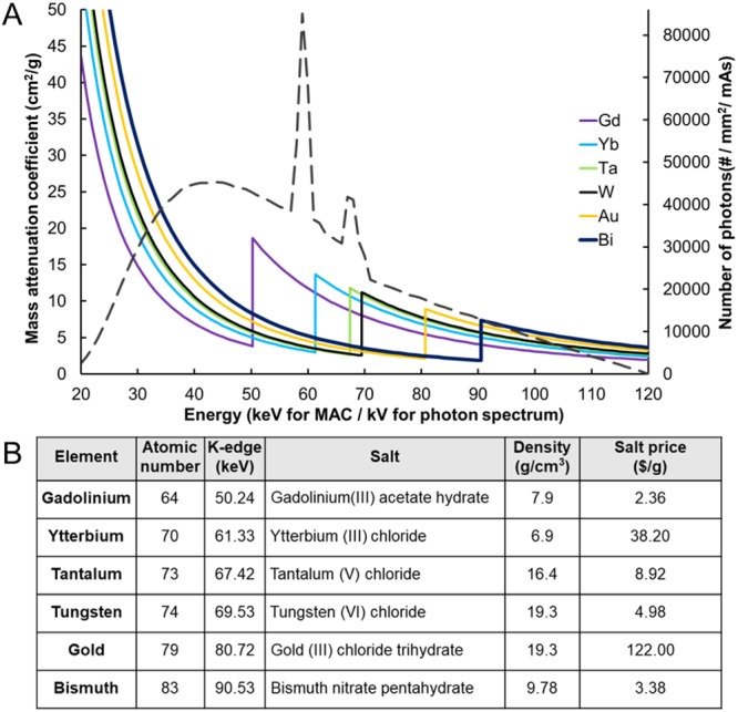
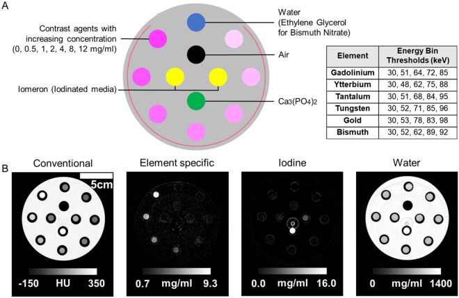
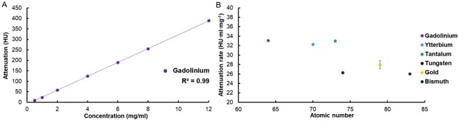
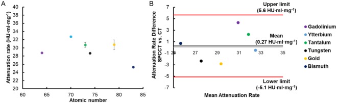
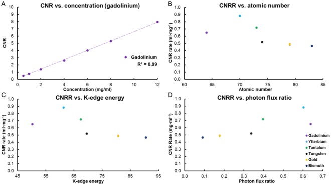
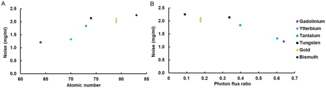
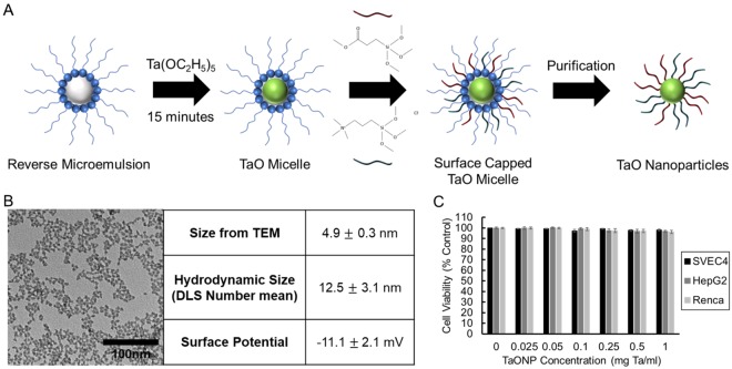
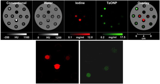
Spectral photon-counting computed tomography (SPCCT) is a rapidly emerging imaging modality that provides energy-dependent information on individual x-ray photons, leading to accurate material decomposition and simultaneous quantification of multiple contrast generating materials. Development of SPCCT-specific contrast agents is needed to overcome the issues with currently used iodinated contrast agents, such as difficulty in differentiation from calcified structures, and yield SPCCT's full promise. In this study, the contrast generation of different elements is investigated using a prototype SPCCT scanner based on a modified clinical CT system and suitable elements for novel contrast agent development for SPCCT imaging are identified. Furthermore, nanoparticles were synthesized from tantalum as a proof of concept spectral photon-counting CT agent and tested for their in vitro cytotoxicity and contrast generation to provide insight into the feasibility of nanoparticle contrast agent development from these elements. We found that gadolinium, ytterbium and tantalum generate high contrast in spectral photon-counting CT imaging and may be suitable elements for contrast agent development for this modality. Our proof of concept results with tantalum-based nanoparticles underscore this conclusion due to their detectability with spectral photon-counting CT, as well as their biocompatibility.


Conflict of interest statement

This work has been supported by a research grant from Philips (Dr. David P. Cormode). Dr. Philippe Coulon and Dr. Ira Blevis are employees of Philips Healthcare, the manufacturer of the scanner.

Figures

Figure 1
Figure 1
(A) Mass attenuation coefficients of various heavy metal elements and x-ray photon intensities at a tube voltage of 120 kV. The x-ray photon intensity spectrum (dotted line) was generated by using Spektr 3.0 (x-ray spectrum modeling software). (B) Characteristics of the six heavy elements used in this study. The salt prices of these elements were obtained from Sigma-Aldrich.
Figure 2
Figure 2
Specifications and photograph of the prototype photon-counting CT scanner used in this study.
Figure 3
Figure 3
(A) Schematic depiction of sample tube locations on the phantom and the energy bin thresholds used for each element. (B) Four images generated from SPCCT imaging of gadolinium. Conventional equivalent CT image, element-specific K-edge image of gadolinium, iodine image and water image (left to right).
Figure 4
Figure 4
(A) Attenuation of gadolinium at a range of concentrations. (B) The attenuation rates of the different elements studied. Error bars represent the standard error of the regression. For data points where the error bars are not visible, this is because the error value is very low and is obscured by the data point.
Figure 5
Figure 5
(A) The attenuation rates of the different elements studied in conventional CT. (B) Bland-Altman analysis of attenuation rate from CT and SPCCT systems. The same key for the elements is used in both A and B.
Figure 6
Figure 6
(A) CNR of gadolinium at a range of concentrations. (B) The CNRR of the different elements studied. (C) The CNRR of the different elements compared with their K-edge energies. (D) The CNRR of the elements compared to the ratio of the number of photons above and below their K-edge energies (based on the photon spectrum in the air shown in Fig. 1A). In B-D the same key is used. CNR and CNRR were calculated from the element specific images.
Figure 7
Figure 7
(A) Noise levels of the element specific images. (B) Noise level of the element specific images of the elements compared to the ratio of the number of photons above and below their K-edge energy.
Figure 8
Figure 8
(A) Schematic depiction of the TaONP synthesis process. (B) TEM of TaONP, size and surface potential of TaONP measured from TEM and DLS. (C) Effect of TaONP on cell viability after 8 hr of incubation.
Figure 9
Figure 9
SPCCT images of the phantom showing differentiation of TaONP ranging from 0 to 12 mg Ta/ml from an iodine contrast agent (2 and 5 mg/ml). Two images on the bottom row are the enlarged iodine and TaONP images centered at tubes with lower element concentrations.

Similar articles

Cited by

References

    1. Kim J, et al. Use of nanoparticle contrast agents for cell tracking with computed tomography. Bioconjug. Chem. 2017;28:1581–1597. doi: 10.1021/acs.bioconjchem.7b00194. - DOI - PMC - PubMed
    1. Cormode DP, et al. Atherosclerotic plaque composition: analysis with multicolor CT and targeted gold nanoparticles. Radiology. 2010;256:774–782. doi: 10.1148/radiol.10092473. - DOI - PMC - PubMed
    1. Leng S, et al. Spectral performance of a whole-body research photon counting detector CT: quantitative accuracy in derived image sets. Phys. Med. Biol. 2017;62:7216–7232. doi: 10.1088/1361-6560/aa8103. - DOI - PMC - PubMed
    1. Pourmorteza A, et al. Abdominal Imaging with Contrast-enhanced Photon-counting CT: First Human Experience. Radiology. 2016;279:239–245. doi: 10.1148/radiol.2016152601. - DOI - PMC - PubMed
    1. Schlomka JP, et al. Experimental feasibility of multi-energy photon-counting K-edge imaging in pre-clinical computed tomography. Phys. Med. Biol. 2008;53:4031–4047. doi: 10.1088/0031-9155/53/15/002. - DOI - PubMed

Publication types