

A Mixture of Atropisomers Enhances Neutral Lipid Degradation in Mammalian Cells with Autophagy Induction
- PMID: 30108268
- PMCID: PMC6092391
- DOI: 10.1038/s41598-018-30679-0
A Mixture of Atropisomers Enhances Neutral Lipid Degradation in Mammalian Cells with Autophagy Induction
Abstract
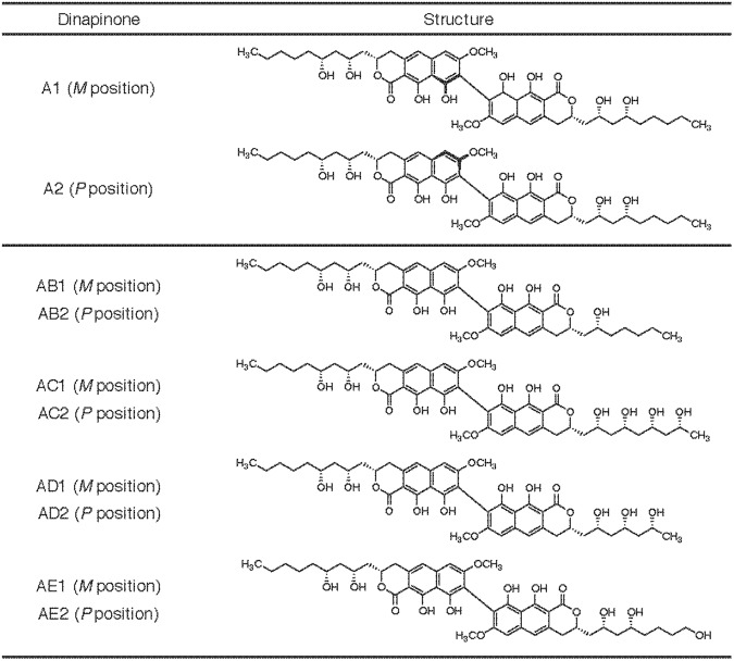
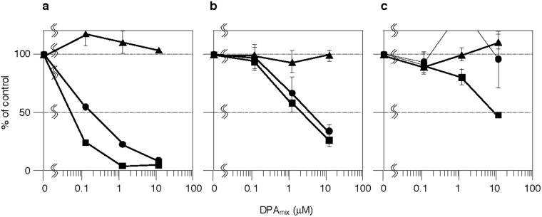
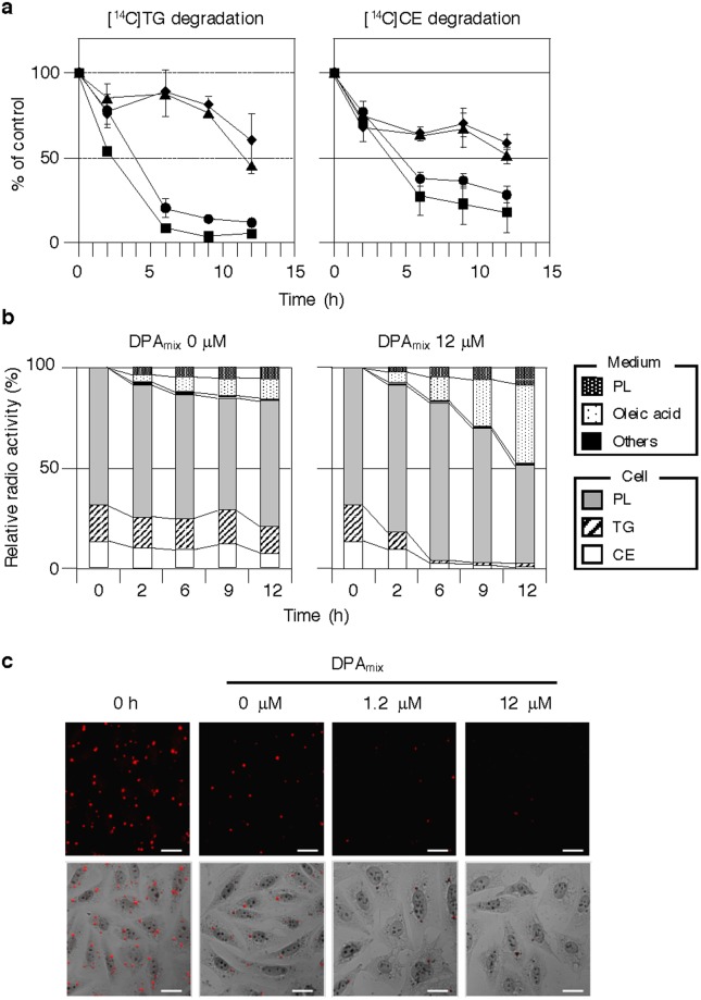
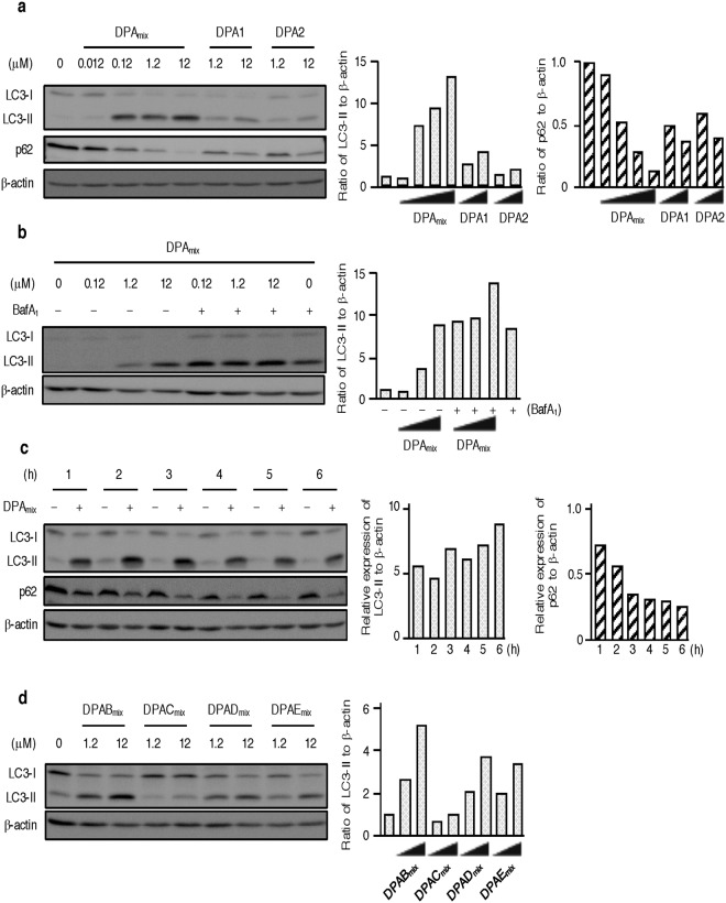
Atropisomers with a biaryl dihydronaphthopyranone structure, dinapinones A1 (DPA1) (M position) and A2 (DPA2) (P position), were isolated from the fungus culture broth of Talaromyces pinophilus FKI-3864 as inhibitors of [14C]neutral lipid ([14C]triacylglycerol (TG) and [14C]cholesteryl ester (CE)) synthesis from [14C]oleic acid in Chinese hamster ovary-K1 (CHO-K1) cells. DPA2 inhibited [14C]TG and [14C]CE synthesis (IC50s, 0.65 and 5.6 μM, respectively), but DPA1 had no inhibitory activity on [14C]TG and [14C]CE synthesis even at 12 μM. However, a 1:1 mixture of DPA1 and DPA2 (DPAmix) had the most potent inhibitory activity on [14C]TG and [14C]CE synthesis (IC50s, 0.054 and 0.18 μM, respectively). The mechanism of action of DPAmix was investigated. DPAmix had no effects on the enzymes involved in neutral lipid synthesis, while DPAmix enhanced the degradation of [14C]neutral lipids with concomitant decrease in cytosolic lipid droplets accumulated in CHO-K1 cells. From analysis of autophagy marker proteins, DPAmix caused dose-dependent induction of microtubule-associated protein light chain 3-II (LC3-II) and degradation of p62. In the autophagic flux assay using bafilomycin A1, DPAmix upregulated autophagosome turnover. These results reveal that DPAmix enhances neutral lipid degradation together with induction of autophagy.
Conflict of interest statement
The authors declare no competing interests.
Figures
Similar articles
-
New dinapinone derivatives, potent inhibitors of triacylglycerol synthesis in mammalian cells, produced by Talaromyces pinophilus FKI-3864.J Antibiot (Tokyo). 2013 Mar;66(3):179-89. doi: 10.1038/ja.2012.127. J Antibiot (Tokyo). 2013. PMID: 23532022
-
Dinapinones, novel inhibitors of triacylglycerol synthesis in mammalian cells, produced by Penicillium pinophilum FKI-3864.J Antibiot (Tokyo). 2011 Jul;64(7):489-94. doi: 10.1038/ja.2011.32. Epub 2011 May 11. J Antibiot (Tokyo). 2011. PMID: 21559025
-
Production of monapinones by fermentation of the dinapinone-producing fungus Penicillium pinophilum FKI-3864 in a seawater-containing medium.J Antibiot (Tokyo). 2011 Jul;64(7):503-8. doi: 10.1038/ja.2011.33. Epub 2011 May 25. J Antibiot (Tokyo). 2011. PMID: 21610716
-
The ménage à trois of autophagy, lipid droplets and liver disease.Autophagy. 2022 Jan;18(1):50-72. doi: 10.1080/15548627.2021.1895658. Epub 2021 Apr 2. Autophagy. 2022. PMID: 33794741 Free PMC article. Review.
-
Reserve lipids and plant autophagy.J Exp Bot. 2020 May 30;71(10):2854-2861. doi: 10.1093/jxb/eraa082. J Exp Bot. 2020. PMID: 32080724 Free PMC article. Review.
References
-
- Xu, S., Zhang, X. & Liu, P. Lipid droplet proteins and metabolic diseases. Biochim. Biophys. Acta, 10.1016/j.bbadis.2017.07.019 (2017). - PubMed
Publication types
MeSH terms
Substances
LinkOut - more resources
Full Text Sources
Other Literature Sources
Miscellaneous
