Review
. 2018 Oct;28(10):969-980.
doi: 10.1038/s41422-018-0078-7. Epub 2018 Aug 14.

Skeletal muscle mitochondrial remodeling in exercise and diseases

Affiliations
Review

Skeletal muscle mitochondrial remodeling in exercise and diseases

Zhenji Gan et al. Cell Res. 2018 Oct.

Abstract

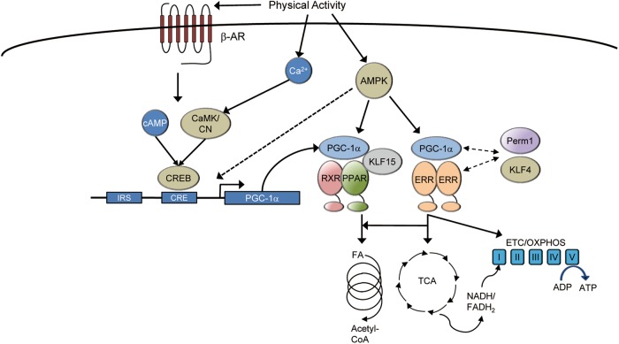
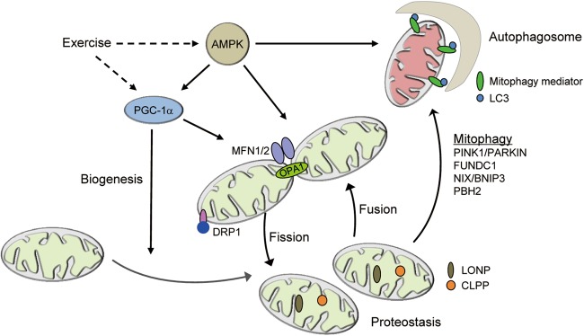
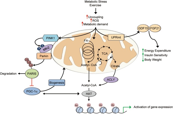
Skeletal muscle fitness and plasticity is an important determinant of human health and disease. Mitochondria are essential for maintaining skeletal muscle energy homeostasis by adaptive re-programming to meet the demands imposed by a myriad of physiologic or pathophysiological stresses. Skeletal muscle mitochondrial dysfunction has been implicated in the pathogenesis of many diseases, including muscular dystrophy, atrophy, type 2 diabetes, and aging-related sarcopenia. Notably, exercise counteracts the effects of many chronic diseases on skeletal muscle mitochondrial function. Recent studies have revealed a finely tuned regulatory network that orchestrates skeletal muscle mitochondrial biogenesis and function in response to exercise and in disease states. In addition, increasing evidence suggests that mitochondria also serve to "communicate" with the nucleus and mediate adaptive genomic re-programming. Here we review the current state of knowledge relevant to the dynamic remodeling of skeletal muscle mitochondria in response to exercise and in disease states.


Conflict of interest statement

The authors declare no competing interests.

Figures

Fig. 1
Fig. 1
Integration of upstream signaling pathways with mitochondrial biogenesis in the muscle. Multiple signaling pathways in the skeletal muscle serve to transmit changes in physical activity or other extracellular signals to mitochondrial biogenesis. Many of these pathways directly activate the peroxisome-proliferator activated receptor γ (PPARγ) coactivator-1α (PGC-1α). PGC-1α interacts directly with its effector nuclear receptors such as the estrogen-related receptor (ERR) and the peroxisome-proliferator activated receptor (PPAR) to regulate genes involved in virtually all aspects of mitochondrial energy metabolism. CaMK calmodulin-dependent kinase, CN calcineurin, AMPK AMP-dependent kinase, β-AR beta adrenergic receptor, IRS insulin response sequence, CRE cAMP response element, CREB cAMP response element binding protein, RXR retinoid X receptor, KLF Krüppel-like factor, FA fatty acid, ETC electron transport chain, OXPHOS oxidative phosphorylation
Fig. 2
Fig. 2
Integration of mitochondrial quality control and biogenesis. Muscle mitochondria undergo extensive turnover and remodeling in response to a variety of physiological stimuli. PGC-1α activates mitochondrial biogenesis. Mitochondrial fission is mediated by DRP1 and its receptors. Mitochondrial fusion is mediated by MFN1/2 for the fusion of the outer membrane and OPA1 for the inner membranes. The main mitochondria matrix proteases such as LONP and CLPP maintain mitochondrial proteostasis and regulate mitochondrial function. The damaged or dysfunctional mitochondria are cleared via mitophagy. Mitophagy is mediated by mitophagy mediators such as PINK1/PARKIN, FUNDC1, NIX/BNIP3, and PBH2 in mammals. Exercise coordinately regulates mitochondrial biogenesis, mitochondrial dynamics, and mitochondrial quality-control program via AMPK and the PGC-1α. Mfn1/2 mitofusin 1/2, Drp1 dynamin-related protein 1, OPA1 optic atrophy 1, LONP Lon peptidase 1, CLPP caseinolytic mitochondrial matrix peptidase proteolytic subunit, PINK1 PTEN-induced putative kinase 1, FUNDC1 FUN14 domain containing 1, Bnip3 BCL2 interacting protein 3
Fig. 3
Fig. 3
Skeletal muscle mitochondria serve as cellular sensors of metabolic demand and stress. Various forms of metabolic stress including exercise triggers responses in muscle mitochondria that result in signals to other cellular compartments or tissues. Increased mitophagy is integrated with biogenesis through activation of PGC-1α, either directly or through mechanisms such as PARIS. Activation of gene expression by PGC-1a also occurs in cooperation with histone acetyltransferases (HAT). Acetyl-CoA generated by increased mitochondrial respiration can donate acetyl groups for histone modifications. Paracrine or endocrine signals can also be generated, at least in part, by activation of the mitochondrial unfolded protein response (UPRmt) that leads to increases in GDF15 and FGF21 production and secretion. PARIS Parkin interacting substrate, ACLY ATP citrate lyase, HAT histone acetyltransferase, Ac acetyl, Ub ubiquitin, FA fatty acid, UPRmt mitochondrial unfolded protein response

References

    1. Glancy B, et al. Mitochondrial reticulum for cellular energy distribution in muscle. Nature. 2015;523:617–620. - PMC - PubMed
    1. Egan B, Zierath JR. Exercise metabolism and the molecular regulation of skeletal muscle adaptation. Cell Metab. 2013;17:162–184. - PubMed
    1. Issemann I, Green S. Activation of a member of the steroid hormone receptor superfamily by peroxisome proliferators. Nature. 1990;347:645–650. - PubMed
    1. Gulick T, Cresci S, Caira T, Moore DD, Kelly DP. The peroxisome proliferator-activated receptor regulates mitochondrial fatty acid oxidative enzyme gene expression. Proc. Natl. Acad. Sci. USA. 1994;91:11012–11016. - PMC - PubMed
    1. Brandt JM, Djouadi F, Kelly DP. Fatty acids activate transcription of the muscle carnitine palmitoyltransferase I gene in cardiac myocytes via the peroxisome proliferator-activated receptor alpha. J. Biol. Chem. 1998;273:23786–23792. - PubMed

Publication types

Substances