

Panic results in unique molecular and network changes in the amygdala that facilitate fear responses
- PMID: 30108314
- PMCID: PMC6410355
- DOI: 10.1038/s41380-018-0119-0
Panic results in unique molecular and network changes in the amygdala that facilitate fear responses
Abstract
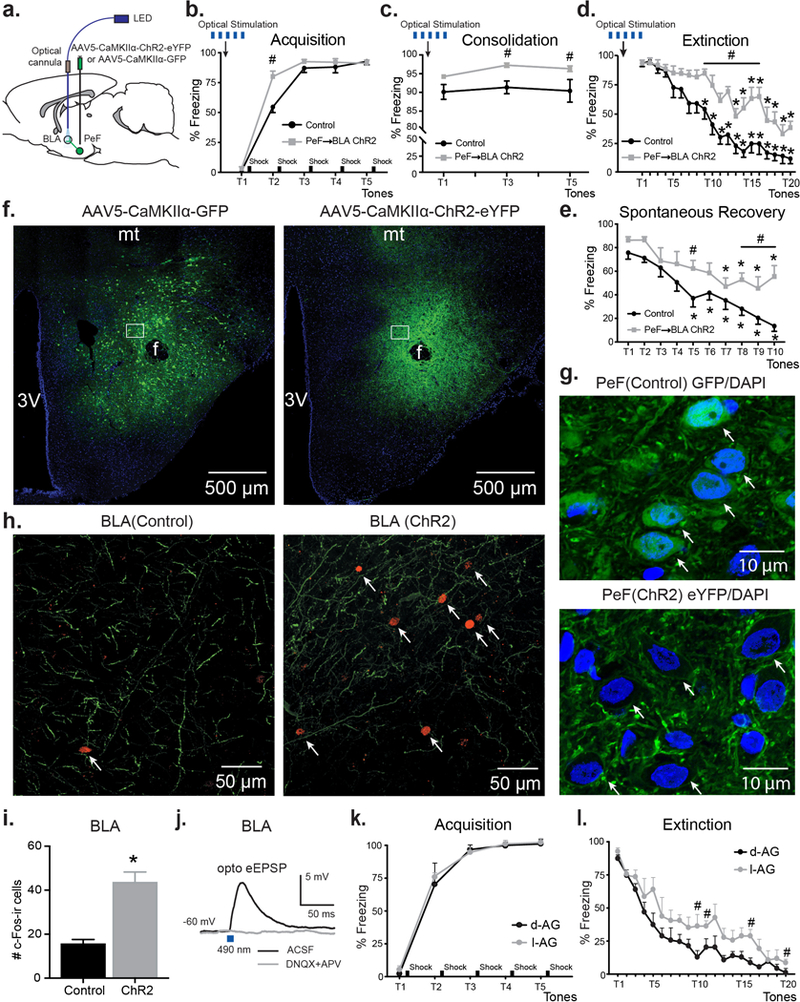
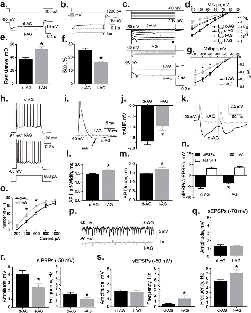
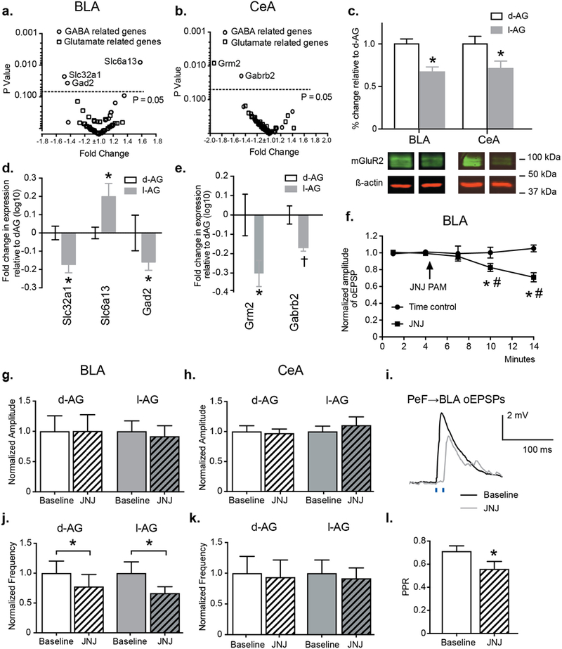
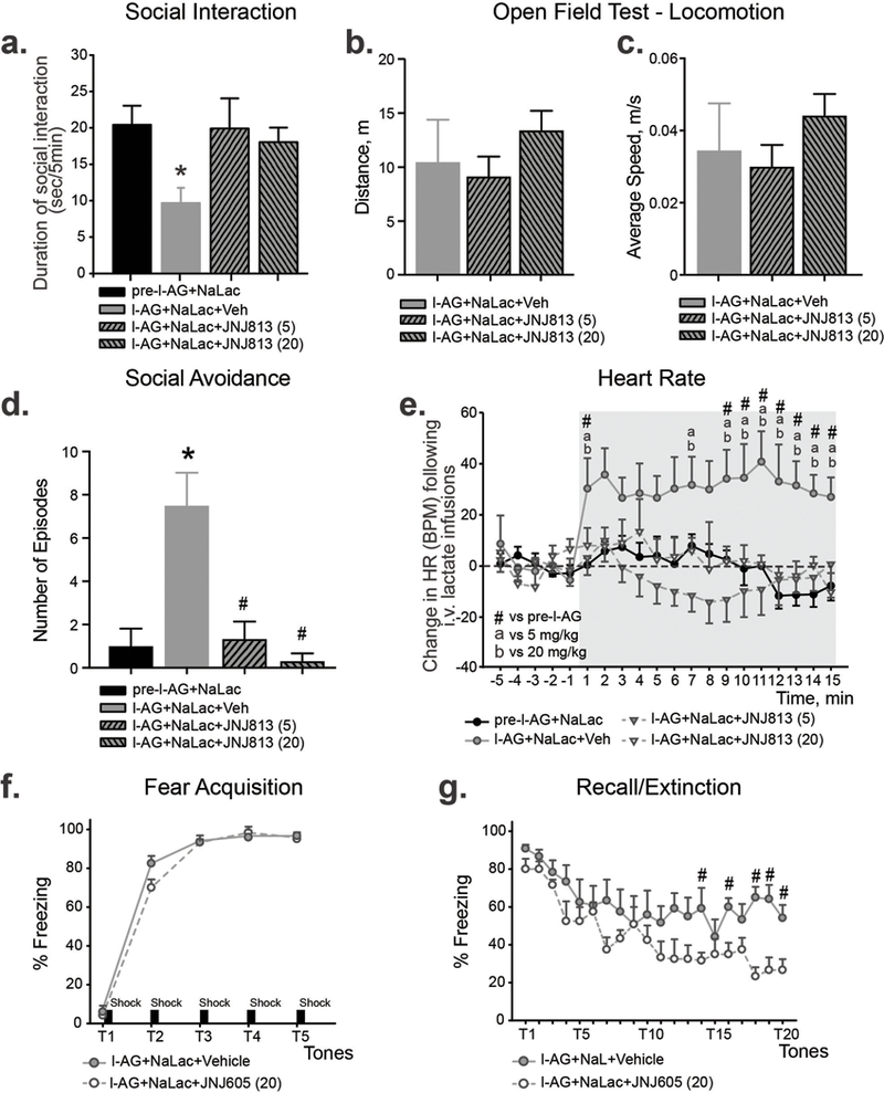
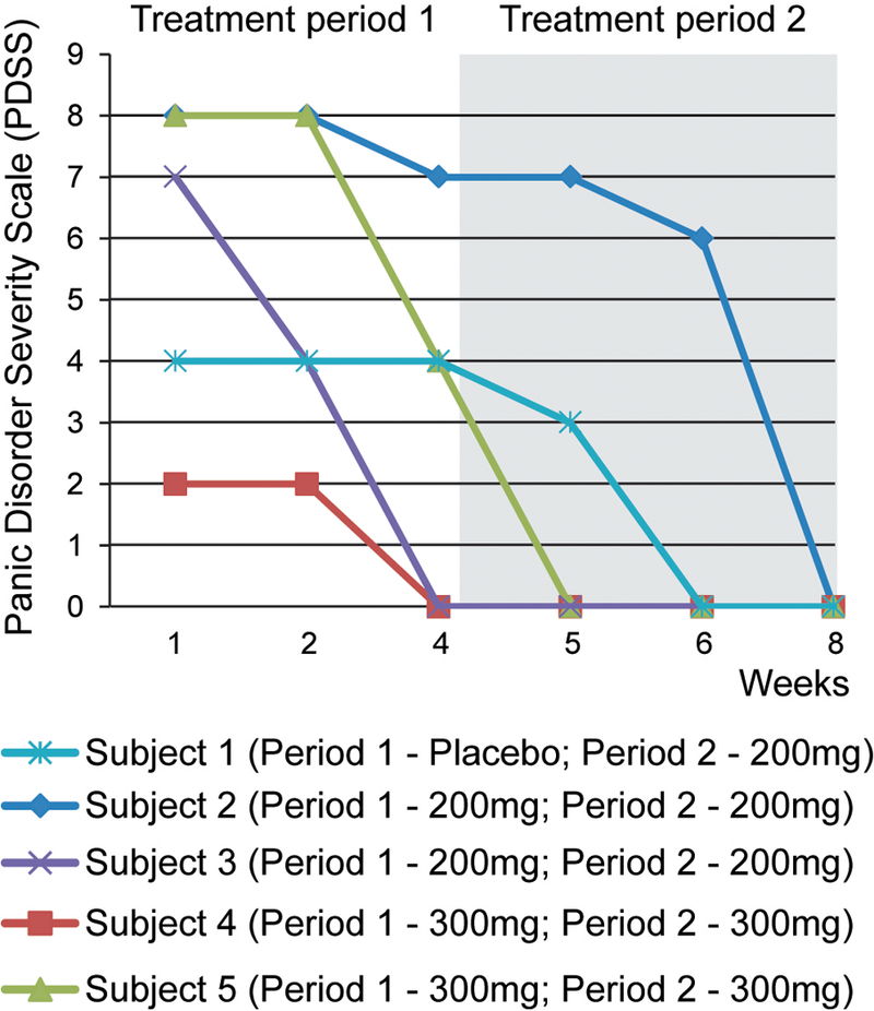
Recurrent panic attacks (PAs) are a common feature of panic disorder (PD) and post-traumatic stress disorder (PTSD). Several distinct brain regions are involved in the regulation of panic responses, such as perifornical hypothalamus (PeF), periaqueductal gray, amygdala and frontal cortex. We have previously shown that inhibition of GABA synthesis in the PeF produces panic-vulnerable rats. Here, we investigate the mechanisms by which a panic-vulnerable state could lead to persistent fear. We first show that optogenetic activation of glutamatergic terminals from the PeF to the basolateral amygdala (BLA) enhanced the acquisition, delayed the extinction and induced the persistence of fear responses 3 weeks later, confirming a functional PeF-amygdala pathway involved in fear learning. Similar to optogenetic activation of PeF, panic-prone rats also exhibited delayed extinction. Next, we demonstrate that panic-prone rats had altered inhibitory and enhanced excitatory synaptic transmission of the principal neurons, and reduced protein levels of metabotropic glutamate type 2 receptor (mGluR2) in the BLA. Application of an mGluR2-positive allosteric modulator (PAM) reduced glutamate neurotransmission in the BLA slices from panic-prone rats. Treating panic-prone rats with mGluR2 PAM blocked sodium lactate (NaLac)-induced panic responses and normalized fear extinction deficits. Finally, in a subset of patients with comorbid PD, treatment with mGluR2 PAM resulted in complete remission of panic symptoms. These data demonstrate that a panic-prone state leads to specific reduction in mGluR2 function within the amygdala network and facilitates fear, and mGluR2 PAMs could be a targeted treatment for panic symptoms in PD and PTSD patients.
Conflict of interest statement
CONFLICT OF INTERESTS
Drs. L Ver Donck, M Ceusters and JM Kent are employees of Janssen. This work was supported by research grants to Indiana University with (Drs. Shekhar, Molosh and Johnson as PIs) from Janssen, but these authors have no other commercial conflicts.
Figures
Similar articles
-
Group II metabotropic glutamate receptors in anxiety circuitry: correspondence of physiological response and subcellular distribution.J Comp Neurol. 2007 Dec 20;505(6):682-700. doi: 10.1002/cne.21525. J Comp Neurol. 2007. PMID: 17948876
-
Group II metabotropic glutamate receptor activation in the basolateral amygdala mediates individual differences in stress-induced changes in rapid eye movement sleep.Prog Neuropsychopharmacol Biol Psychiatry. 2021 Jan 10;104:110014. doi: 10.1016/j.pnpbp.2020.110014. Epub 2020 Jun 10. Prog Neuropsychopharmacol Biol Psychiatry. 2021. PMID: 32534177 Free PMC article.
-
mGluR2/3 in the Lateral Amygdala is Required for Fear Extinction: Cortical Input Synapses onto the Lateral Amygdala as a Target Site of the mGluR2/3 Action.Neuropsychopharmacology. 2015 Dec;40(13):2916-28. doi: 10.1038/npp.2015.145. Epub 2015 May 25. Neuropsychopharmacology. 2015. PMID: 26081171 Free PMC article.
-
From the neurobiology of extinction to improved clinical treatments.Depress Anxiety. 2014 Apr;31(4):279-90. doi: 10.1002/da.22214. Epub 2013 Nov 20. Depress Anxiety. 2014. PMID: 24254958 Free PMC article. Review.
-
Neurochemical and genetic factors in panic disorder: a systematic review.Transl Psychiatry. 2024 Jul 18;14(1):294. doi: 10.1038/s41398-024-02966-0. Transl Psychiatry. 2024. PMID: 39025836 Free PMC article.
Cited by
-
Understanding Emotions: Origins and Roles of the Amygdala.Biomolecules. 2021 May 31;11(6):823. doi: 10.3390/biom11060823. Biomolecules. 2021. PMID: 34072960 Free PMC article. Review.
-
Orexin/hypocretin receptor modulation of anxiolytic and antidepressive responses during social stress and decision-making: Potential for therapy.Brain Res. 2020 Mar 15;1731:146085. doi: 10.1016/j.brainres.2018.12.036. Epub 2018 Dec 24. Brain Res. 2020. PMID: 30590027 Free PMC article. Review.
-
Identification of a novel perifornical-hypothalamic-area-projecting serotonergic system that inhibits innate panic and conditioned fear responses.Transl Psychiatry. 2024 Jan 25;14(1):60. doi: 10.1038/s41398-024-02769-3. Transl Psychiatry. 2024. PMID: 38272876 Free PMC article.
-
Single administration of a psychedelic [(R)-DOI] influences coping strategies to an escapable social stress.Neuropharmacology. 2024 Jul 1;252:109949. doi: 10.1016/j.neuropharm.2024.109949. Epub 2024 Apr 16. Neuropharmacology. 2024. PMID: 38636726 Free PMC article.
-
Advances in translating mGlu2 and mGlu3 receptor selective allosteric modulators as breakthrough treatments for affective disorders and alcohol use disorder.Pharmacol Biochem Behav. 2022 Sep;219:173450. doi: 10.1016/j.pbb.2022.173450. Epub 2022 Aug 18. Pharmacol Biochem Behav. 2022. PMID: 35988792 Free PMC article. Review.
References
-
- Stein DJ, Bouwer C. A neuro-evolutionary approach to the anxiety disorders. JAnxietyDisord 1997; 11(4): 409–429. - PubMed
-
- DSM- V. Diagnostic and Statistical Manual - Fifth Edn. (DSM - V) American Psychiatric Association: Washington, DC, 2013.
-
- Rasche D, Foethke D, Gliemroth J, Tronnier VM. [Deep brain stimulation in the posterior hypothalamus for chronic cluster headache. Case report and review of the literature]. Schmerz 2006; 20(5): 439–444. - PubMed
-
- Wilent WB, Oh MY, Buetefisch C, Bailes JE, Cantella D, Angle C et al. Mapping of microstimulation evoked responses and unit activity patterns in the lateral hypothalamic area recorded in awake humans. Technical note. Journal of neurosurgery 2011; 115(2): 295–300. - PubMed
-
- Wilent WB, Oh MY, Buetefisch CM, Bailes JE, Cantella D, Angle C et al. Induction of panic attack by stimulation of the ventromedial hypothalamus. Journal of neurosurgery 2010; 112(6): 1295–1298. - PubMed
Publication types
MeSH terms
Substances
Grants and funding
LinkOut - more resources
Full Text Sources
Other Literature Sources
Miscellaneous
