. 2018 Jul 31:12:527.
doi: 10.3389/fnins.2018.00527. eCollection 2018.

An Oral Combination of Vitamins A, C, E, and Mg++ Improves Auditory Thresholds in Age-Related Hearing Loss

Affiliations

An Oral Combination of Vitamins A, C, E, and Mg++ Improves Auditory Thresholds in Age-Related Hearing Loss

Juan C Alvarado et al. Front Neurosci. .

Abstract

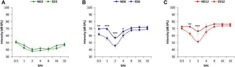
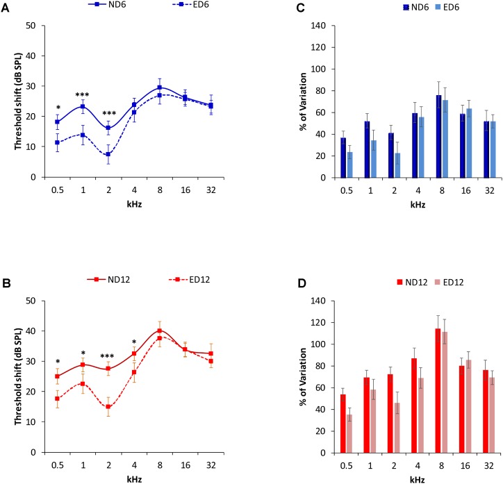
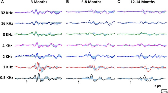
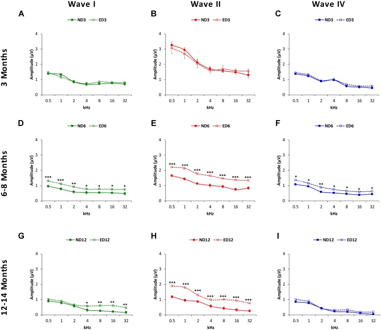
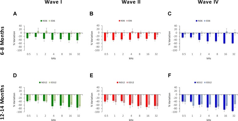
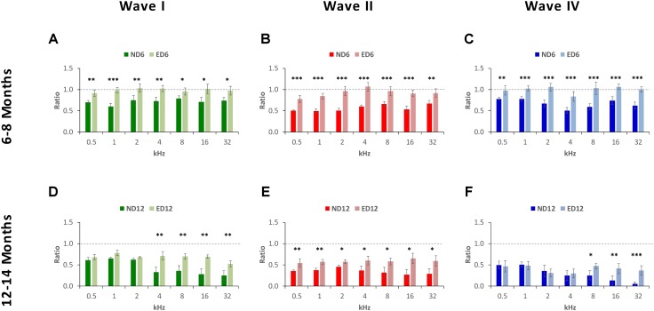
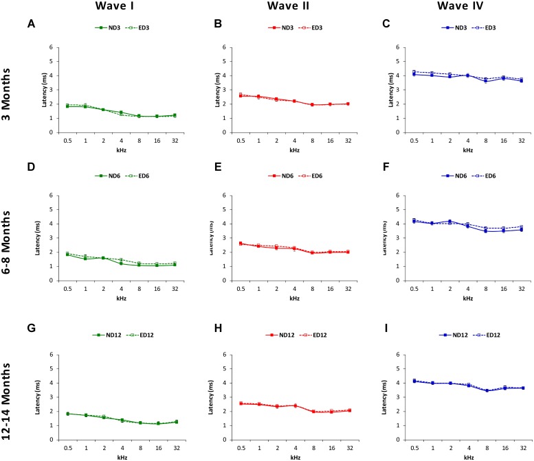
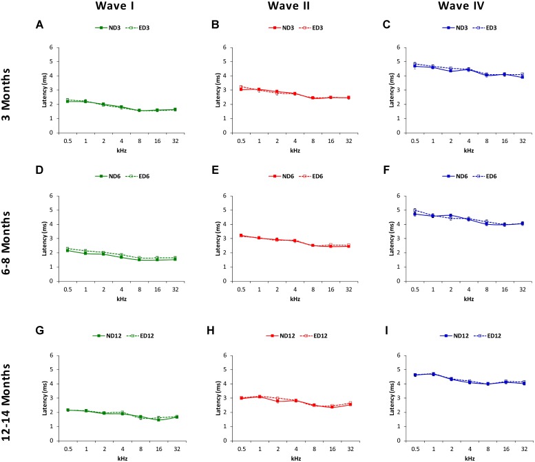
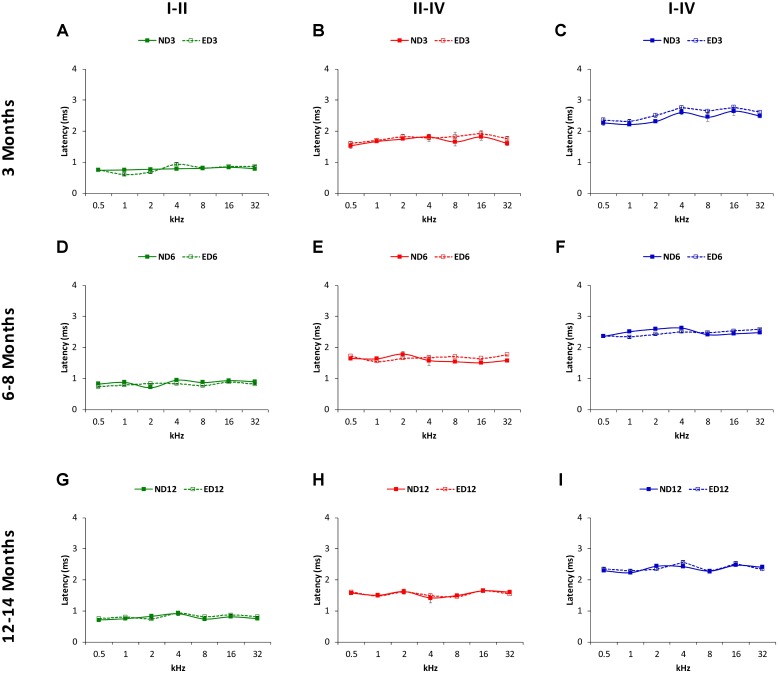
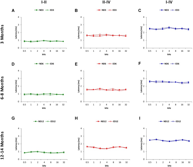
The increasing rate of age-related hearing loss (ARHL), with its subsequent reduction in quality of life and increase in health care costs, requires new therapeutic strategies to reduce and delay its impact. The goal of this study was to determine if ARHL could be reduced in a rat model by administering a combination of antioxidant vitamins A, C, and E acting as free radical scavengers along with Mg++, a known powerful cochlear vasodilator (ACEMg). Toward this goal, young adult, 3 month-old Wistar rats were divided into two groups: one was fed with a diet composed of regular chow ("normal diet," ND); the other received a diet based on chow enriched in ACEMg ("enhanced diet," ED). The ED feeding began 10 days before the noise stimulation. Auditory brainstem recordings (ABR) were performed at 0.5, 1, 2, 4, 8, 16, and 32 kHz at 3, 6-8, and 12-14 months of age. No differences were observed at 3 months of age, in both ND and ED animals. At 6-8 and 12-14 months of age there were significant increases in auditory thresholds and a reduction in the wave amplitudes at all frequencies tested, compatible with progressive development of ARHL. However, at 6-8 months threshold shifts in ED rats were significantly lower in low and medium frequencies, and wave amplitudes were significantly larger at all frequencies when compared to ND rats. In the oldest animals, differences in the threshold shift persisted, as well as in the amplitude of the wave II, suggesting a protective effect of ACEMg on auditory function during aging. These findings indicate that oral ACEMg may provide an effective adjuvant therapeutic intervention for the treatment of ARHL, delaying the progression of hearing impairment associated with age.

Keywords: age-related hearing loss; aging; auditory brainstem response; hearing loss; magnesium; micronutrients; presbycusis; vitamins.


Figures

FIGURE 1
FIGURE 1
Line graphs illustrating auditory thresholds at different frequencies in ND and ED rats at 3 (A), 6–8 (B), and 12–14 (C) months of age. In both ND and ED animals, mean threshold values rose as the age of the animals increased (B,C). However, at lower and medium frequencies the mean auditory thresholds in the ED6 (B) and ED12 (C) groups, were lower, closer to normal thresholds, than those observed in the ND group.
FIGURE 2
FIGURE 2
Auditory threshold shifts (line graphs) and percentage of variation (bar graphs) between ND and ED rats at 6–8 (A,C) and 12–14 months (B,D). At 6–8 (A) and at 12–14 (B) months of age, threshold shifts in ED animals were smaller than those in ND animals in the lower and medium frequencies. No significant differences were observed at higher frequencies. The percentage of variation of the threshold shifts at 6–8 months of age (C), were smaller in ED rats compared to those in ND rats, being 13.35, 17.74, and 18.77%, at 0.5, 1, and 2 kHz respectively. In the remaining frequencies, differences ranged from –4.64 to 4.28% (C). In 12–14 month-old animals (D), the percentage of variation of the threshold shifts in ED rats were smaller than those in ND rats. Percentages were 18.43, 11.08, 26.19, and 18.13% at 0.5, 1, 2, and 4 kHz; respectively, while at higher frequencies the differences ranged from –6.94 to 5.56%. p < 0.05, ∗∗∗p < 0.001.
FIGURE 3
FIGURE 3
Line graphs showing examples of ABR recordings from ND (closed dashed lines) and ED (open solid lines) rats at all ages evaluated. In ND and ED rats at 3 months of age (A), traces showed the characteristic 4 to 5 evoked waves after stimulus onset. No differences were apparent between groups. At 6–8 months of age (B), there was a reduction in the amplitude of all waves at all frequencies, although it was more evident in ND rats. At 12–14 month of age (C), despite the fact that reduction in the wave amplitudes was even more pronounced than that seen at 6–8 months, amplitudes in ND animals were still smaller than in ED rats. Arrows indicate stimulus onset.
FIGURE 4
FIGURE 4
Line graphs depicting wave amplitudes (in μV) plotted as a function of frequency in ND (solid lines) and ED (dashed lines) rats at the different ages evaluated. At 3 months of age (A–C), the mean values of the largest waves (I, II, and IV) were similar in both groups, with larger wave amplitudes in the lower frequencies and smaller at medium and higher frequencies, being wave II the largest of all. In 6–8-month-old rats (D–F), the mean amplitudes of all waves were reduced, but values in ED rats were significantly larger compared to ND rats. At 12–14 months (G–I), while the reduction in the mean amplitudes of all waves persisted, in the ED group values of wave II at all frequencies (H) and of wave I at higher frequencies (G) were still larger than those observed in ND animals. No differences were observed in wave IV (I). p < 0.05, ∗∗p < 0.01, ∗∗∗p < 0.001.
FIGURE 5
FIGURE 5
Bar graphs illustrating the percentage of variation in the wave amplitudes in older rats relative to the control condition (3-month-old rats). At 6–8 months of age, the percentage of variation in the wave amplitudes in ND rats was greater than in ED rats. These values in ND rats ranged from –23.48 to –42.11% for wave I (A), from –32.00% to –47.07% for wave II (B) and from –26.31 to 60.51% for wave IV (C), while in ED rats they ranged from –8.36 to 11.44% for wave I (A), from –11.22 to 7.81% for wave II (B), and from –10.30 to 8.60% for wave IV (C). At 12–14 months of age, while variations relative to controls were larger, differences between both groups were reduced. In ND rats, these values fluctuated from –36.52 to –71.58% for wave I (D), from –35.71 to –71.19% for wave II (E) and from –39.91 to 94.30% for wave IV (F). Meanwhile, in ED rats, they ranged from –32.67 to 67.84% for wave I (D), from 31.46 to 65.95% for wave II (E) and from –41.78 to 68.40% for wave IV (F).
FIGURE 6
FIGURE 6
Bar graphs illustrating the wave amplitude ratio in older animals relative to the control condition. In ND rats at 6–8 months of age, the wave amplitude ratios for waves I (A), II (B), and IV (C) were smaller at all frequencies when compared to ED rats. In ND rats at 12–14 months of age, compared to ED rats, ratios were still smaller for wave II (E) at all frequencies assessed and for waves I (D) and for wave IV (F) at the higher frequencies. p < 0.05, ∗∗p < 0.01, ∗∗∗p < 0.001.
FIGURE 7
FIGURE 7
Line graphs illustrating absolute latencies of the positive peaks (ms) of waves I, II and IV plotted as a function of frequency in ND and ED rats. At 3 months of age (A–C), the mean values for the absolute positive latencies were similar between ND and ED rats. At 6–8 months of age (D–F), absolute latency times were longer, compared to 3 month-old rats. However, there were no differences between ND and ED rats in any wave at any frequency assessed. Similarly, although the absolute latency times were even longer at 12–14 months of age (G–I), still no differences were observed in any wave at any frequency in the positive latency times when ND and ED rats were compared.
FIGURE 8
FIGURE 8
Line graphs illustrating absolute negative latencies (in ms) of waves I, II, and IV plotted as a function of frequency in ND and ED rats. The mean values for the absolute negative latencies in ND3 and ED3 rats (A–C) did not differ signficantly between them. Negative absolute latency times were longer at 6–8 months of age (D–F), but there were no differences between ND6 and ED6 animals in any wave at any frequency assessed. Despite of the longer absolute negative latency times at 12–14 months of age (G–I), still no differences were observed in any wave at any frequency when ND12 and ED12 rats where compared.
FIGURE 9
FIGURE 9
Line graphs illustrating interpeak positive latencies (in ms) plotted as a function of frequency in ND and ED rats. Regardless the age of the animal, the mean values of the interpeak positive latencies observed at 3 (A–C), 6–8 (D–F), and 12–14 (G–I) months were similar in ND and ED rats at all frequencies evaluated.
FIGURE 10
FIGURE 10
Line graphs illustrating interpeak negative latencies (in ms) plotted as a function of frequency in ND and ED rats. Similar to interpeak positive latencies, no significant differences were observed at any stimulus frequency, when ND3 vs. ED3 (A–C), ND6 vs. ED6 (D–F) and ND12 vs. ED12 (G–I) were compared.

References

    1. Alvarado J. C., Fuentes-Santamaría V., Gabaldón-Ull M. C., Blanco J. L., Juiz J. M. (2014). Wistar rats: a forgotten model of age-related hearing loss. Front. Aging Neurosci. 6:29. 10.3389/fnagi.2014.00029 - DOI - PMC - PubMed
    1. Alvarado J. C., Fuentes-Santamaría V., Gabaldón-Ull M. C., Jareño-Flores T., Miller J. M., Juiz J. M. (2016). Noise-induced “Toughening” effect in wistar rats: enhanced auditory brainstem responses are related to calretinin and nitric oxide synthase upregulation. Front. Neuroanat. 10:19 10.3389/fnana.2016.00019 - DOI - PMC - PubMed
    1. Alvarado J. C., Fuentes-Santamaría V., Jareño-Flores T., Blanco J. L., Juiz J. M. (2012). Normal variations in the morphology of auditory brainstem response (ABR) waveforms: a study in wistar rats. Neurosci. Res. 73 302–311. 10.1016/j.neures.2012.05.001 - DOI - PubMed
    1. Alvarado J. C., Fuentes-Santamaría V., Melgar-Rojas P., Gabaldón-Ull M. C., Juiz J. M. (2015a). Short-Duration and Long-Term Noise Overstimulation Could Increase Hearing Loss in Aging Wistar Rat. SENC Communications Abstracts Book. Available at: https://www.congresomovil.com/senc2015
    1. Alvarado J. C., Fuentes-Santamaría V., Melgar-Rojas P., Valero M. L., Gabaldón-Ull M. C., Miller J. M., et al. (2015b). Synergistic effects of free radical scavengers and cochlear vasodilators: a new otoprotective strategy for age-related hearing loss. Front. Aging Neurosci. 7:86. 10.3389/fnagi.2015.00086 - DOI - PMC - PubMed

LinkOut - more resources