. 2018 Jul 31:12:200.
doi: 10.3389/fncel.2018.00200. eCollection 2018.

PolyQ Tract Toxicity in SCA1 is Length Dependent in the Absence of CAG Repeat Interruption

Affiliations

PolyQ Tract Toxicity in SCA1 is Length Dependent in the Absence of CAG Repeat Interruption

Suran Nethisinghe et al. Front Cell Neurosci. .

Abstract

Spinocerebellar ataxia type 1 (SCA1) is an autosomal dominant neurodegenerative disorder caused by an expansion of a polyglutamine tract within the ATXN1 gene. Normal alleles have been reported to range from 6 to 35 repeats, intermediate alleles from 36 to 38 repeats and fully penetrant pathogenic alleles have at least 39 repeats. This distribution was based on relatively few samples and the narrow intermediate range makes the accuracy of the repeat sizing crucial for interpreting and reporting diagnostic tests, which can vary between laboratories. Here, we examine the distribution of 6378 SCA1 chromosomes and identify a very late onset SCA1 family with a fully penetrant uninterrupted pathogenic allele containing 38 repeats. This finding supports the theory that polyQ toxicity is related to the increase of the length of the inherited tracts and not as previously hypothesized to the structural transition occurring above a specific threshold. In addition, the threshold of toxicity shifts to a shorter polyQ length with the increase of the lifespan in SCA1. Furthermore, we show that SCA1 intermediate alleles have a different behavior compared to the other polyglutamine disorders as they do not show reduced penetrance when uninterrupted. Therefore, the pathogenic mechanism in SCA1 is distinct from other cytosine-adenine-guanine (CAG) repeat disorders. Accurately sizing repeats is paramount in precision medicine and can be challenging particularly with borderline alleles. We examined plasmids containing cloned CAG repeat tracts alongside a triplet repeat primed polymerase chain reaction (TP PCR) CAG repeat ladder to improve accuracy in repeat sizing by fragment analysis. This method accurately sizes the repeats irrespective of repeat composition or length. We also improved the model for calculating repeat length from fragment analysis sizing by fragment analyzing 100 cloned repeats of known size. Therefore, we recommend these methods for accurately sizing repeat lengths and restriction enzyme digestion to identify interruptions for interpretation of a given allele's pathogenicity.

Keywords: CAG repeat; PolyQ; SCA1; ataxia; genetic counseling; neurodegeneration.


Figures

Figure 1
Figure 1
Frequency distribution of spinocerebellar ataxia type 1 (SCA1) alleles performed on a UK cohort at the Neurogenetics Unit, National Hospital for Neurology and Neurosurgery, London. Six-thousand three-hundred and seventy-eight discrete chromosomes were analyzed for SCA1 (A). The alleles are normally distributed with the most frequent allele having 31 repeats. Three-hundred and sixty-seven chromosomes were sized in the intermediate range (35–38 repeats) (B), whilst there were 54 chromosomes in the pathogenic range (39–71 repeats) (C). All intermediate and expanded alleles were digested with SfaNI to identify interruptions. Only one allele (0.3% of intermediate alleles) was uninterrupted and had 38 repeats. The most frequent pathogenic allele had 39 repeats (n = 10) with the next most frequent allele having 52 repeats (n = 6).
Figure 2
Figure 2
Identification of the lowest reported SCA1 pathogenic allele to date. Pedigree showing the proband (II:3) and affected individuals (I:2, II:1 and II:2) with a autosomal dominant cerebellar ataxia (ADCA) type 1 phenotype (A). DNA was available from individuals II:1, II:3, II:4 and II:5, with the pathogenic allele being sized at 38 repeats in both affected individuals (II:1 and II:3). Sagittal 1.5T T1-weighted brain magnetic resonance imaging (MRI) of the Proband’s eldest sister (II:1) at 88 years old, showing mild midline cerebellar atrophy with the volume of medulla and pons preserved (B). Other diagnostic tests for the proband (II:3) proved negative, suggesting the SCA1 allele of 38 repeats is pathogenic (C).
Figure 3
Figure 3
Fragment analysis alongside a triplet repeat primed polymerase chain reaction (TP PCR) cytosine-adenine-guanine (CAG) repeat ladder accurately sizes cloned SCA1 alleles irrespective of repeat sequence or length. Fragment analysis traces for cloned SCA1 alleles of 40 and 41 repeats, with and without cytosine-adenine-thymine (CAT) interruptions (A). Fragment analysis traces for cloned SCA1 alleles of 65 repeats with different interruption configurations (B). Fragment analysis traces for long, interrupted SCA1 alleles with total lengths of 71 and 83 repeats (C). Fragment analysis trace for a SCA1 allele with a point mutation resulting in an atypical AAG interruption (D). Individual fragment analysis traces are available as Supplementary Material (Supplementary Datasheet S1).
Figure 4
Figure 4
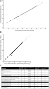
Improving the accuracy of calculating repeat length from fragment analysis size data. One-hundred cloned SCA1 alleles of known sequence were used as templates for fragment analysis by capillary electrophoresis. The fragment analyzed size was plotted against the actual size of the CAG repeat determined by sequencing (A). The data tightly fit a linear model (Number of repeats = [(Fragment analyzed size − 135.8487)/2.8669]; R2 = 0.99675), with one significant outlier. Comparing the proposed model (white squares) with the original model (Number of repeats = [(Fragment analyzed size − 139)/3] + 3; black squares), the proposed model points fall much closer to the identity line suggesting a better representation of the sequenced repeat lengths by the fragment sizing data (B). To validate the model, plasmids that were previously sized alongside a TP PCR CAG repeat ladder (Figure 3) were fragment analyzed and the number of repeats calculated using either the original model or the proposed model (C). Both models are able to accurately size clones in the range 40–48 repeats, however the proposed model can accurately size clones of 65 and 83 repeats.

References

    1. Currier R. D., Glover G., Jackson J. F., Tipton A. C. (1972). Spinocerebellar ataxia: study of a large kindred: I. General information and genetics. Neurology 22, 1040–1043. 10.1212/wnl.22.9.1040 - DOI - PubMed
    1. Döhlinger S., Hauser T.-K., Borkert J., Luft A. R., Schulz J. B. (2008). Magnetic resonance imaging in spinocerebellar ataxias. Cerebellum 7, 204–214. 10.1007/s12311-008-0025-0 - DOI - PubMed
    1. Giunti P., Sweeney M. G., Spadaro M., Jodice C., Novelletto A., Malaspina P., et al. . (1994). The trinucleotide repeat expansion on chromosome 6p (SCA1) in autosomal dominant cerebellar ataxias. Brain 117, 645–649. 10.1093/brain/117.4.645 - DOI - PubMed
    1. Goldfarb L. G., Vasconcelos O., Platonov F. A., Lunkes A., Kipnis V., Kononova S., et al. . (1996). Unstable triplet repeat and phenotypic variability of spinocerebellar ataxia type 1. Ann. Neurol. 39, 500–506. 10.1002/ana.410390412 - DOI - PubMed
    1. Greenfield J. G. (1954). The Spino-Cerebellar Degenerations. Springfield, IL: Blackwell Scientific Publications.

LinkOut - more resources