. 2018 Jul 31:10:208.
doi: 10.3389/fnagi.2018.00208. eCollection 2018.

The Aβ Containing Brain Extracts Having Different Effects in Alzheimer's Disease Transgenic Caenorhabditis elegans and Mice

Affiliations

The Aβ Containing Brain Extracts Having Different Effects in Alzheimer's Disease Transgenic Caenorhabditis elegans and Mice

Yufang Yang et al. Front Aging Neurosci. .

Abstract

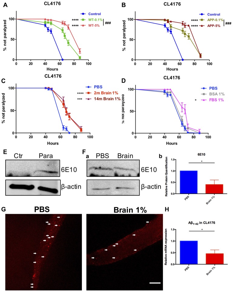
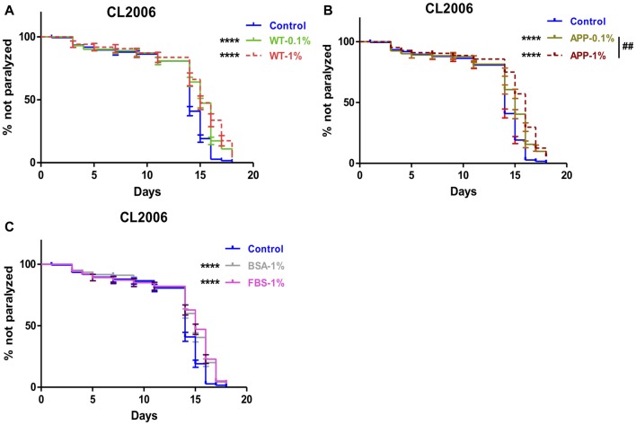
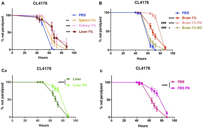
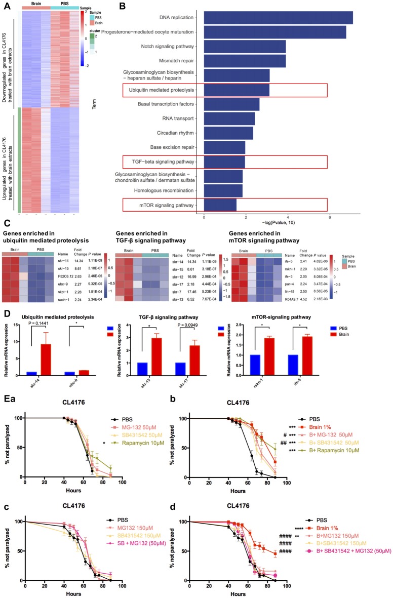
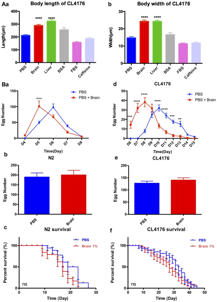
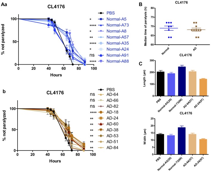
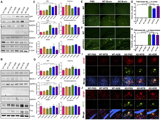
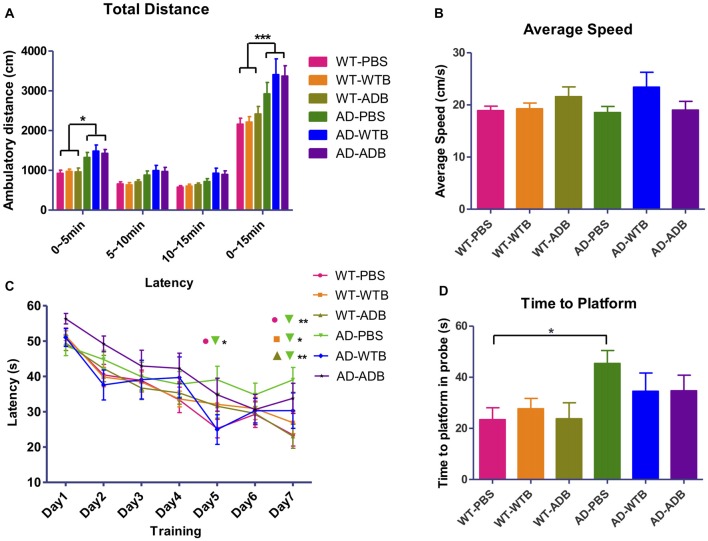
Background: The deposition of β-sheet rich amyloid in senile plaques is a pathological hallmark of Alzheimer's disease (AD), which is thought to cause neuronal dysfunction. Previous studies have strongly implicated that intracerebral infusion of brain extract containing aggregated β-amyloid (Aβ) is able to induce cerebral amyloidosis thus causing neuronal damage and clinical abnormalities in rodents and nonhuman primates, which are reminiscent of a prion-like mechanism. Prion disease has been documented in cases of prion-contaminated food consumption. Methods: We investigated whether cerebral transmission of Aβ was possible via oral administration of Aβ-rich brain extract in non-susceptible and susceptible host mice by immunohistochemistry, western blotting and behavior tests. Also brain extracts were supplied to AD transgenic Caenorhabditis elegans, and paralysis curve were conducted, following detection of Aβ amyloid. RNA sequencing of nematodes was applied then inhibitors for relevant dysregulated genes were used in the paralysis induction. Results: The oral treatment of AD brain extract or normal brain extract neither aggravated nor mitigated the Aβ load, glial activation or the abnormal behaviors in recipient Amyloid precursor protein/presenilin 1 (APP/PS1) mice. Whereas, a significant improvement of AD pathology was detected in worms treated with Aβ-rich or normal brain extracts, which was attributable to the heat-sensitive components of brain extracts. Transcriptome sequencing of CL4176 nematodes suggested that brain extracts could delay worm paralysis through multiple pathways, including ubiquitin mediated proteolysis and Transforming growth factor β (TGF-β) signaling pathway. Inhibitors of the ubiquitin proteasome system and the TGF-β signaling pathway significantly blocked the suppressive effects of brain extracts on worm paralysis. Conclusions: Our results suggest that systemic transmissible mechanisms of prion proteopathy may not apply to β amyloid, at least in terms of oral administration. However, brain extracts strongly ameliorated AD pathology in AD transgenic nematodes partially through TGF-β signaling pathway and ubiquitin mediated proteolysis, which indicated that some natural endogenous components in the mammalian tissues could resist Aβ toxicity.

Keywords: APP/PS1 transgenic mice; Alzheimer’s disease; RNA-sequencing (RNA-seq); oral administration; transgenic AD C. elegans; β-amyloid.


Figures

Figure 1
Figure 1
Beta-amyloid (Aβ)-induced paralysis was delayed in Alzheimer’s disease (AD) transgenic nematode CL4176 treated with brain extracts. (A) The effect of feeding brain extracts from wild type (WT) mice at 0.1% and 5% concentration. (B) The effect of feeding brain extracts from AD transgenic mice at 0.1% and 5% concentration. (C) Brain extracts from 2-month-old mice and 14-month-old mice showing similar anti-paralysis effects in CL4176 worms. (D) Different effects between 1% fetal bovine serum (FBS) and 1% bovine serum albumin (BSA) on paralysis. Data were analyzed using a paired log rank survival test. Sixty worms were counted per group. The level of significance was shown in brackets (*P < 0.05, ***P < 0.001, ****P < 0.0001, ###P < 0.001). (E) Protein levels of toxic Aβ1–42 detected by western blot with antibody 6E10 in CL4176 worms. Transgenic worms were harvested at 40 h with or without temperature induction. (F) The effects of 1% brain extracts on Aβ expression. (a) Worms were harvested at 64 h after giving extracts and applied to western blot analysis. (b) Statistics of Aβ1–42 expression levels. Samples were collected from three independent experiments. β-actin served as the internal control. Data were analyzed by unpaired Student’s t test. The level of significance was shown in brackets (*P < 0.05). (G) Representative fluorescence images for Aβ1–42 in CL4176 worms. Worms were collected at 64 h after giving extracts. The toxic Aβ aggregates, stained by 6E10 (red) and indicated with white arrows, were significantly decreased in worms treated with brain extracts. Scale bar: 10 μm. (H) The relative mRNA expression level of Aβ1–42 in CL4176 worms with or without treating brain extracts. Worms were harvested at 64 h after giving extracts and collected from three independent experiments. Data were analyzed by unpaired t-test with equal SD. The level of significance was shown in brackets (*P < 0.05), n = 3.
Figure 2
Figure 2
Aβ-induced paralysis was delayed in AD transgenic nematode CL2006 treated with brain extracts. (A) The effect of feeding brain extracts from WT mice at 0.1% and 1% concentration. (B) The effect of feeding brain extracts from AD transgenic mice at 0.1% and 1% concentration. (C) The effects of feeding 1% BSA and 1% FBS. Data were analyzed using a paired log rank survival test. The level of significance was shown in brackets (****P < 0.0001, ##P < 0.01).
Figure 3
Figure 3
The effects of brain extracts, peripheral tissue extracts and FBS with extra treatment in CL4176. (A) The effects of extracts from spleen, kidney and liver on paralysis at 1% concentration. (B) The compromised effect of brain extracts after boiling plus centrifugation and the enhanced protective effects of brain extracts after PK plus boiling treatment. (C) The enhanced protective effects of liver extracts (a) and FBS (b) after PK treatment. Data were analyzed using a paired log rank survival test. *P < 0.05, **P < 0.01, ***P < 0.001, ****P < 0.0001, ###P < 0.001.
Figure 4
Figure 4
Multiple pathways were involved in the suppressive effects of brain extracts on worm paralysis. (A) The heat map of total dysregulated genes in brain extracts-treated CLL4176 worms (P < 0.01, fold change ≥2). (B) KEGG pathway analysis of genes significantly upregulated in CL4176 treated with brain extracts (P < 0.05). (C) The heat map showing upregulation of genes that were involved in three KEGG pathways: ubiquitin mediated proteolysis, TGF-β signaling pathway and mammalian target of rapamycin (mTOR) signaling pathway. (D) The mRNA expression levels of skr-14, ubc-9, skr-15, skr-17, rskn-1, ife-5, components of the above KEGG pathways, were verified by real time polymerase chain reaction (RT-PCR) with worm collected from another three sets of independent experiments, *P < 0.05, with unpaired t-test. (E) The effects of brain effects on worm paralysis under the intervention of inhibitors for the three upregulated pathways in CL4176 worms. (a) The effects of the inhibitors at low doses on CL4176 worms in Phosphate buffer saline (PBS). (b) The effect of brain extracts with the low doses of inhibitors. (c) The effects of the inhibitors at high doses on CL4176 worms in PBS. (d) The effect of brain extracts with high doses of inhibitors. MG-132: inhibitor of ubiquitin mediated proteolysis; SB431542: inhibitor of Transforming growth factor β (TGF-β) signaling pathway: Rapamycin: inhibitor of mTOR signaling pathway. *P < 0.05, **P < 0.01, ***P < 0.001, ****P < 0.0001, compared with PBS group; #P < 0.05, ##P < 0.01, ####P < 0.0001, compared with Brain group.
Figure 5
Figure 5
The effect of brain extracts on the growth of CL4176 and WT N2 worms. (A) The effect of brain extracts on body sizes in CL4176 worms. (a) The body length of worms. (b) The body width of worms. Data were analyzed with one-way ANOVA, Tukey’s multiple comparisons post hoc that each group was compared with the PBS group. (B) The effect of brain extracts on fertility and lifespan of worms. (a–c) The egg number in each spawning day, total egg number and life span of N2 worms. (d–f) The egg number in each spawning day, total egg number, and life span of AD transgenic CL4176 worms. Egg numbers by days were analyzed with two-way ANOVA, Tukey’s multiple comparisons post hoc. Total egg numbers were analyzed with unpaired Student’s t-test. Life spans were presented as survival curves and analyzed with a paired log rank test. **P < 0.01; ***P < 0.001, ****P < 0.0001.
Figure 6
Figure 6
The effects of serums from normal subjects and AD patients on the paralysis in CL4176. (A) The effects of serum from normal subjects (a) and AD patients (b) in CL4176 worms. (B) Comparable median time of paralysis in CL4176 worms by feeding serum from normal subjects and AD patients. (C) No growth-promoting effects by feeding serum. Paralysis data were analyzed using a paired log rank survival test. Median time of paralysis was analyzed with two-tailed unpaired Student’s t-test. Body size was analyzed with One-way ANOVA, Tukey’s multiple comparisons post hoc. *P < 0.05; **P < 0.001; ****P < 0.0001, ns: P > 0.05, no significant difference.
Figure 7
Figure 7
Aβ load and neuroinflammation in 11-month-old mice with gavage. The depicted western blot indicates the expression levels of amyloid precursor protein (APP), β secretase (BACE1), C-terminal fragments (CTFs), total Tau and phosphorylated Tau in the cortex (A) and hippocampus (B) of 11-month-old mice with different treatments at the age of 6 months old. The relative expression levels of proteins in the cortex (C) and hippocampus (D) were shown in the diagram (n = 3–5 for APP and CTFs and n = 3 for BACE1). GAPDH or β-actin served as the internal control. The Aβ load and amyloid plaques in the cortex (E) and hippocampus (F) of transgenic AD and WT mice were stained with Thioflavin S. The toxic Aβ1–42 level in cortex (G) and hippocampus (H) were examined with the ELISA assay. Representative fluorescent images of glial fibrillary acidic protein (GFAP; red), Aβ (green) and merged with 4′,6-diamidino-2-phenylindole DAPI; blue) for the cortex (I) and hippocampus (J) of the indicated mouse strains with three different treatments were depicted. The astrocytes marked by GFAP expression were morphologically activated in the transgenic mice but had no difference among three treatments. Scale bar: 100 μm. All data were analyzed with one-way ANOVA, Tukey’s multiple comparisons post hoc. *P < 0.5, **P < 0.01, ***P < 0.001.
Figure 8
Figure 8
Feeding brain extracts at the age of 6 months did not alter the behaviors of mice. (A) The diagram summarizing the total ambulatory distance (cm) for each of the six groups (n = 9–22 per group) in the open-field test for the three 5 min blocks in 15 min observation. (B) The diagram representing the average swimming speed for different groups (n = 14–25) in Morris Water Maze test. (C) The average escape latency (four trails per day) for seven consecutive days and (D) Time for reaching the former platform location at the first time in the probe test. Diagrams in (A,C) were analyzed with two-way ANOVA, Tukey’s multiple comparisons post hoc. Diagrams in (B,D) were analyzed with One-way ANOVA, Tukey’s multiple comparisons post hoc. Significance level between certain groups are indicated with brackets (*P < 0.05, **P < 0.01, ***P < 0.001).

Similar articles

References

    1. An W. L., Cowburn R. F., Li L., Braak H., Alafuzoff I., Iqbal K., et al. (2003). Up-regulation of phosphorylated/activated p70 S6 kinase and its relationship to neurofibrillary pathology in Alzheimer’s disease. Am. J. Pathol. 163, 591–607. 10.1016/s0002-9440(10)63687-5 - DOI - PMC - PubMed
    1. Baptista M. S., Duarte C. B., Maciel P. (2012). Role of the ubiquitin-proteasome system in nervous system function and disease: using C. elegans as a dissecting tool. Cell. Mol. Life Sci. 69, 2691–2715. 10.1007/s00018-012-0946-0 - DOI - PMC - PubMed
    1. Bian M., Yu M., Yang S., Gao H., Huang Y., Deng C., et al. (2008). Expression of Cbl-interacting protein of 85 kDa in MPTP mouse model of Parkinson’s disease and 1-methyl-4-phenyl-pyridinium ion-treated dopaminergic SH-SY5Y cells. Acta Biochim. Biophys. Sin. 40, 505–512. 10.1111/j.1745-7270.2008.00423.x - DOI - PubMed
    1. Bonardi C., de Pulford F., Jennings D., Pardon M. C. (2011). A detailed analysis of the early context extinction deficits seen in APPswe/PS1dE9 female mice and their relevance to preclinical Alzheimer’s disease. Behav. Brain Res. 222, 89–97. 10.1016/j.bbr.2011.03.041 - DOI - PubMed
    1. Brenner S. (1974). The genetics of Caenorhabditis elegans. Genetics 77, 71–94. - PMC - PubMed

LinkOut - more resources