. 2018 Jul 31:9:856.
doi: 10.3389/fphys.2018.00856. eCollection 2018.

Vascular Smooth Muscle Contractile Function Declines With Age in Skeletal Muscle Feed Arteries

Affiliations

Vascular Smooth Muscle Contractile Function Declines With Age in Skeletal Muscle Feed Arteries

John W Seawright et al. Front Physiol. .

Abstract

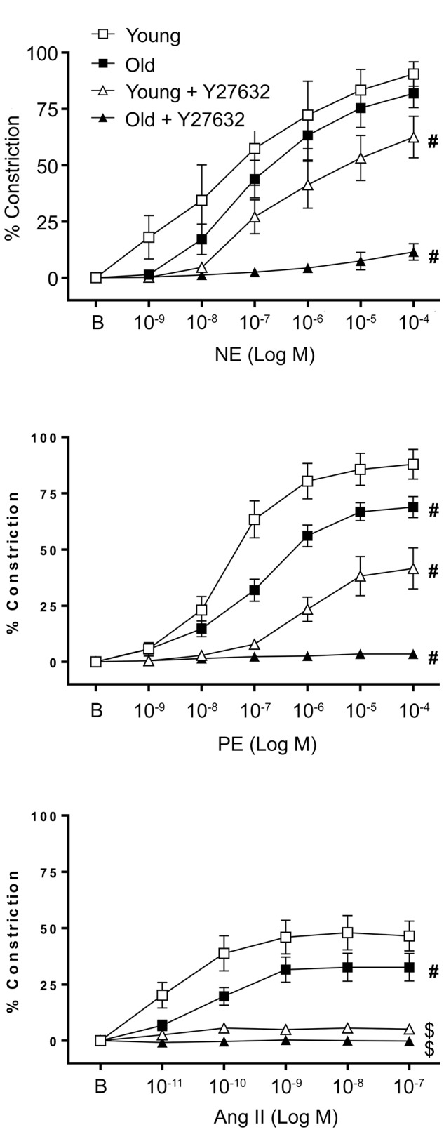
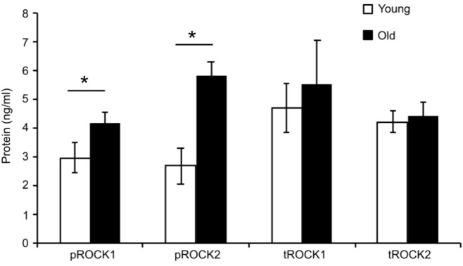
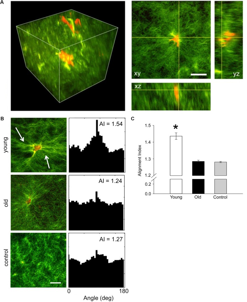
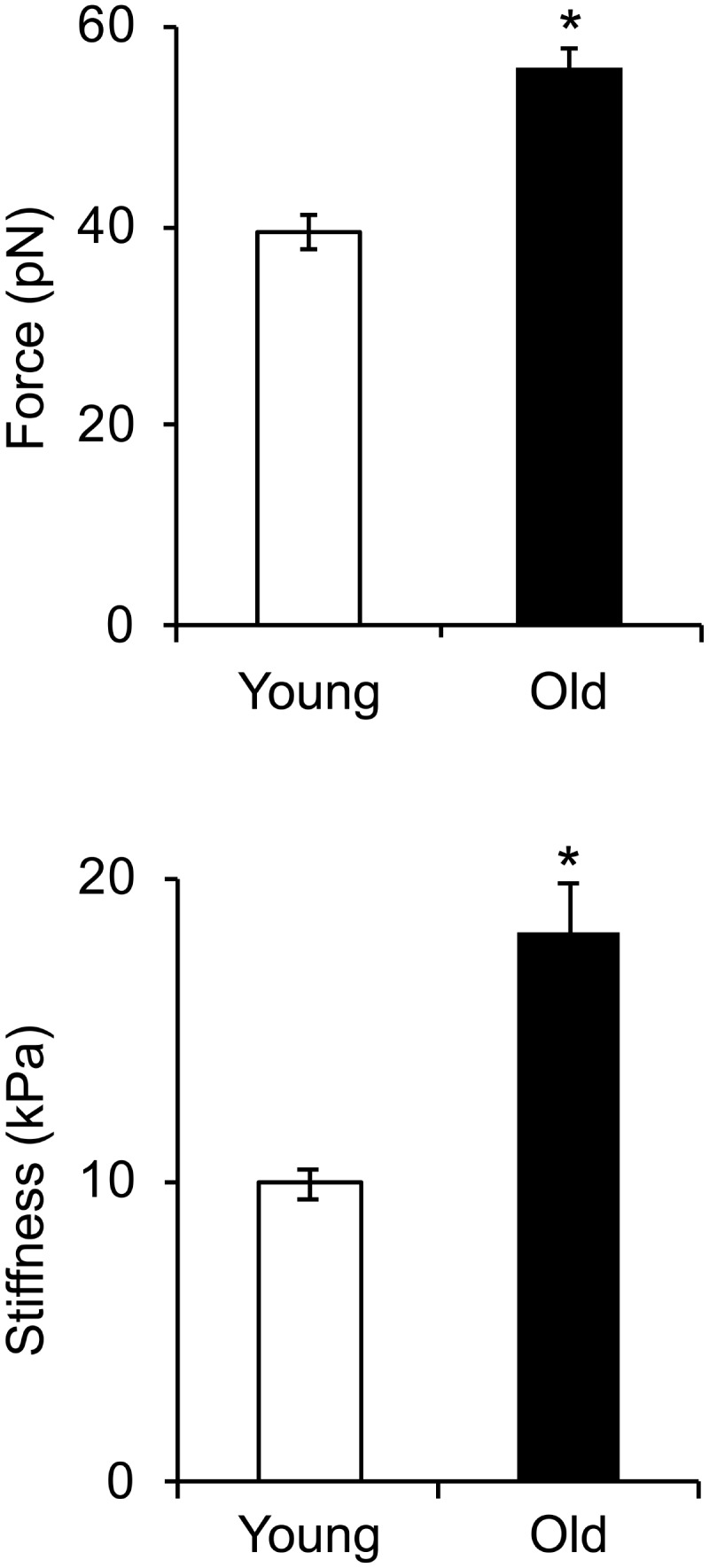
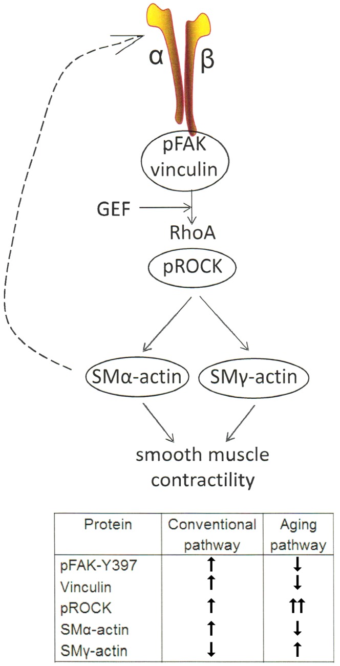
Aging induces a progressive decline in vasoconstrictor responses in central and peripheral arteries. This study investigated the hypothesis that vascular smooth muscle (VSM) contractile function declines with age in soleus muscle feed arteries (SFA). Contractile function of cannulated SFA isolated from young (4 months) and old (24 months) Fischer 344 rats was assessed by measuring constrictor responses of denuded (endothelium removed) SFA to norepinephrine (NE), phenylephrine (PE), and angiotensin II (Ang II). In addition, we investigated the role of RhoA signaling in modulation of VSM contractile function. Structural and functional characteristics of VSM cells were evaluated by fluorescence imaging and atomic force microscopy (AFM). Results indicated that constrictor responses to PE and Ang II were significantly impaired in old SFA, whereas constrictor responses to NE were preserved. In the presence of a Rho-kinase inhibitor (Y27632), constrictor responses to NE, Ang II, and PE were significantly reduced in young and old SFA. In addition, the age-group difference in constrictor responses to Ang II was eliminated. ROCK1 and ROCK2 content was similar in young and old VSM cells, whereas pROCK1 and pROCK2 were significantly elevated in old VSM cells. Aging was associated with a reduction in smooth muscle α-actin stress fibers and recruitment of proteins to cell-matrix adhesions. Old VSM cells presented an increase in integrin adhesion to the matrix and smooth muscle γ-actin fibers that was associated with increased cell stiffness. In conclusion, our results indicate that VSM contractile function declined with age in SFA. The decrement in contractile function was mediated in part by RhoA/ROCK signaling. Upregulation of pROCK in old VSM cells was not able to rescue contractility in old SFA. Collectively, these results indicate that changes at the VSM cell level play a central role in the reduced contractile function of aged SFA.

Keywords: Rho-kinase; aging; atomic force microscopy; vascular smooth muscle; vasoconstriction.


Figures

FIGURE 1
FIGURE 1
Effect of aging on vascular smooth muscle contractile responses to NE, PE, and Ang II was assessed in denuded SFA. To evaluate the role of ROCK, VSM constrictor responses were further assessed in the presence of Y27632. N = 6–12 rats per group. B represents the baseline diameter at a pressure of 90 cm H2O before the first dose of NE, PE, or Ang II. Data shown are mean ± SE. Significance was evaluated at p < 0.05. #Concentration-response curve was significantly different from all other curves; $Concentration-response curve was significantly different from young and old SFA in the absence of Y27632.
FIGURE 2
FIGURE 2
Elisa assay values for total and phospho-ROCK proteins. While total ROCK does not change with age, there was a significant increase in old cells for both isoforms in the active form pROCK1 and pROCK2. N = 4. Data shown as mean ± SD. Significance was evaluated at p < 0.05.
FIGURE 3
FIGURE 3
(A) Representative three-dimensional volumetric imaging of the collagen gel and 2D projections in xy, xz, and yz planes. Collagen gel was pseudo-colored in green (SHG signal), while auto-fluorescence signal from VSM cells was pseudo-colored in red. (B) Representative projections of the 3D volumetric images and their alignment angle distributions and alignment index (AI), respectively. Young VSM cells exert force on the collagen matrix inducing higher remodeling of the collagen fibers (see arrows). In contrast, old VSM cells have reduced local matrix remodeling. (C) The distribution of collagen fiber angle alignment index for young cells shows a significant increase in respect to control (collagen gel without cells), while no change was recorded for old cells. N = 8–10 tri-dimensional volumetric images per condition. Scale bar represents 50 μm. Data shown as mean ± SD. Significance was evaluated at p < 0.05.
FIGURE 4
FIGURE 4
Age effects on the architecture of VSM cells. Representative (A) confocal images of SMα- and SMγ-actin, (B) TIRF images of vinculin and pFAK-Y397, and (C) confocal images of phalloidin in VSM cells isolated from young and old Fischer 344 rats. SMα-actin forms actin bundles at cell edges, while SMγ-actin forms finer fibers toward the cell interior without strong fibers at cell edges. Scale bar represents 10 μm. (D) Quantitative analysis of fluorescence images are presented as old to young ratios of relative protein area to respective cell area. N = 7–11 cells per condition. Values different from a ratio of 1 are statistically significant (p < 0.05).
FIGURE 5
FIGURE 5
Integrin α5β1-fibronectin adhesion force spectroscopy measurements. Cell stiffness and adhesion force measurements were performed on young and old cells. Cell stiffness and adhesion force of integrin α5β1 binding to fibronectin significantly increased with age. N ∼ 4,000 individual measurements for each condition. Significance was evaluated at p < 0.05.
FIGURE 6
FIGURE 6
One of the main mechanotransduction pathways in VSM cell contractility is the RhoA/ROCK regulated integrin-actomyosin axis. Mechanical signals (e.g., pressure, matrix stiffness) presented to VSM cells through integrins stimulate the activity of guanine nucleotide exchange factors (GEF) which in turn activate RhoA pathway, including ROCK which enhances actomyosin contractility. Consequently, actomyosin-induced contractility promotes VSM cell stiffening. This effect feeds back (dashed arrow) into changing cytoskeletal tension and integrin mechanosensing at the membrane level. Integrin α5β1 is known to be involved in regulating cellular and vessel contractility (Martinez-Lemus et al., 2005a). In young cells, actomyosin apparatus is strong and the VSM cell is able to properly regulate its stiffness and adhesion to the matrix in response to mechanical signals. In aging, however, the decrease in SMα-actin stress fiber formation and alteration of cell-matrix adhesions induces a reduced VSM cell contractility and deficient mechanosensing. We suggest that this dysregulation induces compensatory effects by up-regulating ROCK and SMγ-actin which increases cell stiffness without regaining contractile properties of the cell (see table inset).

References

    1. Arnoldi R., Hiltbrunner A., Dugina V., Tille J. C., Chaponnier C. (2013). Smooth muscle actin isoforms: a tug of war between contraction and compliance. Eur. J. Cell Biol. 92 187–200. 10.1016/j.ejcb.2013.06.002 - DOI - PubMed
    1. Bai Y., Lee P. F., Gibbs H. C., Bayless K. J., Yeh A. T. (2014). Dynamic multicomponent engineered tissue reorganization and matrix deposition measured with an integrated nonlinear optical microscopy-optical coherence microscopy system. J. Biomed. Opt. 19:36014. 10.1117/1.JBO.19.3.036014 - DOI - PMC - PubMed
    1. Briones A. M., Salaices M., Vila E. (2007). Mechanisms underlying hypertrophic remodeling and increased stiffness of mesenteric resistance arteries from aged rats. J. Gerontol. A Biol. Sci. Med. Sci. 62 696–706. 10.1093/gerona/62.7.696 - DOI - PubMed
    1. Chaponnier C., Gabbiani G. (2016). Monoclonal antibodies against muscle actin isoforms: epitope identification and analysis of isoform expression by immunoblot and immunostaining in normal and regenerating skeletal muscle. F1000Res. 5:416. 10.12688/f1000research.8154.2 - DOI - PMC - PubMed
    1. Chaudhuri S., Nguyen H., Rangayyan R. M., Walsh S., Frank C. B. (1987). A fourier domain directional filtering method for analysis of collagen alignment in ligaments. IEEE Trans. Biomed. Eng. 34 509–518. 10.1109/TBME.1987.325980 - DOI - PubMed

LinkOut - more resources