Review
. 2016 Oct 19;8(1):21-52.
doi: 10.1039/c6md00432f. eCollection 2017 Jan 1.

Hypoxia inducible factor down-regulation, cancer and cancer stem cells (CSCs): ongoing success stories

Affiliations
Review

Hypoxia inducible factor down-regulation, cancer and cancer stem cells (CSCs): ongoing success stories

Anthony R Martin et al. Medchemcomm. .

Abstract

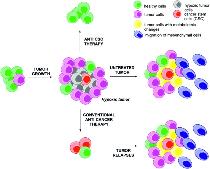
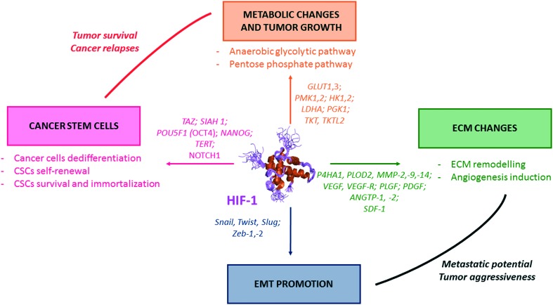
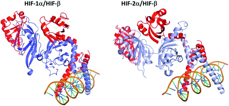
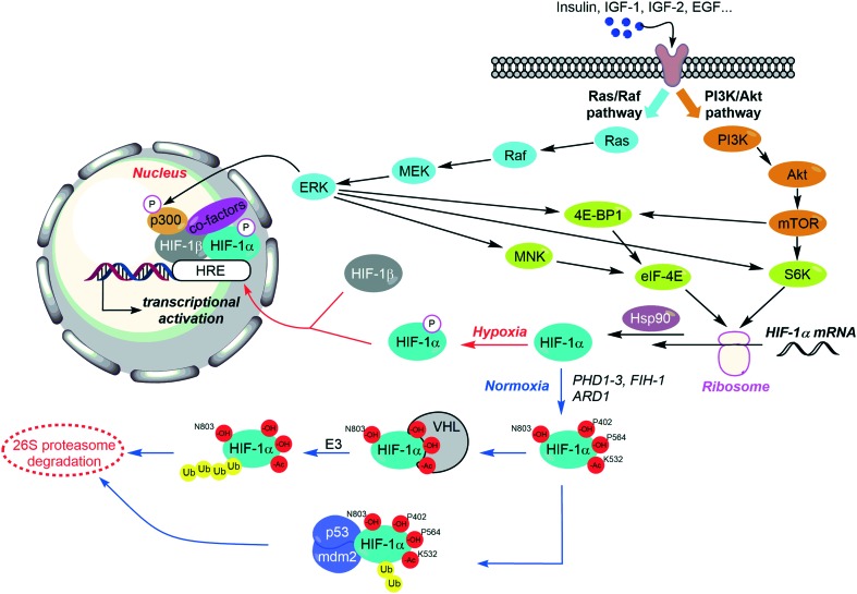
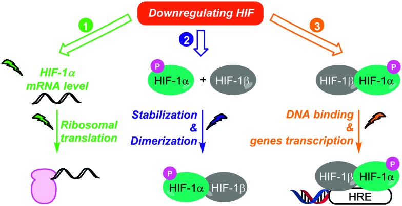
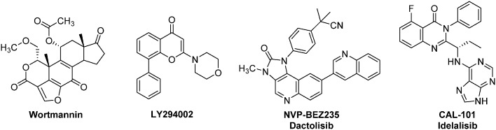
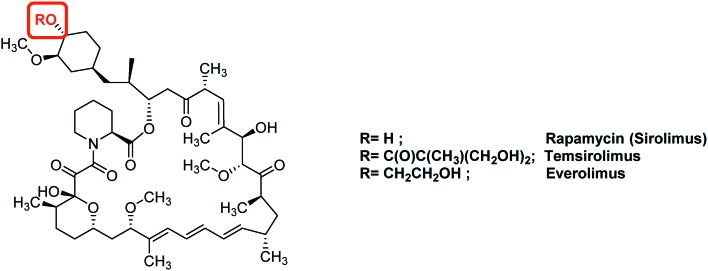
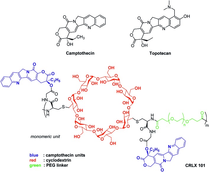
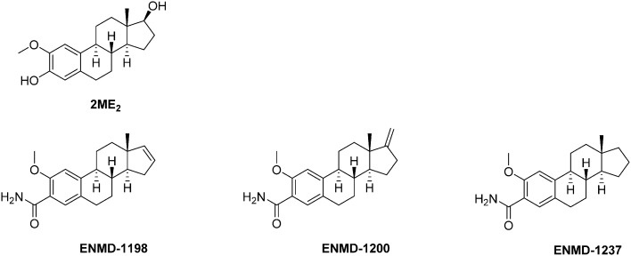
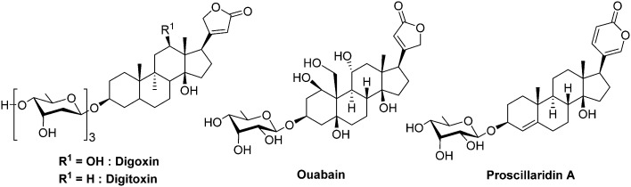
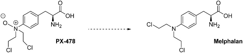
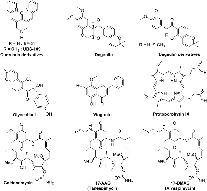
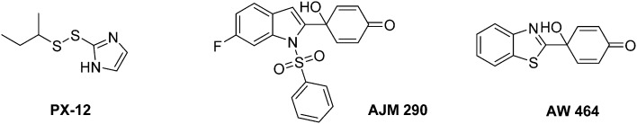
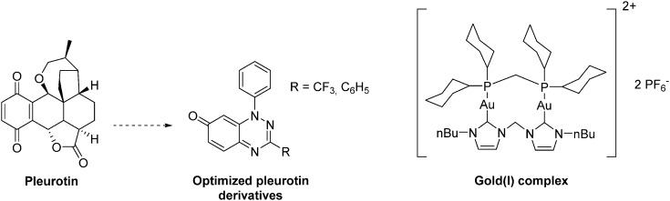
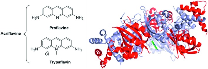
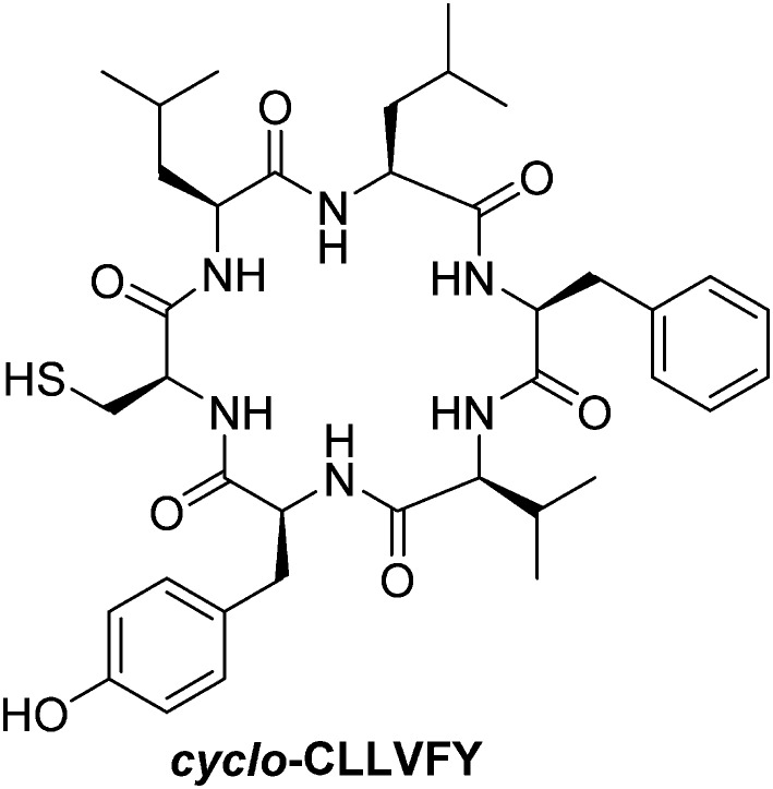
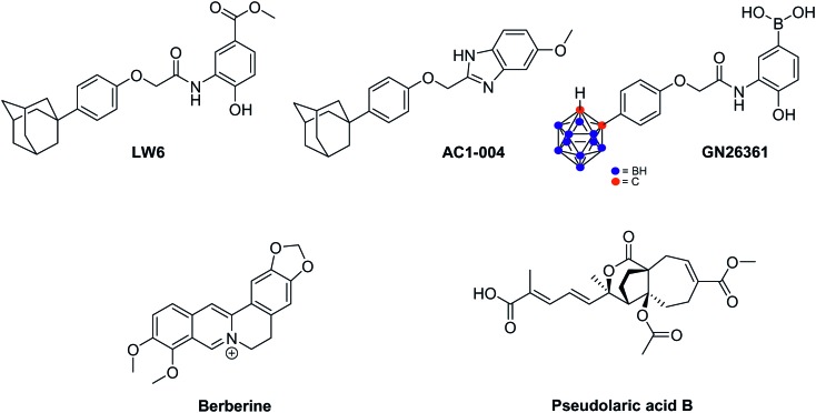
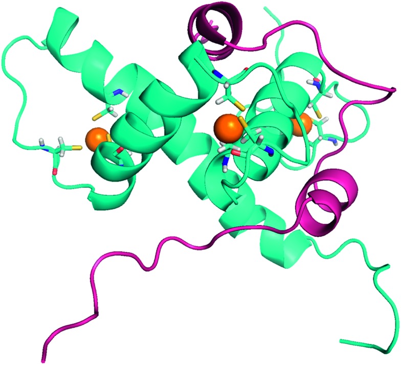
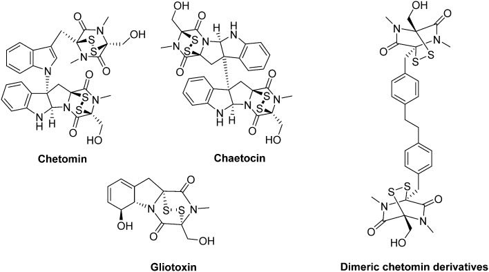
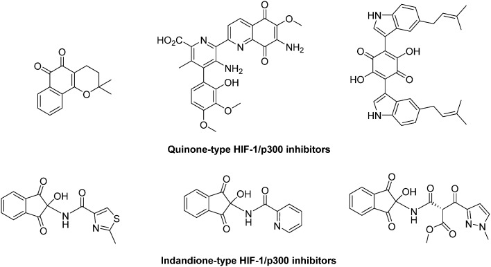
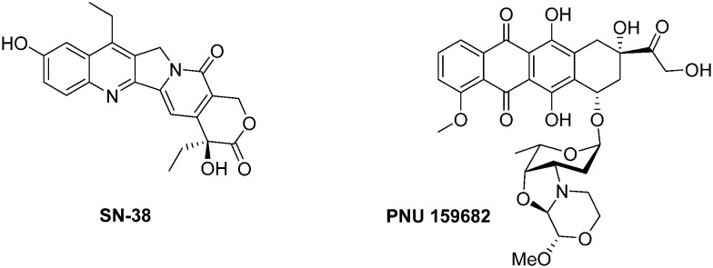
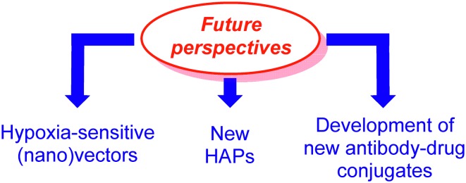
In cancers, hypoxia inducible factor 1 (HIF-1) is an over-expressed transcription factor, which regulates a large set of genes involved in tumour vascularization, metastases, and cancer stem cells (CSCs) formation and self-renewal. This protein has been identified as a relevant target in oncology and several HIF-1 modulators are now marketed or in advanced clinical trials. The purpose of this review is to summarize the advances in the understanding of its regulation and its inhibition, from the medicinal chemist point of view. To this end, we selected in the recent literature relevant examples of "hit" compounds, including small-sized organic molecules, pseudopeptides and nano-drugs, exhibiting in vitro and/or in vivo both anti-HIF-1 and anti-tumour activities. Whenever possible, a particular emphasis has been dedicated to compounds that selectively target CSCs.


Figures

Fig. 1
Fig. 1. The aggressiveness potential of hypoxic tumors and the cancer stem cells (CSCs) postulate. The tumour growth generates poorly vascularized areas resulting in the induction of an hypoxic stress. If untreated (middle panel), the hypoxic tumor cells (grey) will develop an adaptive response in three points to overcome this stress: (a) metabolic changes (yellow), (b) phenotypic changes resulting in an increase of the metastatic potential of the tumour (the epithelial mesenchymal transition, EMT, resulting in the migration of the mesenchymal cells, blue) and (c) improvement of cell dedifferentiation and promotion of cancer stem cells (CSCs, red). If treated by conventional anti-cancer agents (bottom panel), hypoxic tumour will always relapse within few months, due to the persistence of the untreated CSCs. In this two cases, the relapsed tumours are heterogeneous cells composed by a mixture of mutated cells; this lead to a poor clinical prognosis. By a marked contrast, the combination of a specific anti-CSCs therapy with conventional anti-cancer drugs or surgery, generally leads to a better outcome (top panel).
Fig. 2
Fig. 2. The pivotal role of HIF-1 in tumour survival and aggressiveness. HIF-1 is a transcription factor which induces the transcription a large set of genes, the hypoxia response elements (HRE). Some of them are summarized in this figure. One can divide the HRE in four different classes, and representative examples are depicted herein. (i) HRE promoting metabolic switches (orange); (ii) HRE promoting CSCs formation and self-renewal (pink); (iii) HRE promoting changes in the extra cellular matrix (ECM) (green); and (iv) HRE promoting the epithelial mesenchymal transition (EMT) (blue).
Fig. 3
Fig. 3. Comparison of the 3D structures of the HIF-1α/HIF-β and the HIF-2α/HIF-β heterodimers bound to DNA. Dark blue: HIF-1α; light blue: HIF-2α; red: HIF-β (ARNT); orange: the DNA double helix bound to the dimeric form of HIF. These two heterodimers have very close 3D structures, since HIF-1α and HIF-2α share 80% of sequence homology. HIF-1α/HIF-β: PDB code 4ZPR; HIF-2α/HIF-β: PDB code ; 4ZPK.
Fig. 4
Fig. 4. The complex regulation of HIF-1. HIF-1 is activated by growth factors and cytokines through two main signalling pathways: PI3K/Akt (orange) and MAPK (blue). Nonetheless, in normoxic tissues, successive hydroxylations lead to (i) HIF-1 degradation by the 26S proteasome or to (ii) the inhibition of its transcriptional activity (blue arrows). Otherwise, interaction with the chaperone protein Hsp90 (for its dimerization) and co-factors such as p300/CEB (for the activation of its transcriptional activity) are also required.
Fig. 5
Fig. 5. Three strategies to downregulate HIF-1. These three strategies include (i) the inhibition of HIF-1α biosynthesis (green); (ii) its destabilization leading to its proteasomal degradation (blue); and (iii) the inhibition of its transcriptional activity (orange).
Fig. 6
Fig. 6. Representative examples of anti-HIF therapeutic agents targeting the Akt/mTOR signalling pathway: selected PI3K inhibitors.
Fig. 7
Fig. 7. Representative examples of anti-HIF therapeutic agents targeting the Akt/mTOR signalling pathway: mTOR inhibitors.
Fig. 8
Fig. 8. Representative examples of anti-HIF therapeutic agents targeting the topoisomerases.
Fig. 9
Fig. 9. Representative examples therapeutic agents targeting HIF-1α expression.
Fig. 10
Fig. 10. Representative examples therapeutic agents targeting the microtubule network and consequently affecting HIF-1α expression.
Fig. 11
Fig. 11. Cardiac glycosides affecting HIF-1α expression.
Fig. 12
Fig. 12. PX-478 and its metabolite (melphalan).
Fig. 13
Fig. 13. Representative examples of Hsp90 inhibitors exhibiting an anti-HIF-1α activity.
Fig. 14
Fig. 14. Ganetespib: structure and binding mode. Structure of the molecule (left panel), X-ray crystal of ganetespib bound to the N-Terminal ATP binding pocket of Hsp90 (middle panel, PDB code: 3TUH) and structural features highlighting the H-bond network and the hydrophobic interactions (right panel).
Fig. 15
Fig. 15. Representative examples of HDAC inhibitors exhibiting an anti-HIF-1α activity. All the compounds represented herein are marketed for their therapeutic uses, excepted Dacinostat.
Fig. 16
Fig. 16. Representative examples of sirtuin inhibitors exhibiting an anti-HIF-1α activity.
Fig. 17
Fig. 17. Schematic overview of Thioredoxin reductase (TrxR)-mediated thioredoxin-1 (Trx-1) reduction and the mode of action of some selected inhibitors. The NADPH-dependent reduction of Trx-1, a protein overexpressed in a set of cancer tissues, is mediated by TrxR. Inhibitors of either TrxR and Trx-1 have been developed and exert an action on the activation and stabilization of HIF-1α. See text for details.
Fig. 18
Fig. 18. Representative examples of thioredoxin-1 (Trx-1) inhibitors exhibiting an anti-HIF-1α activity.
Fig. 19
Fig. 19. Representative examples of thioredoxin reductase (TrxR) inhibitors exhibiting an anti-HIF-1α activity.
Fig. 20
Fig. 20. The acridines mixture of Acriflavine is an inhibitor of the HIF dimerization. Left panel: Structure of the two components of acriflavine; Right panel: crystallographic structure of proflavine bound to HIF-2α/HIF-β (PDB code: 4ZPH; blue: HIF-2α ; red: HIF-β ; green: proflavine).
Fig. 21
Fig. 21. cyclo-CCLVFY, a cyclopentapeptide inhibitor of HIF-1 dimerization.
Fig. 22
Fig. 22. Miscellaneous compounds destabilizing HIF-1α.
Fig. 23
Fig. 23. 3D-structure of HIF-1α bound to p300 (PDB: 1L3E). The C-TAD domain of HIF-1α (blue ribbon) is characterized by three short helices, wrapping around the CH1 domain of p300 (pink ribbon). CH1 adopts a triangular geometry, and encompasses three cations Zn2+, responsible for its structural stability. Importantly, unbounded C-TAD is not structured.
Fig. 24
Fig. 24. Chetomin and its derivatives.
Fig. 25
Fig. 25. Quinone and indandione derivatives which antagonize the HIF-1/p300 interaction.
Fig. 26
Fig. 26. α-Helix mimics as antagonists of the HIF-1α/p300 interaction. A: Structure of HIF-1α bound to p300 (PDB: 1L8C), highlighting two relevant α-helix involved in the binding of the two partner proteins and their corresponding sequences (blue: HIF-1α; pink: p300; grey: Zn atoms); B: peptidomimetic designed by Henchey (ref. 154), encompassing a hydrogen bon surrogate motive (red); C: oligoamides designed by Burslem (ref. 155) to specifically mimic helix 2 or helix 3. See text for details.
Fig. 27
Fig. 27. Series of natural products, antagonists of the HIF-1/p300 interaction.
Fig. 28
Fig. 28. Anthracyclines as inhibitors of the HIF-1 transcriptional activity.
Fig. 29
Fig. 29. Two cyclopeptides inhibitors of the HIF-1 transcriptional activity. A: Echinomycin; B: crystallographic structure of echinomycin bound to HRE DNA sequence (PDB: 2ADW) featuring the intercalated carboxyquinoxaline cores (red circle); C: triostin A.
Fig. 30
Fig. 30. A. Chemical bases of the hypoxia-activated agents; B. structure of Q39.
Fig. 31
Fig. 31. Structure of two HIF-1 inhibitors used to design ADC directed towards CSCs.
Fig. 32
Fig. 32. The three main axes for the development of new anti-HIF compounds.
None
Anthony R. Martin
None
Cyril Ronco
None
Luc Demange
None
Rachid Benhida

References

    1. Semenza G. L., Wilson W. R., Hay M. P. Biochim. Biophys. Acta. Nat. Rev. Cancer. 2016;2011;186311:382. 393. - PMC - PubMed
    1. Liu J., Liu Y., Bu W., Bu J., Sun Y., Du J., Shi J., Vaupel P., Höckel M., Mayer A. J. Am. Chem. Soc. Antioxid. Redox Signaling. 2014;2007;1369:9701. 1221. - PubMed
    1. Bhatt A. N., Chauhan A., Khanna S., Rai Y., Singh S., Soni R., Kalra N., Dwarakanath B. S., Emara M., Allalunis-Turner J., Camaj P., Carsten J., Krebs S., DeToni E. N., Blum H., Jauch K.-W., Nelson P. J., Bruns C. J., Bao B., Aamir A., Kong D., Ali S., Azmi A. S., Li Y., Banerjee S., Padhye S., Sarkar F. H., Hill R. P., Marie-Egyptienne D. T., Hedley D. W., Axelson H., Fredlund E., Ovenberger M., Landberg G., Påhlman S., Jögi A., Øra I., Nilsson H., Poellinger L., Axelson H., Påhlman S. BMC Cancer. Oncol. Rep. Mol. Cancer Res. PLoS One. Semin. Radiat. Oncol. Semin. Cell Dev. Biol. Cancer Lett. 2015;2014;2014;2012;2009;2005;2003;15311271916197:335. 1947, 421, e43726, 106, 554, 145. - PMC - PubMed
    1. Asgari Y., Zabihinpour Z., Salehzadeh-Yazdi A., Schreiber F., Masoudi-Nejad A., Chen X., Qian Y., Wu S., Zhang W., Zhang S.-L., Hu X., Tam K. Y. Genomics. Free Radical Biol. Med. Int. J. Biol. Sci. 2015;2015;2015;1057911:275. 253, 1390. - PubMed
    1. Sancho P., Barneda D., Heeschen C., Elf S., Li R., Xia S., Pan Y., Shan C., Wu S., Lonial S., Gaddh M., Arellano M. L., Khoury H. J., Khuri F. R., Lee B. H., Boggon T. J., Fan J., Chen J., Chen Y., Xu Q., Ji D., Wei Y., Chen H., Li T., Wan B., Yuan L., Huang R., Chen G. Br. J. Cancer. Oncogene. Tumour Biol. 2016;2016;2016;11437:1305. 6027. doi: 10.1038/onc.2016.196. - DOI - PMC - PubMed