

Upconversion fluorescent and X-ray-sensitive bifunctional nanoprobes for assessing the penetrability of inorganic nanoparticles in the digestive system
- PMID: 30108818
- PMCID: PMC6071934
- DOI: 10.1039/c6md00703a
Upconversion fluorescent and X-ray-sensitive bifunctional nanoprobes for assessing the penetrability of inorganic nanoparticles in the digestive system
Abstract
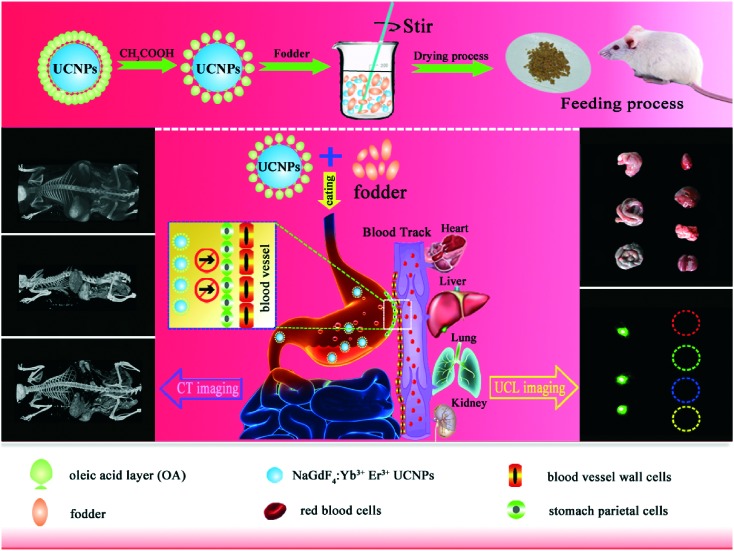
Nanotechnology is receiving increasing attention due to its fantastic advantages and potential applications in nanofood and nanomedicine. However, the safety of touching manufactured nanoparticles is still uncertain for human beings. Here, we track inorganic nanoparticles in the digestive system of the mouse through upconversion fluorescence and X-ray imaging, and try to demonstrate whether or not the inorganic nanoparticles will penetrate the digestive system to enter the blood system. Lanthanide-doped upconversion nanoparticles, which can convert infrared light to visible light and are simultaneously sensitive to X-rays, were selected as model inorganic nanoparticles. The investigation clarifies that even the ultrathin nanoparticles (∼5 nm) could not penetrate the digestive tract to enter the bloodstream or surrounding tissues, but were gradually excreted out. Our results help assess the safety of inorganic nanoparticles potentially used in nanofood and nanomedicine.
Figures


Similar articles
-
Emerging ≈800 nm Excited Lanthanide-Doped Upconversion Nanoparticles.Small. 2017 Feb;13(6). doi: 10.1002/smll.201602843. Epub 2016 Dec 16. Small. 2017. PMID: 27982542 Review.
-
Synergistic dual-modality in vivo upconversion luminescence/X-ray imaging and tracking of amine-functionalized NaYbF(4):Er nanoprobes.ACS Appl Mater Interfaces. 2014 Mar 26;6(6):3839-46. doi: 10.1021/am500383m. Epub 2014 Mar 14. ACS Appl Mater Interfaces. 2014. PMID: 24597514
-
Lanthanide-Activated Nanoparticles: A Toolbox for Bioimaging, Therapeutics, and Neuromodulation.Acc Chem Res. 2020 Nov 17;53(11):2692-2704. doi: 10.1021/acs.accounts.0c00513. Epub 2020 Oct 26. Acc Chem Res. 2020. PMID: 33103883 Review.
-
Direct visualization of gastrointestinal tract with lanthanide-doped BaYbF5 upconversion nanoprobes.Biomaterials. 2013 Oct;34(30):7444-52. doi: 10.1016/j.biomaterials.2013.06.060. Epub 2013 Jul 9. Biomaterials. 2013. PMID: 23849344
-
Engineered lanthanide-doped upconversion nanoparticles for biosensing and bioimaging application.Mikrochim Acta. 2022 Feb 17;189(3):109. doi: 10.1007/s00604-022-05180-1. Mikrochim Acta. 2022. PMID: 35175435 Review.
Cited by
-
An optical strategy for detecting hypochlorite in vitro and cells with high selectivity and stability based on a lanthanide-doped upconversion probe.RSC Adv. 2022 Nov 3;12(49):31608-31616. doi: 10.1039/d2ra05414k. eCollection 2022 Nov 3. RSC Adv. 2022. PMID: 36380959 Free PMC article.
-
Bioimaging with Upconversion Nanoparticles.Adv Photonics Res. 2022 Dec;3(12):2200098. doi: 10.1002/adpr.202200098. Epub 2022 Sep 9. Adv Photonics Res. 2022. PMID: 36686152 Free PMC article.
References
LinkOut - more resources
Full Text Sources
Other Literature Sources