Review
. 2017 May 16;8(7):1378-1392.
doi: 10.1039/c7md00109f. eCollection 2017 Jul 1.

Advances in indoleamine 2,3-dioxygenase 1 medicinal chemistry

Affiliations
Review

Advances in indoleamine 2,3-dioxygenase 1 medicinal chemistry

Alice Coletti et al. Medchemcomm. .

Abstract

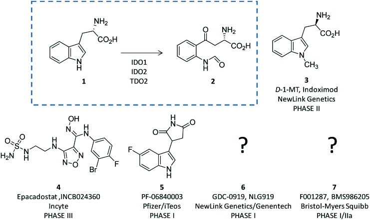
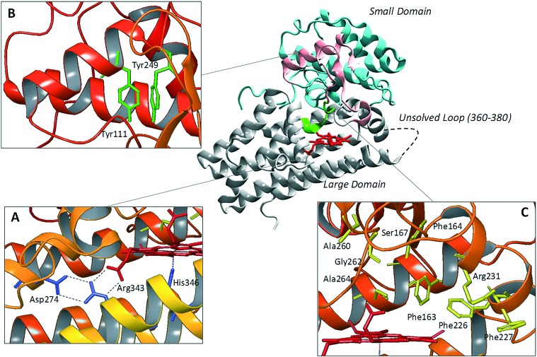
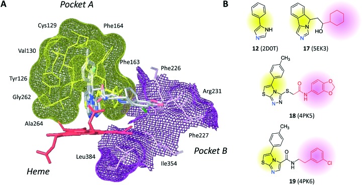
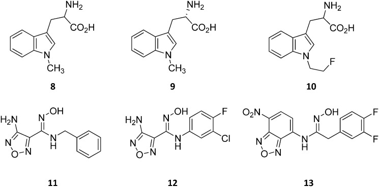
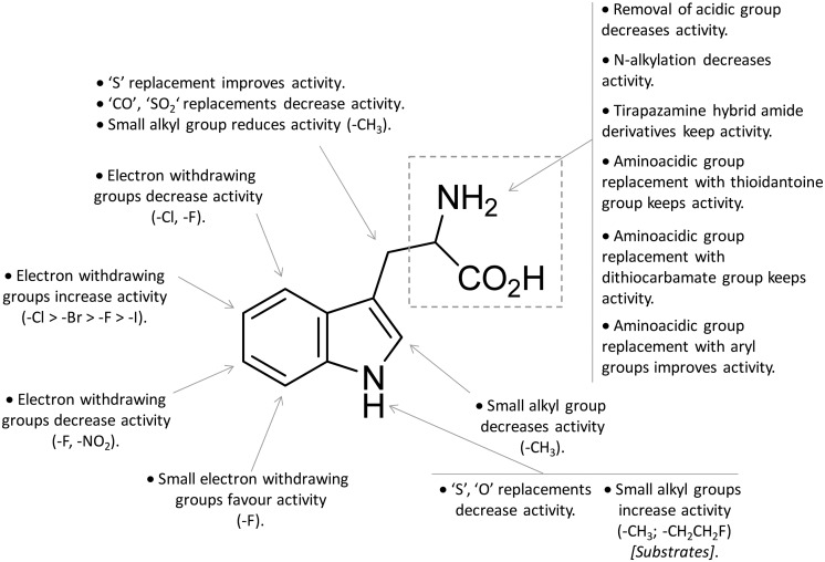
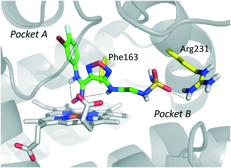
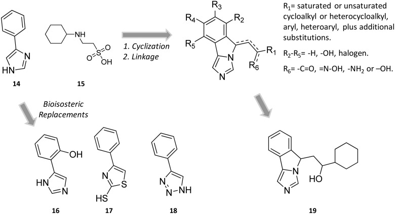
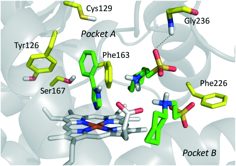
Indoleamine 2,3-dioxygenase 1 (IDO1) mediates multiple immunoregulatory processes including the induction of regulatory T cell differentiation and activation, suppression of T cell immune responses and inhibition of dendritic cell function, which impair immune recognition of cancer cells and promote tumor growth. On this basis, this enzyme is widely recognized as a valuable drug target for the development of immunotherapeutic small molecules in oncology. Although medicinal chemistry has made a substantial contribution to the discovery of numerous chemical classes of potent IDO1 inhibitors in the past 20 years, only very few compounds have progressed in clinical trials. In this review, we provide an overview of the current understanding of structure-function relationships of the enzyme, and discuss structure-activity relationships of selected classes of inhibitors that have shaped the hitherto few successes of IDO1 medicinal chemistry. An outlook opinion is also given on trends in the design of next generation inhibitors of the enzyme.


Figures

Fig. 1
Fig. 1. The chemical structures of the substrate (1) and product (2) of the rate limiting step of the kynurenine pathway are shown in the dashed box. The chemical structures of drug candidates targeting IDO1 (3–6) are shown outside of the box. The structures of compounds 5 and 6 are undisclosed.
Fig. 2
Fig. 2. Structure of IDO1 and structure–function relationships depicting residues involved in holding the heme group (box A), ITIM mediated signaling functions (box B), and catalytic activity (box C).
Fig. 3
Fig. 3. (A) The catalytic cleft of IDO1 is shaped with two pockets: pocket A (yellow surface) is mostly composed of aromatic and hydrophobic residues, while pocket B (magenta surface) is made of one positively charged residue and aromatic residues. (B) Co-crystallized inhibitors (14, 19–21) with shadow colors indicating the part of the chemical structure occupying pocket A (yellow) and/or pocket B (magenta).
Fig. 4
Fig. 4. Chemical structures of slow substrates (8–10) and competitive inhibitors (11–13).
Fig. 5
Fig. 5. Structure–activity relationships of indole-based inhibitors originating from ref. 42, 44, 57 and 59–63.
Fig. 6
Fig. 6. Structure–activity relationships of hydroxyamidine-based inhibitors originating from ref. 64 and 65.
Fig. 7
Fig. 7. Proposed binding mode of epacadostat (4) to IDO1 as a result of the docking study reported in ref. 65. Intra-molecular hydrogen bonds of 4 are shown with dashed black lines.
Fig. 8
Fig. 8. Chemical structures of non-competitive/uncompetitive inhibitors (14, 16–19) and relative structure-based design strategies. The Markush structure of NLG919 analogues is taken from ref. 78.
Fig. 9
Fig. 9. Binding mode of 4-PI (14, shown in green carbon atom sticks) to IDO1 resulting from crystallographic studies (pdb code: ; 2D0T). Key residues for structure–activity relationships are labeled and shown in yellow carbon atom sticks. CHES (15) molecules binding to the catalytic cleft are also shown in green carbon atom sticks.
Fig. 10
Fig. 10. Structure–activity relationships of 4-PI-based inhibitors originating from ref. 73–76.
Fig. 11
Fig. 11. Binding mode of the NLG919 analogue (19, shown in green carbon atom sticks) to IDO1 resulting from crystallographic studies (pdb code: ; 5EK3). Key residues are labeled and shown in yellow carbon atom sticks. Inter- and intra-molecular hydrogen bonds of 19 are shown with dashed black lines.
Fig. 12
Fig. 12. Structure–activity relationships of NLG919-based inhibitors originating from ref. 27.
Fig. 13
Fig. 13. Chemical structures of IDO1 inhibitors (20–25) with a hypothetical non-competitive and/or uncompetitive mechanism of inhibition.
Fig. 14
Fig. 14. Binding mode of imidazothiazole derivative 21 (shown in green carbon atom sticks) to IDO1 resulting from crystallographic studies (pdb code: ; 4PK6). Phe226 and Arg231 are labeled and shown in sticks. Ligand-induced conformational changes are highlighted with dashed arrows from positions observed in the Amg-1 (20) bound crystal structure of IDO1 (Phe226 and Arg231 shown in yellow atom sticks, pdb code: ; 4PK6) to actual positions in the ligand 21 bound complex (Phe226 and Arg231 shown in green atom sticks, pdb code: ; 4PK5).

References

    1. Dounay A. B., Tuttle J. B., Verhoest P. R. J. Med. Chem. 2015;58:8762–8782. - PubMed
    1. Macchiarulo A., Camaioni E., Nuti R., Pellicciari R. Amino Acids. 2009;37:219–229. - PubMed
    1. Ball H. J., Yuasa H. J., Austin C. J., Weiser S., Hunt N. H. Int. J. Biochem. Cell Biol. 2009;41:467–471. - PubMed
    1. Pantouris G., Serys M., Yuasa H. J., Ball H. J., Mowat C. G. Amino Acids. 2014;46:2155–2163. - PubMed
    1. Rafice S. A., Chauhan N., Efimov I., Basran J., Raven E. L. Biochem. Soc. Trans. 2009;37:408–412. - PubMed