

Polyhydroxyalkanoates as biomaterials
- PMID: 30108887
- PMCID: PMC6084198
- DOI: 10.1039/c7md00252a
Polyhydroxyalkanoates as biomaterials
Abstract
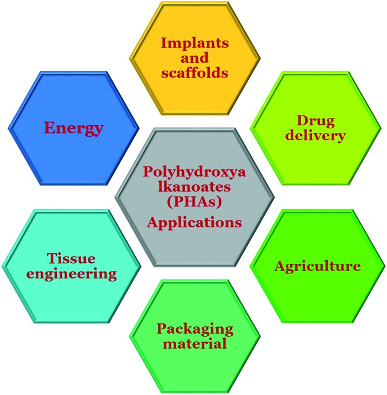
Polyhydroxyalkanoates (PHAs) are biopolymers synthesized by bacteria under unbalanced growth conditions. These biopolymers are considered as potential biomaterials for future applications because they are biocompatible, biodegradable, and easy to produce and functionalize with strong mechanical strength. Currently, PHAs are being extensively innovated for biomedical applications due to their prerequisite properties. The wide range of biomedical applications includes drug delivery systems, implants, tissue engineering, scaffolds, artificial organ constructs, etc. In this article we review the utility of PHAs in various forms (bulk/nano) for biomedical applications so as to bring about the future vision for PHAs as biomaterials for the advancement of research and technology.
Figures


Similar articles
-
Biomedical applications of microbially engineered polyhydroxyalkanoates: an insight into recent advances, bottlenecks, and solutions.Appl Microbiol Biotechnol. 2019 Mar;103(5):2007-2032. doi: 10.1007/s00253-018-09604-y. Epub 2019 Jan 15. Appl Microbiol Biotechnol. 2019. PMID: 30645689 Review.
-
Review of Hybrid Materials Based on Polyhydroxyalkanoates for Tissue Engineering Applications.Polymers (Basel). 2021 May 26;13(11):1738. doi: 10.3390/polym13111738. Polymers (Basel). 2021. PMID: 34073335 Free PMC article. Review.
-
Exploiting Polyhydroxyalkanoates for Biomedical Applications.Polymers (Basel). 2023 Apr 19;15(8):1937. doi: 10.3390/polym15081937. Polymers (Basel). 2023. PMID: 37112084 Free PMC article. Review.
-
Additive manufacturing of polyhydroxyalkanoates (PHAs) biopolymers: Materials, printing techniques, and applications.Mater Sci Eng C Mater Biol Appl. 2021 Aug;127:112216. doi: 10.1016/j.msec.2021.112216. Epub 2021 May 29. Mater Sci Eng C Mater Biol Appl. 2021. PMID: 34225868 Review.
-
Polyhydroxyalkanoates (PHAs) as Biomaterials in Tissue Engineering: Production, Isolation, Characterization.Materials (Basel). 2022 Feb 14;15(4):1410. doi: 10.3390/ma15041410. Materials (Basel). 2022. PMID: 35207952 Free PMC article. Review.
Cited by
-
Genotypic and Phenotypic Detection of Polyhydroxyalkanoate Production in Bacterial Isolates from Food.Int J Mol Sci. 2023 Jan 8;24(2):1250. doi: 10.3390/ijms24021250. Int J Mol Sci. 2023. PMID: 36674766 Free PMC article.
-
Exploring Schwann Cell Behavior on Electrospun Polyhydroxybutyrate Scaffolds with Varied Pore Sizes and Fiber Thicknesses: Implications for Neural Tissue Engineering.Polymers (Basel). 2023 Dec 6;15(24):4625. doi: 10.3390/polym15244625. Polymers (Basel). 2023. PMID: 38139877 Free PMC article.
-
The Synthesis, Characterization and Applications of Polyhydroxyalkanoates (PHAs) and PHA-Based Nanoparticles.Polymers (Basel). 2021 Sep 27;13(19):3302. doi: 10.3390/polym13193302. Polymers (Basel). 2021. PMID: 34641118 Free PMC article. Review.
-
Active biodegradable packaging films modified with grape seeds lignin.RSC Adv. 2020 Aug 7;10(49):29202-29213. doi: 10.1039/d0ra04074f. eCollection 2020 Aug 5. RSC Adv. 2020. PMID: 35521111 Free PMC article.
-
Predicting the Mechanical Response of Polyhydroxyalkanoate Biopolymers Using Molecular Dynamics Simulations.Polymers (Basel). 2022 Jan 17;14(2):345. doi: 10.3390/polym14020345. Polymers (Basel). 2022. PMID: 35054751 Free PMC article.
References
-
- Quirino R. L., Garrison T. F., Kessler M. R. Green Chem. 2014;16:1700–1715.
-
- Khanna S., Srivastava A. K. Process Biochem. 2005;40:607–619.
-
- Liu H., Jiang N., Mao H., Wang Z. Gongcheng Suliao Yingyong. 2014;42:131–134.
-
- Li Z. B., Loh X. J. Wiley Interdiscip. Rev.: Nanomed. Nanobiotechnol. 2017;9(3) - PubMed
-
- Tomizawa S., Hyakutake M., Saito Y., Agus J., Mizuno K., Abe H., Tsuge T. Biomacromolecules. 2011;12:2660–2666. - PubMed
Publication types
LinkOut - more resources
Full Text Sources
Other Literature Sources