. 2017 Jul 7;8(9):1788-1796.
doi: 10.1039/c7md00311k. eCollection 2017 Sep 1.

MAO inhibitory activity of bromo-2-phenylbenzofurans: synthesis, in vitro study, and docking calculations

Affiliations

MAO inhibitory activity of bromo-2-phenylbenzofurans: synthesis, in vitro study, and docking calculations

G L Delogu et al. Medchemcomm. .

Abstract

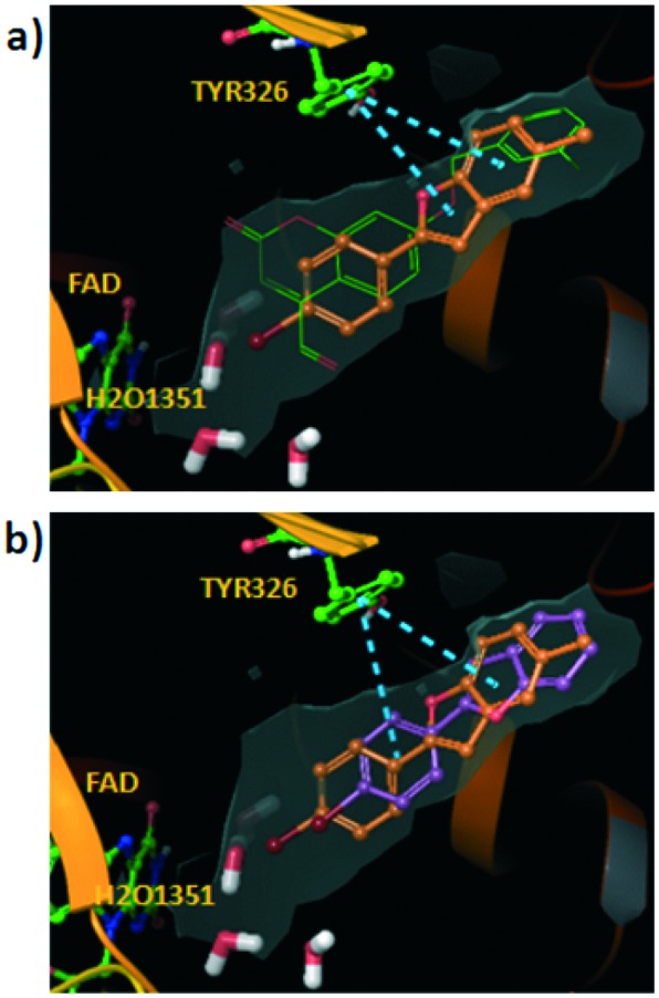
Monoamine oxidase (MAO) is an enzyme responsible for metabolism of monoamine neurotransmitters which play an important role in brain development and function. This enzyme exists in two isoforms, and it has been demonstrated that MAO-B activity, but not MAO-A activity, increases with aging. MAO inhibitors show clinical value because besides the monoamine level regulation they reduce the formation of by-products of the MAO catalytic cycle, which are toxic to the brain. A series of 2-phenylbenzofuran derivatives was designed, synthesized and evaluated against hMAO-A and hMAO-B enzymes. A bromine substituent was introduced in the 2-phenyl ring, whereas position 5 or 7 of the benzofuran moiety was substituted with a methyl group. Most of the tested compounds inhibited preferentially MAO-B in a reversible manner, with IC50 values in the low micro or nanomolar range. The 2-(2'-bromophenyl)-5-methylbenzofuran (5) was the most active compound identified (IC50 = 0.20 μM). In addition, none of the studied compounds showed cytotoxic activity against the human neuroblastoma cell line SH-SY5Y. Molecular docking simulations were used to explain the observed hMAO-B structure-activity relationship for this type of compounds.


Figures

Scheme 1
Scheme 1. Synthesis of 2-phenylbenzofuran derivatives via a Wittig reaction. Reagents and conditions: a) NaBH4, EtOH, 0 °C to rt, 2 h; b) PPh3 HBr, CH3CN, 82 °C, 2 h; c) toluene, Et3N, 110 °C, 2 h.
Fig. 1
Fig. 1. Cytotoxic activity after 24 h incubation with compounds 1–12 at 1 μM concentration (1a) or 10 μM concentration (1b) on SH-SY5Y cells. Cell viability was measured as MTT reduction and data were normalized as % of control treated with 1% DMSO. Results are expressed as mean ± S.E.M from at least 5 different cultures. No statistical differences were found with the control group using the one-way ANOVA followed by Dunnett's test.
Fig. 2
Fig. 2. Hypothetical binding modes for the studied compounds extracted from the docking in the hMAO-B. The co-crystallized coumarin c17 (; 2V60) is also shown for comparative purposes (green carbons). The hydrophobic surface in the pocket is represented in grey color. π–π stacking interactions with residue Tyr326 are represented in blue dashed lines. Ribbons in the protein are partially omitted for clarity. a) Binding mode for compound 3 (grey carbons). b) Pose determined for compound 1 (pink carbons). c) Pose extracted from docking for compound 5 (turquoise carbons). d) Residue interaction contributions for the binding with compound 5 (sum of Coulomb, van der Waals and hydrogen bond energies).
Fig. 3
Fig. 3. Chemical structure of c17 (7-(3-chlorobenzyloxy)-4-carboxaldehydecoumarin).
Fig. 4
Fig. 4. a) Hypothetical binding mode calculated for compound 7 inside the hMAO-B (the co-crystallized coumarin c17 in green color is shown for comparative purposes. Blue dashed lines represent π–π stacking interactions). However, for this binding mode to occur, compound 7 should displace the water molecule HOH1351 present in the crystallized hMAO-B protein. This fact could negatively affect the complex formation. Although different water molecules in the pocket are shown, the pose was extracted from docking performed using protocol 1 with only the water molecule HOH1159 retained in the pocket. b) Comparison between the poses determined by docking for compounds 7 and 11. Compound 11 is shifted towards the hydrophobic entrance cavity and the bromine substituent is farther from the water molecules placed in the catalytic site.

References

    1. Ramsay R. R. Curr. Top. Med. Chem. 2012;12:2189. - PubMed
    1. Finberg J. P. Pharmacol. Ther. 2014;143:133. - PubMed
    1. Fowler J. S., Volkow N. D., Wang G. J., Logan J., Pappas N., Shea C., MacGregor R. Neurobiol. Aging. 1997;18:431. - PubMed
    1. Naoi M., Maruyama W. Curr. Pharm. Des. 2010;16:2799. - PubMed
    1. Cohen G. J. Neural Transm., Suppl. 1990;32:229. - PubMed

LinkOut - more resources