. 2017 Jul 13;8(9):1810-1816.
doi: 10.1039/c7md00193b. eCollection 2017 Sep 1.

Synthesis and biological evaluation of chalcone-linked pyrazolo[1,5- a]pyrimidines as potential anticancer agents

Affiliations

Synthesis and biological evaluation of chalcone-linked pyrazolo[1,5- a]pyrimidines as potential anticancer agents

Chandrakant Bagul et al. Medchemcomm. .

Abstract

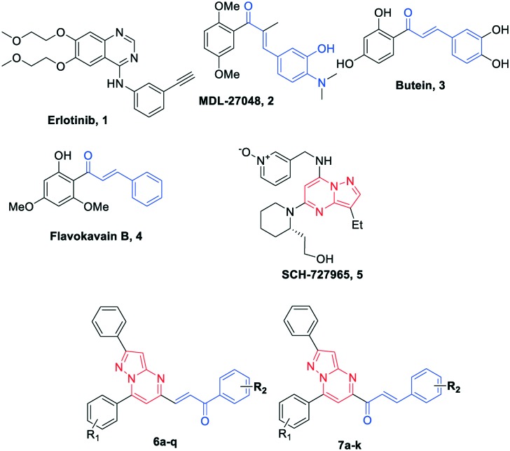
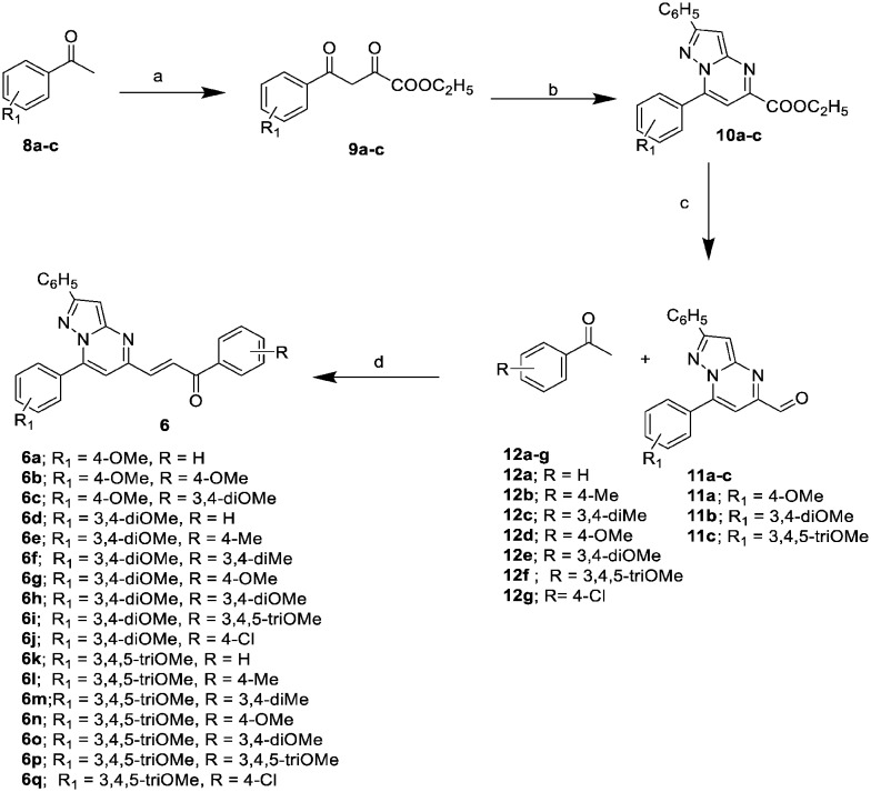
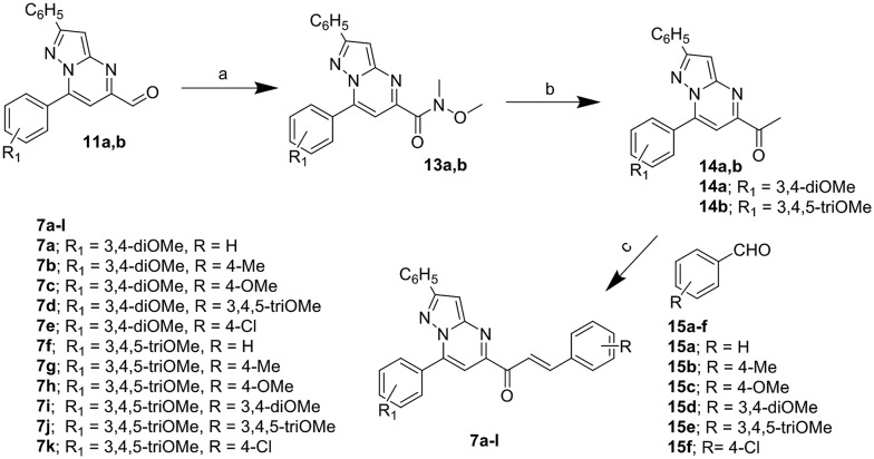
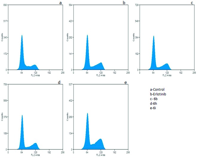
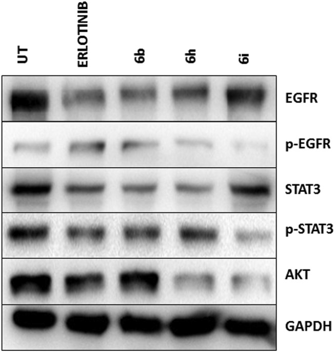
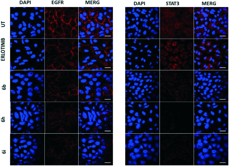
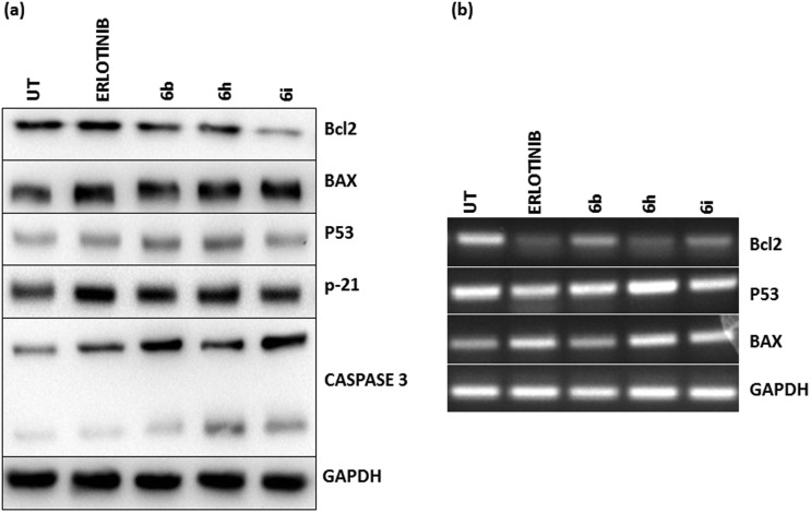
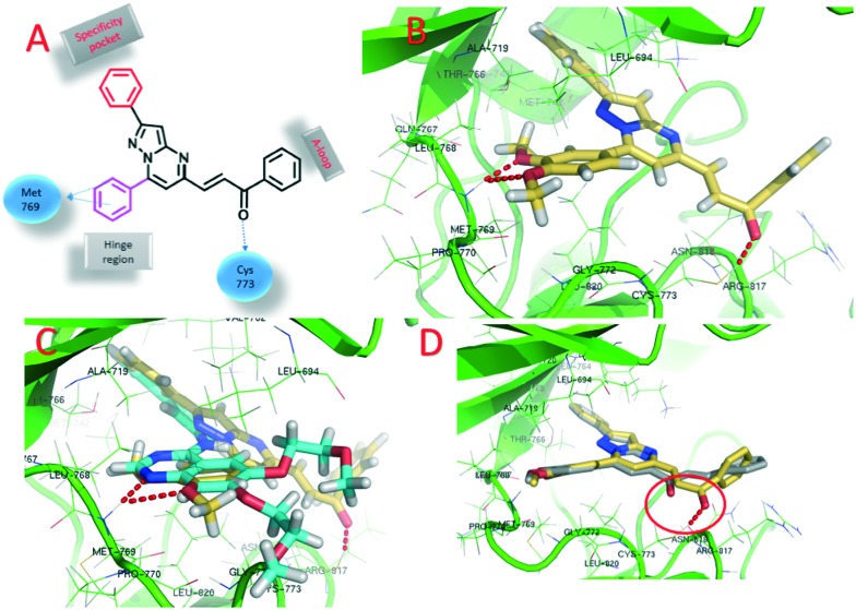
A series of pyrazolo[1,5-a]pyrimidines substituted at C5 with 1-phenylprop-2-en-1-one (6a-q) and 3-phenylprop-2-en-1-one (7a-k) was synthesized and evaluated for antiproliferative activity. Among them, 6h was found to be the most active compound against the MDA-MB-231 cell line with an IC50 of 2.6 μM . The antiproliferative activity of this series of compounds ranged from 2.6 to 34.9 μM against A549 (lung cancer), MDA-MB-231 (breast cancer) and DU-145 (prostate cancer) cell lines. FACS analysis revealed that these hybrids arrest the cell cycle at the subG1 phase. Western blot analysis and an immunofluorescence assay showed the inhibition of the EGFR and STAT3 axis, which plays an important role in cell survival and apoptosis. Western blot and RT-PCR analyses that displayed an increase in apoptotic proteins such as p53, p21 and Bax and a decrease in the anti-apoptotic proteins Bcl-2 and procaspase-9 confirmed the ability of these hybrids to trigger cell death by apoptosis. Molecular docking studies described the binding of these hybrids to the ATP binding site of EGFR.


Figures

Fig. 1
Fig. 1. Chemical structures of EGFR inhibitors: 1 (erlotinib), chalcone scaffold containing compounds 2 (MDL-27048), 3 (butein) and 4 (flavokavain B), pyrazolo[1,5-a]pyrimidine containing scaffold compound 5 (SCH-727965) and designed chalcone-linked pyrazolo[1,5-a]pyrimidines (6a–q and 7a–k).
Scheme 1
Scheme 1. Synthesis of pyrazolo[1,5-a]pyrimidines (6a–q); reagents and conditions: a) diethyl oxalate, NaOEt, EtOH, rt, 12 h, (yield 80–85%); b) 3-amino-5-phenyl-pyrazole, conc. HCl (cat.), EtOH, reflux, 2–4 h, (yield 90–95%); c) DIBAL-H, CH2Cl2, –78 °C, 2 h, (yield 78–80%); d) Ba(OH)2, MeOH, rt, 4–6 h, (yield 71–91%).
Scheme 2
Scheme 2. Synthesis of pyrazolo[1,5-a]pyrimidines (7a–k); reagents and conditions: a) N,O-dimethylhydroxylamine hydrochloride, Al(CH3)3, dry dichloromethane, 0–25 °C, 2 h, (yield 77–79%); b) methylmagnesium bromide, dry tetrahydrofuran, 0 °C, 1 h, (yield 85–92%); c) barium hydroxide, methanol, rt, 6 h, (yield 79–92%).
Fig. 2
Fig. 2. Flow cytometric analysis displaying G2/M arrest in cells post-treatment. A549 cells subjected to compound treatment for 24 h at 2 μM concentration were collected, fixed and stained for FACS analysis. Untreated cells were employed as a negative control.
Fig. 3
Fig. 3. Identification of the affected signaling pathway using western blot analysis. A549 cells were treated with 6b, 6h and 6i at 2 μM final concentration and were collected 24 h post-treatment. The total protein lysate was isolated and analyzed by western blotting using the corresponding antibody. Compounds 6b and 6h revealed significantly reduced levels of p-EGFR, p-STAT3 and AKT. Erlotinib was utilized as a positive control, whereas untreated cells were considered as a negative control.
Fig. 4
Fig. 4. Regulation of EGFR and STAT3 in cells 24 h post-treatment. A549 cells were fixed, stained and observed for 24 h post-treatment with compounds 6b, 6h and 6i at a 2 μM final concentration under a confocal microscope (FLOW VIEW FV 1000 series). The Cy3 dye was used to stain both EGFR and STAT3, whereas DAPI was used as a nuclear stain. Compounds 6h and 6i showed profound inhibitory effects on the levels of EGFR and STAT3.
Fig. 5
Fig. 5. Analysis of pro-apoptotic and anti-apoptotic proteins 24 h post-treatment. (a) The total lysate was isolated from A549 cells after 24 h of treatment with compounds 6b, 6h and 6i at 2 μM final concentrations and was analyzed by western blotting using a corresponding antibody. Compounds 6b and 6i show obvious up-regulation of apoptotic proteins including Bax, p53, p21 and caspase-3, whereas down-regulation of the anti-apoptotic protein Bcl2 was observed. Erlotinib was employed as a positive control and untreated cells were used as a negative control. (b) RNA was isolated from cells using the Trizol reagent and cDNA was synthesized. End point RT-PCR results showed p53 and Bax upregulation in cells treated with compounds 6b and 6h and 6i and a significant reduction in the levels of anti-apoptotic protein Bcl2.
Fig. 6
Fig. 6. Docking poses on EGFR. A) 2D structure of a chalcone-linked pyrazolo[1,5-a]pyrimidine with observed interactions with EGFR; B) binding pose of 6j (yellow color, EGFR is shown in green and hydrogen bonds are shown by the red dotted lines); C) superimposed pose of 6j and erlotinib; D) superimposed pose of 6j and 7a showing the difference in hydrogen bonding between the carbonyl carbons and Cys773.

Similar articles

Cited by

References

    1. Torre L. A., Bray F., Siegel R. L., Ferlay J., Lortet-Tieulent J., Jemal A. Ca-Cancer J. Clin. 2015;65:87–108. - PubMed
    1. Bray F., Jemal A., Grey N., Ferlay J., Forman D. Lancet Oncol. 2012;13:790–801. - PubMed
    1. Zugazagoitia J., Guedes C., Ponce S., Ferrer I., Molina-Pinelo S., Paz-Ares L. Clin. Ther. 2016;38:1551–1566. - PubMed
    1. Biemar F., Foti M. Cancer Biol. Med. 2013;10:183. - PMC - PubMed
    1. Backes A., Zech B., Felber B., Klebl B., Müller G. Expert Opin. Drug Discovery. 2008;3:1427–1449. - PubMed