Review
. 2017 Nov 22;9(1):27-43.
doi: 10.1039/c7md00474e. eCollection 2018 Jan 1.

Synthesis and biological evaluation of rapamycin-derived, next generation small molecules

Affiliations
Review

Synthesis and biological evaluation of rapamycin-derived, next generation small molecules

Shiva Krishna Reddy Guduru et al. Medchemcomm. .

Abstract

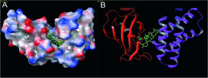
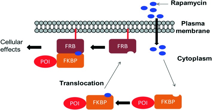
Over the years, rapamycin has attracted serious attention due to its remarkable biological properties and as a potent inhibitor of the mammalian target of rapamycin (mTOR) protein through its binding with FKBP-12. Several efficient strategies that utilize synthetic and biosynthetic approaches have been utilized to develop small molecule rapamycin analogs or for synthesizing hybrid compounds containing a partial rapamycin structure to improve pharmacokinetic properties. Herein, we report selected case studies related to the synthesis of rapamycin-derived compounds and hybrid molecules to explore their biological properties.


Figures

Fig. 1
Fig. 1. Structure of rapamycin and its analogs.
Fig. 2
Fig. 2. (a) mTOR domain structure: mTOR consists of tandem HEAT repeats, a central FAT domain, a FRB domain, a catalytic kinase domain and a FATC domain. Rapamycin binds with its intracellular receptor, FKBP12, and the resulting complex then binds with the FRB domain of mTOR. The binding of rapamycin–FKBP12 to the FRB domain disrupts the association of mTOR with the mTORC1 specific component, Raptor, and thus decouples mTORC1 from its substrates, thereby blocking mTORC1 signaling. (b) mTORC1 and mTORC2 composition and regulation: mTORC1 contains mTOR, Raptor, PRAS40, mLST8 (also known as GβL) and Deptor which regulate cell growth through S6K and 4E-BP1 effectors. mLST8 attaches to the mTOR kinase domain in both complexes, where it seems to be essential for their assembly. Deptor acts as an inhibitor of both mTORC1 and mTORC2 complexes. Other protein partners differ between these two complexes. mTORC2 contains Protor, Sin1 and Rictor which regulate the prosurvival kinase Akt/PKB and PDK1.
Fig. 3
Fig. 3. The structure of a ternary complex of FKBP12–rapamycin–FRB: (A) surface model and (B) ribbon model; red color – FKBP12, green color (small molecule) – rapamycin and purple color – FRB (PDB code: 1NSG). Molecular model images were generated using the Maestro v9.6 software package (Schrodinger LLC).
Scheme 1
Scheme 1. Nicolaou's approach to rapamycin synthesis.
Scheme 2
Scheme 2. Schreiber's approach to rapamycin and C32-(S)-dihydrorapamycin synthesis.
Scheme 3
Scheme 3. Danishefsky's approach to the total synthesis of rapamycin.
Scheme 4
Scheme 4. Smith's approach to the total synthesis of rapamycin.
Scheme 5
Scheme 5. Ley's approach to rapamycin synthesis.
Scheme 6
Scheme 6. Rapamycin structure, 20-thiarapamycin and 15-deoxo-19-sulfoxylrapamycin.
Scheme 7
Scheme 7. (a) Re-establishment of pre-rapamycin 7.2 production in S. hygroscopicus MG2-10 by feeding the pseudostarter unit 4.1. (b and c) Exclusive production of 7.4–7.8 as analogs of pre-rapamycin by feeding cyclohexanecarboxylic acid, cyclohex-1-enecarboxylic acid, and cycloheptanecarboxylic acid to S. hygroscopicus MG2-10.
Scheme 8
Scheme 8. The corresponding pre-rapamycin analogs obtained by a mutasynthesis approach.
Scheme 9
Scheme 9. Molecular design and retrosynthetic analysis of FKBP12 ligands RAP-P (9.5–9.7).
Fig. 4
Fig. 4. Schematic representation of CID experiment in living cells: one protein component (here FRB) anchoring on a plasma membrane at the desired site of the protein of action takes place. The other protein component (FKBP) is attached to the protein of interest (POI). In the absence of a dimerizer (rapamycin), the POI–FKBP complex freely diffuses into the cytoplasm. Upon addition of a dimerizer (rapamycin) to the cells, it passes through the plasma membrane and first binds to FKBP forming the FKBP–rapamycin complex. Then, the FKBP–rapamycin complex moves towards the plasma membrane along with the POI and binds to FRB to form the FKBP–rapamycin–FRB complex. POI translocation to the desired functional site (in this case the plasma membrane) induces or inhibits cellular function.
Scheme 10
Scheme 10. C16-substituted derivatives of rapamycin.
Scheme 11
Scheme 11. Solid-phase synthesis of rapalogs (11a–n).
Scheme 12
Scheme 12. Arya's approach to rapamycin fragment-derived macrocycles 12.1.
Scheme 13
Scheme 13. Synthesis of rapamycin fragment-derived macrocycles 12.1(a–d).

Similar articles

Cited by

References

    1. Sehgal S. N., Baker H., Vezina C. J. Antibiot. 1975;28:727–732. - PubMed
    1. Vezina C., Kudelski A., Sehgal S. N. J. Antibiot. 1975;28:721–726. - PubMed
    1. Benjamin D., Colombi M., Moroni C., Hall M. N. Nat. Rev. Drug Discovery. 2011;10:868–880. - PubMed
    1. Schmelzle T., Hall M. N. Cell. 2000;103:253–262. - PubMed
    1. Yang H., Rudge D. G., Koos J. D., Vaidialingam B., Yang H. J., Pavletich N. P. Nature. 2013;497:217–223. - PMC - PubMed

LinkOut - more resources