

Impact of the springtail's cuticle nanotopography on bioadhesion and biofilm formation in vitro and in the oral cavity
- PMID: 30109045
- PMCID: PMC6083677
- DOI: 10.1098/rsos.171742
Impact of the springtail's cuticle nanotopography on bioadhesion and biofilm formation in vitro and in the oral cavity
Abstract
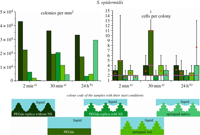
Springtails (Collembola) have a nanostructured cuticle. To evaluate and to understand anti-biofouling properties of springtail cuticles' morphology under different conditions, springtails, shed cuticles and cuticle replicates were studied after incubation with protein solutions and bacterial cultures using common in vitro models. In a second step, they were exposed to human oral environment in situ in order to explore potential application in dentistry. In vitro, the cuticular structures were found to resist wetting by albumin solutions for up to 3 h and colonization by Staphylococcus epidermidis was inhibited. When exposed in the oral cavity, initial pellicle formation was of high heterogeneity: parts of the surface were coated by adsorbed proteins, others remained uncoated but exhibited locally attached, 'bridging', proteinaceous membranes spanning across cavities of the cuticle surface; this unique phenomenon was observed for the first time. Also the degree of bacterial colonization varied considerably. In conclusion, the springtail cuticle partially modulates bioadhesion in the oral cavity in a unique and specific manner, but it has no universal effect. Especially after longer exposure, the nanotextured surface of springtails is masked by the pellicle, resulting in subsequent bacterial colonization, and, thus, cannot effectively avoid bioadhesion in the oral cavity comprehensively. Nevertheless, the observed phenomena offer valuable information and new perspectives for the development of antifouling surfaces applicable in the oral cavity.
Keywords: bioadhesion; collembolan; hexapods; pellicle; saliva; springtail.
Conflict of interest statement
We declare we have no competing interests.
Figures


Similar articles
-
Bioadhesion in the oral cavity and approaches for biofilm management by surface modifications.Clin Oral Investig. 2020 Dec;24(12):4237-4260. doi: 10.1007/s00784-020-03646-1. Epub 2020 Oct 27. Clin Oral Investig. 2020. PMID: 33111157 Free PMC article. Review.
-
Bioadhesion on Textured Interfaces in the Human Oral Cavity-An In Situ Study.Int J Mol Sci. 2022 Jan 21;23(3):1157. doi: 10.3390/ijms23031157. Int J Mol Sci. 2022. PMID: 35163081 Free PMC article.
-
The oral cavity--a key system to understand substratum-dependent bioadhesion on solid surfaces in man.Clin Oral Investig. 2009 Jun;13(2):123-39. doi: 10.1007/s00784-008-0243-3. Epub 2009 Jan 10. Clin Oral Investig. 2009. PMID: 19137331 Review.
-
Collembola cuticles and the three-phase line tension.Beilstein J Nanotechnol. 2017 Aug 18;8:1714-1722. doi: 10.3762/bjnano.8.172. eCollection 2017. Beilstein J Nanotechnol. 2017. PMID: 28875109 Free PMC article.
-
Increase in egg resistance to desiccation in springtails correlates with blastodermal cuticle formation: Eco-evolutionary implications for insect terrestrialization.J Exp Zool B Mol Dev Evol. 2021 Dec;336(8):606-619. doi: 10.1002/jez.b.22979. Epub 2020 Jul 10. J Exp Zool B Mol Dev Evol. 2021. PMID: 32649025
Cited by
-
Recent Progress on Bioinspired Antibacterial Surfaces for Biomedical Application.Biomimetics (Basel). 2022 Jul 4;7(3):88. doi: 10.3390/biomimetics7030088. Biomimetics (Basel). 2022. PMID: 35892358 Free PMC article. Review.
-
Quantification of the Adhesion Strength of Candida albicans to Tooth Enamel.Microorganisms. 2021 Oct 25;9(11):2213. doi: 10.3390/microorganisms9112213. Microorganisms. 2021. PMID: 34835339 Free PMC article.
-
Bioadhesion in the oral cavity and approaches for biofilm management by surface modifications.Clin Oral Investig. 2020 Dec;24(12):4237-4260. doi: 10.1007/s00784-020-03646-1. Epub 2020 Oct 27. Clin Oral Investig. 2020. PMID: 33111157 Free PMC article. Review.
-
Bioadhesion on Textured Interfaces in the Human Oral Cavity-An In Situ Study.Int J Mol Sci. 2022 Jan 21;23(3):1157. doi: 10.3390/ijms23031157. Int J Mol Sci. 2022. PMID: 35163081 Free PMC article.
-
Viability of Salmonella Typhimurium biofilms on major food-contact surfaces and eggshell treated during 35 days with and without water storage at room temperature.Poult Sci. 2020 Sep;99(9):4558-4565. doi: 10.1016/j.psj.2020.05.055. Epub 2020 Jun 26. Poult Sci. 2020. PMID: 32868000 Free PMC article.
References
-
- Dunger W. 2008. Tiere im boden (animals in soil). Hohe Börde, Germany: Westarp Wissenschaften.
-
- Bellinger PF, Christiansen KA, Janssens F. 2016. http://www.collembola.org.
-
- Hensel R, Helbig R, Aland S, Voigt A, Neinhuis C, Werner C. 2013. Tunable nano-replication to explore the omniphobic characteristics of springtail skin. Npg Asia Mater. 5, e37 (10.1038/am.2012.66) - DOI
Associated data
LinkOut - more resources
Full Text Sources
Other Literature Sources
